Myofibroblast

Last updated
Myofibroblast
Testicle-histology-boar.jpg
Histological section through testicular parenchyma of a boar. 1 Lumen of tubulus seminiferus contortus, 2 spermatids, 3 spermatocytes, 4 spermatogonia, 5 Sertoli cell, 6 Myofibroblasts, 7 Leydig cells, 8 capillaries
Details
Identifiers
Latin myofibroblastus
MeSH D058628
TH H2.00.03.0.01013
Anatomical terms of microanatomy

A myofibroblast is a cell phenotype that was first described as being in a state between a fibroblast and a smooth muscle cell.

Contents

Structure

Myofibroblasts are contractile web-like fusiform cells that are identifiable by their expression of α-smooth muscle actin within their cytoplasmic stress fibers. [1]

In the gastrointestinal and genitourinary tracts, myofibroblasts are found subepithelially in mucosal surfaces. Here they not only act as a regulator of the shape of the crypts and villi, but also act as stem-niche cells in the intestinal crypts and as parts of atypical antigen-presenting cells. They have both support as well as paracrine function in most places.

Location

Myofibroblasts were first identified in granulation tissue during skin wound healing. [2] Typically, these cells are found in granulation tissue, scar tissue (fibrosis) and the stroma of tumours. They also line the gastrointestinal tract, wherein they regulate the shapes of crypts and villi.

Markers

Myofibroblasts usually stain for the intermediate filament vimentin, which is a general mesenchymal marker, α-smooth muscle actin (human gene = ACTA2 ), and for palladin, which is a cytoskeletal actin scaffold protein. They are positive for other smooth muscle markers, such as intermediate filament type desmin in some tissues, but may be negative for desmin in other tissues. Similar heterogeneous positivity may exist for almost every smooth muscle marker except probably a few which are positive only in contractile smooth muscles like metavinculin and smoothelin.

Myofibroblasts upregulate the expression of fibronectin, collagens, and hyaluronic acid during and after their differentiation from fibroblasts. Among these, the EDA isoform of fibronectin (EDA-FN), and collagen type I (COL1A1/COL1A2) are typical markers of myofibroblast-dependent synthesis of pro-fibrotic extracellular matrix.

Some myofibroblasts (especially if they have a stellate form) may also be positive for GFAP.

Development

There are many possible ways of myofibroblast development:

  1. Partial smooth muscle differentiation of a fibroblastic cell
  2. Activation of a stellate cell (e.g. hepatic Ito cells or pancreatic stellate cells).
  3. Loss of contractile phenotype (or acquisition of "synthetic phenotype") of a smooth muscle cell.
  4. Direct myofibroblastic differentiation of a progenitor cell resident in a stromal tissue.
  5. Homing and recruitment of a circulating mesenchymal precursor which can directly differentiate as above or indirectly differentiate through the other cell types as intermediates.
  6. Epithelial to mesenchymal transdifferentiation (EMT) of an epithelial cell.

Perhaps the most studied pathway of myofibroblast formation is TGF-beta1 dependent differentiation from fibroblast cells. Activation of the TGF-beta receptor 1 and TGF-beta receptor 2 leads to induction of the canonical SMAD2/SMAD3 pathway. [3] Together with the co-activation of the non-canonical EGFR pathway, these events lead to upregulation of the ACTA2 gene and subsequent alpha smooth muscle actin protein production. Several regulators of the myofibroblast differentiation pathway have been described, including hyaluronan and CD44 co-receptor activation of EGFR. [4]

Primary culture of cardiac fibroblasts stimulated with TGF-beta to differentiate them to myofibroblasts. Images taken at different post-stimulus times. Diferenciacion de fibroblastos a miofibroblastos cardiacos.jpg
Primary culture of cardiac fibroblasts stimulated with TGF-beta to differentiate them to myofibroblasts. Images taken at different post-stimulus times.

Function

In many organs like liver, lung, and kidneys, they are primarily involved in fibrosis. In the wound tissue they are implicated in wound strengthening by extracellular collagen fiber deposition and then wound contraction by intracellular contraction and concomitant alignment of the collagen fibers by integrin-mediated pulling on to the collagen bundles. Pericytes and renal mesangial cells are some examples of modified myofibroblast-like cells.

Myofibroblasts may interfere with the propagation of electrical signals [5] controlling heart rhythm, [6] leading to arrhythmia in both patients who have suffered a heart attack and in foetuses. Ursodiol is a promising drug for this condition. [7]

Wound healing

Myofibroblasts can contract by using smooth muscle type actin-myosin complex, rich in a form of actin called alpha-smooth muscle actin. These cells are then capable of speeding wound repair by contracting the edges of the wound.

Early work on wound healing showed that granulation tissue taken from a wound could contract in vitro (or in an organ bath) in a similar fashion to smooth muscle, when exposed to substances that cause smooth muscle to contract, such as adrenaline or angiotensin.

More recently it has been shown that fibroblasts can transform into myofibroblasts with photobiomodulation.

After healing is complete, these cells are lost through apoptosis and it has been suggested that in several fibrotic diseases (for example liver cirrhosis, kidney fibrosis, retroperitoneal fibrosis) that this mechanism fails to work, leading to persistence of the myofibroblasts, and consequently expansion of the extracellular matrix (fibrosis) with contraction.

Similarly, in wounds that fail to resolve and become keloids or hypertrophic scars, myofibroblasts may persist, rather than disappearing by apoptosis. [8]

See also

Related Research Articles

<span class="mw-page-title-main">Scar</span> Area of fibrous tissue that replaces normal skin after an injury

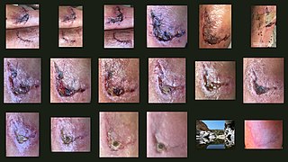
A scar is an area of fibrous tissue that replaces normal skin after an injury. Scars result from the biological process of wound repair in the skin, as well as in other organs, and tissues of the body. Thus, scarring is a natural part of the healing process. With the exception of very minor lesions, every wound results in some degree of scarring. An exception to this are animals with complete regeneration, which regrow tissue without scar formation.

<span class="mw-page-title-main">Extracellular matrix</span> Network of proteins and molecules outside cells that provides structural support for cells

In biology, the extracellular matrix (ECM), also called intercellular matrix (ICM), is a network consisting of extracellular macromolecules and minerals, such as collagen, enzymes, glycoproteins and hydroxyapatite that provide structural and biochemical support to surrounding cells. Because multicellularity evolved independently in different multicellular lineages, the composition of ECM varies between multicellular structures; however, cell adhesion, cell-to-cell communication and differentiation are common functions of the ECM.

<span class="mw-page-title-main">Healing</span> Process of the restoration of health

With physical trauma or disease suffered by an organism, healing involves the repairing of damaged tissue(s), organs and the biological system as a whole and resumption of (normal) functioning. Medicine includes the process by which the cells in the body regenerate and repair to reduce the size of a damaged or necrotic area and replace it with new living tissue. The replacement can happen in two ways: by regeneration in which the necrotic cells are replaced by new cells that form "like" tissue as was originally there; or by repair in which injured tissue is replaced with scar tissue. Most organs will heal using a mixture of both mechanisms.

<span class="mw-page-title-main">Smooth muscle</span> Involuntary non-striated muscle

Smooth(soft) muscle is an involuntary non-striated muscle, so-called because it has no sarcomeres and therefore no striations. It is divided into two subgroups, single-unit and multiunit smooth muscle. Within single-unit muscle, the whole bundle or sheet of smooth muscle cells contracts as a syncytium.

<span class="mw-page-title-main">Wound healing</span> Series of events that restore integrity to damaged tissue after an injury

Wound healing refers to a living organism's replacement of destroyed or damaged tissue by newly produced tissue.

Mesangial cells are specialised cells in the kidney that make up the mesangium of the glomerulus. Together with the mesangial matrix, they form the vascular pole of the renal corpuscle. The mesangial cell population accounts for approximately 30-40% of the total cells in the glomerulus. Mesangial cells can be categorized as either extraglomerular mesangial cells or intraglomerular mesangial cells, based on their relative location to the glomerulus. The extraglomerular mesangial cells are found between the afferent and efferent arterioles towards the vascular pole of the glomerulus. The extraglomerular mesangial cells are adjacent to the intraglomerular mesangial cells that are located inside the glomerulus and in between the capillaries. The primary function of mesangial cells is to remove trapped residues and aggregated protein from the basement membrane thus keeping the filter free of debris. The contractile properties of mesangial cells have been shown to be insignificant in changing the filtration pressure of the glomerulus.

<span class="mw-page-title-main">Fibrosis</span> Excess connective tissue in healing

Fibrosis, also known as fibrotic scarring, is a pathological wound healing in which connective tissue replaces normal parenchymal tissue to the extent that it goes unchecked, leading to considerable tissue remodelling and the formation of permanent scar tissue.

<span class="mw-page-title-main">Platelet-derived growth factor</span> Signaling glycoprotein regulating cell proliferation

Platelet-derived growth factor (PDGF) is one among numerous growth factors that regulate cell growth and division. In particular, PDGF plays a significant role in blood vessel formation, the growth of blood vessels from already-existing blood vessel tissue, mitogenesis, i.e. proliferation, of mesenchymal cells such as fibroblasts, osteoblasts, tenocytes, vascular smooth muscle cells and mesenchymal stem cells as well as chemotaxis, the directed migration, of mesenchymal cells. Platelet-derived growth factor is a dimeric glycoprotein that can be composed of two A subunits (PDGF-AA), two B subunits (PDGF-BB), or one of each (PDGF-AB).

Haptotaxis is the directional motility or outgrowth of cells, e.g. in the case of axonal outgrowth, usually up a gradient of cellular adhesion sites or substrate-bound chemoattractants. These gradients are naturally present in the extracellular matrix (ECM) of the body during processes such as angiogenesis or artificially present in biomaterials where gradients are established by altering the concentration of adhesion sites on a polymer substrate.

Durotaxis is a form of cell migration in which cells are guided by rigidity gradients, which arise from differential structural properties of the extracellular matrix (ECM). Most normal cells migrate up rigidity gradients.

A fibrocyte is an inactive mesenchymal cell, that is, a cell showing minimal cytoplasm, limited amounts of rough endoplasmic reticulum and lacks biochemical evidence of protein synthesis.

<span class="mw-page-title-main">Hepatic stellate cell</span>

Hepatic stellate cells (HSC), also known as perisinusoidal cells or Ito cells, are pericytes found in the perisinusoidal space of the liver, also known as the space of Disse. The stellate cell is the major cell type involved in liver fibrosis, which is the formation of scar tissue in response to liver damage, in addition these cells store and concentrate vitamin A.

<span class="mw-page-title-main">ACTA2</span> Protein-coding gene in the species Homo sapiens

ACTA2 is an actin protein with several aliases including alpha-actin, alpha-actin-2,aortic smooth muscle or alpha smooth muscle actin. Actins are a family of globular multi-functional proteins that form microfilaments. ACTA2 is one of 6 different actin isoforms and is involved in the contractile apparatus of smooth muscle. ACTA2 is extremely highly conserved and found in nearly all mammals.

<span class="mw-page-title-main">Integrin alpha 11</span> Protein-coding gene in the species Homo sapiens

Integrin alpha-11 is a protein that, in humans, is encoded by the ITGA11 gene.

<span class="mw-page-title-main">Desmoplasia</span> Growth of fibrous or connective tissue

In medicine, desmoplasia is the growth of fibrous connective tissue. It is also called a desmoplastic reaction to emphasize that it is secondary to an insult. Desmoplasia may occur around a neoplasm, causing dense fibrosis around the tumor, or scar tissue (adhesions) within the abdomen after abdominal surgery.

Pancreatic stellate cells (PaSCs) are classified as myofibroblast-like cells that are located in exocrine regions of the pancreas. PaSCs are mediated by paracrine and autocrine stimuli and share similarities with the hepatic stellate cell. Pancreatic stellate cell activation and expression of matrix molecules constitute the complex process that induces pancreatic fibrosis. Synthesis, deposition, maturation and remodelling of the fibrous connective tissue can be protective, however when persistent it impedes regular pancreatic function.

Cenderitide is a natriuretic peptide developed by the Mayo Clinic as a potential treatment for heart failure. Cenderitide is created by the fusion of the 15 amino acid C-terminus of the snake venom dendroaspis natriuretic peptide (DNP) with the full C-type natriuretic peptide (CNP) structure. This peptide chimera is a dual activator of the natriuretic peptide receptors NPR-A and NPR-B and therefore exhibits the natriuretic and diuretic properties of DNP, as well as the antiproliferative and antifibrotic properties of CNP.

Dermal fibroblasts are cells within the dermis layer of skin which are responsible for generating connective tissue and allowing the skin to recover from injury. Using organelles, dermal fibroblasts generate and maintain the connective tissue which unites separate cell layers. Furthermore, these dermal fibroblasts produce the protein molecules including laminin and fibronectin which comprise the extracellular matrix. By creating the extracellular matrix between the dermis and epidermis, fibroblasts allow the epithelial cells of the epidermis to affix the matrix, thereby allowing the epidermal cells to effectively join together to form the top layer of the skin.

A cancer-associated fibroblast (CAF) is a cell type within the tumor microenvironment that promotes tumorigenic features by initiating the remodelling of the extracellular matrix or by secreting cytokines. CAFs are a complex and abundant cell type within the tumour microenvironment; the number cannot decrease, as they are unable to undergo apoptosis.

<span class="mw-page-title-main">Invasion (cancer)</span> Direct extension and penetration by cancer cells into neighboring tissues

Invasion is the process by which cancer cells directly extend and penetrate into neighboring tissues in cancer. It is generally distinguished from metastasis, which is the spread of cancer cells through the circulatory system or the lymphatic system to more distant locations. Yet, lymphovascular invasion is generally the first step of metastasis.

References

  1. Tai, Yifan; Woods, Emma L.; Dally, Jordanna; Kong, Deling; Steadman, Robert; Moseley, Ryan; Midgley, Adam C. (August 2021). "Myofibroblasts: Function, Formation, and Scope of Molecular Therapies for Skin Fibrosis". Biomolecules. 11 (8): 1095. doi: 10.3390/biom11081095 . PMC   8391320 . PMID   34439762.
  2. Majno, G.; Gabbiani, G.; Hirschel, B. J.; Ryan, G. B.; Statkov, P. R. (1971-08-06). "Contraction of Granulation Tissue in vitro: Similarity to Smooth". Science. 173 (3996): 548–Muscle550. Bibcode:1971Sci...173..548M. doi:10.1126/science.173.3996.548. ISSN   0036-8075. PMID   4327529. S2CID   36685378.
  3. Evans RA, Tian YC, Steadman R, Phillips AO (January 2003). "TGF-beta1-mediated fibroblast-myofibroblast terminal differentiation-the role of Smad proteins". Experimental Cell Research. 282 (2): 90–100. doi:10.1016/S0014-4827(02)00015-0. PMID   12531695.
  4. Midgley AC, Rogers M, Hallett MB, Clayton A, Bowen T, Phillips AO, Steadman R (May 2013). "Transforming growth factor-β1 (TGF-β1)-stimulated fibroblast to myofibroblast differentiation is mediated by hyaluronan (HA)-facilitated epidermal growth factor receptor (EGFR) and CD44 co-localization in lipid rafts". The Journal of Biological Chemistry. 288 (21): 14824–38. doi: 10.1074/jbc.M113.451336 . PMC   3663506 . PMID   23589287.
  5. Quinn TA, Camelliti P, Rog-Zielinska EA, Siedlecka U, Poggioli T, O'Toole ET, Knöpfel T, Kohl P (December 2016). "Electrotonic coupling of excitable and nonexcitable cells in the heart revealed by optogenetics". Proceedings of the National Academy of Sciences of the United States of America. 113 (51): 14852–14857. Bibcode:2016PNAS..11314852Q. doi: 10.1073/pnas.1611184114 . PMC   5187735 . PMID   27930302.
  6. Gourdie RG, Dimmeler S, Kohl P (September 2016). "Novel therapeutic strategies targeting fibroblasts and fibrosis in heart disease". Nature Reviews. Drug Discovery. 15 (9): 620–38. doi:10.1038/nrd.2016.89. PMC   5152911 . PMID   27339799.
  7. BBC News
  8. Frangogiannis NG (2017). "The extracellular matrix in myocardial injury, repair, and remodeling". J Clin Invest. 127 (5): 1600–1612. doi:10.1172/JCI87491. PMC   5409799 . PMID   28459429.