Nasodigitoacoustic syndrome

Last updated

Nasodigitoacoustic syndrome
Specialty Medical genetics   OOjs UI icon edit-ltr-progressive.svg

Nasodigitoacoustic syndrome, also called Keipert syndrome, is a rare congenital syndrome first described by J.A. Keipert and colleagues in 1973. The syndrome is characterized by a misshaped nose, broad thumbs and halluces (the big toes), brachydactyly, sensorineural hearing loss, facial features such as hypertelorism (unusually wide-set eyes), and developmental delay.

Contents

It is believed to be inherited in an X-linked recessive manner, which means a genetic mutation causing the disorder is located on the X chromosome, and while two copies of the mutated gene must be inherited for a female to be born with the disorder, just one copy is sufficient to cause a male to be born with the disorder. Nasodigitoacoustic syndrome is likely caused by a mutated gene located on the X chromosome between positions Xq22.2–q28.

The incidence of the syndrome has not been determined, but it is considered to affect less than 200,000 people in the United States, and no greater than 1 per 2,000 in Europe. It is similar to Keutel, Muenke, Rubinstein and Teunissen–Cremers syndrome. [1] [2] [3] [4] [5]

Characteristics

The distal, or terminal phalanges (orange) are at the end of the fingers and toes. Scheme human hand bones-en.svg
The distal, or terminal phalanges (orange) are at the end of the fingers and toes.

Nasodigitoacoustic syndrome is congenital and is characterized by a number of nasal, facial and cranial features. These include a broad and high, sometimes depressed nasal bridge (top of the nose, between the eyes) and a flattened nasal tip. [2] [6] [7] This can give the nose a shortened, arch-like appearance. [8] Hypertelorism (unusually wide-set eyes), [4] prominent frontal bones and supraorbital ridge (the eyebrow ridge), bilateral epicanthic folds (an extra flap of skin over the eyelids), a broad forehead and an overall enlarged head circumference have also been observed. A bulging of the upper lip with an exaggerated cupid's bow shape, [9] and maxillary hypoplasia (underdevelopment of the upper jaw) with retraction have also been reported. [2] [7] [10]

Several anomalies affecting the digits (fingers and toes) have been observed with the syndrome. A broadening of the thumbs and big toes (halluces) was reported in two brothers. The broadening was apparent in all distal phalanges of the fingers, although the pinkies were unaffected yet appeared to be clinodactylic (warped, or bent toward the other fingers). [2] Additional reports described this broadness of the thumbs and big toes, with brachydactyly (shortness) in the distal phalanges of the other digits except the pinkies in affected individuals. On X-rays of a two-year-old boy with the disorder, the brachydactyly was shown to be caused by shortening of epiphyses (joint-ends) of the distal phalanges. [7] [11] The broadness and brachydactyly of the big toes in particular may give them a stunted, rounded and stub-like appearance. [8]

The auditory, or "acoustic" abnormalities observed with the syndrome include sensorineural hearing loss and hoarseness. Two affected Turkish brothers with a mild form of this hearing loss, and a hoarse voice were reported. A laryngoscopic examination of both brothers revealed swelling of the vocal cords, and a malformed epiglottis. [6] Sensorineural-associated hearing impairment and hoarseness was also observed in a 10-year-old girl and her father, [10] and in a number of other cases. [3] [7]

Other characteristics seen with the syndrome include developmental delay, growth retardation, pulmonary stenosis (an obstruction of blood-flow from the right ventricle of the heart to the pulmonary artery) with associated dyspnea (shortness of breath), and renal agenesis (failure of the kidneys to develop during the fetal period). Undescended testes, hyperactivity and aggressive behavior have also been noted. [2] [3] [4]

Genetics

Schematic of the human X chromosome Chromosome X.svg
Schematic of the human X chromosome
Nasodigitoacoustic syndrome has an X-linked recessive pattern of inheritance. X-linked recessive.svg
Nasodigitoacoustic syndrome has an X-linked recessive pattern of inheritance.

Nasodigitoacoustic syndrome is thought to be caused by a mutation in a gene on the X chromosome. A 2007 study concluded, based on analysis of microsatellite markers (small gene sequences found in common among individuals having the same ethnicity, ancestry or genetic disease) of the family described by Keipert, that this gene was likely located on the long arm of the X chromosome between positions Xq22.2–q28. This is not definitive, however, and no specific gene has been named. [3]

The syndrome is strongly believed to be inherited in an X-linked recessive manner. [3] When a female carries a mutated gene on one of her two copies of the X chromosome, there is a 50% chance of passing the mutation on to her children. Much like her, a daughter inheriting this mutation will be a carrier, but will not herself have the associated disease. However, a son who inherits the mutation will have the disease; this is because males have only one copy of the X chromosome and therefore could only express the disease mutation.

This form of inheritance for Nasodigitoacoustic syndrome is not yet absolute, though, as a girl has been reported with the disorder. It is suggested that further analysis is needed for the inheritance to be formally established. [7] [10]

The mutations causing this syndrome have been mapped to the glypican 4 ( GPC4 ) gene. [12] This gene is located on the long arm of chromosome X (Xq26.2).

Diagnosis

The constellation of anomalies seen with nasodigitoacoustic syndrome result in a distinct diagnosis. The diagnostic criteria for the disorder are broad distal phalanges of the thumbs and big toes, accompanied by a broad and shortened nose, sensorineural hearing loss and developmental delay, with predominant occurrence in males. [4] [9]

Classification

Nasodigitoacoustic syndrome is similar to several syndromes that share its features. [4] [7] Brachydactyly of the distal phalanges, sensorineural deafness and pulmonary stenosis are common with Keutel syndrome. [13] In Muenke syndrome, developmental delay, distal brachydactyly and sensorineural hearing loss are reported; features of Teunissen-Cremers syndrome include nasal aberrations and broadness of the thumbs and big toes, also with brachydactyly. [14] [15] Broad thumbs and big toes are primary characteristics of Rubinstein syndrome. [16]

Management

A number of features found with nasodigitoacoustic syndrome can be managed or treated. Sensorineural hearing loss in humans may be caused by a loss of hair cells (sensory receptors in the inner ear that are associated with hearing). This can be hereditary and/or within a syndrome, as is the case with nasodigitoacoustic syndrome, [4] or attributed to infections such as viruses. For the management of sensorineural hearing loss, hearing aids have been used. Treatments, depending upon the cause and severity, may include a pharmacological approach (i.e., the use of certain steroids), or surgical intervention, like a cochlear implant. [17] [18] [19]

Pulmonary, or pulmonic stenosis is an often congenital narrowing of the pulmonary valve; it can be present in nasodigitoacoustic-affected infants. [4] Treatment of this cardiac abnormality can require surgery, or non-surgical procedures like balloon valvuloplasty (widening the valve with a balloon catheter). [20]

History and epidemiology

The syndrome was initially described in 1973 by James A. Keipert and associates. They reported of two brothers with broad distal phalanges, sensorineural hearing loss, and facial features consistent with what would become known as Keipert or "nasodigitoacoustic" syndrome. [2] [4] Although no specific rate of incidence has been determined, the syndrome is considered a rare disease by both the Office of Rare Diseases (ORDR) at the National Institutes of Health, and Orphanet. This suggests, respectively, that nasodigitoacoustic syndrome affects less than 200,000 people in the U.S., or affects no greater than 1 per 2,000 people in Europe. [5]

Related Research Articles

Genetic disorder Health problem caused by one or more abnormalities in the genome

A genetic disorder or genetic brain injury (GBI), is a health problem caused by one or more abnormalities in the genome. It can be caused by a mutation in a single gene (monogenic) or multiple genes (polygenic) or by a chromosomal abnormality. Although polygenic disorders are the most common, the term is mostly used when discussing disorders with a single genetic cause, either in a gene or chromosome. The mutation responsible can occur spontaneously before embryonic development, or it can be inherited from two parents who are carriers of a faulty gene or from a parent with the disorder. When the genetic disorder is inherited from one or both parents, it is also classified as a hereditary disease. Some disorders are caused by a mutation on the X chromosome and have X-linked inheritance. Very few disorders are inherited on the Y chromosome or mitochondrial DNA.

Tietz syndrome Congenital gisorder

Tietz syndrome, also called Tietz albinism-deafness syndrome or albinism and deafness of Tietz, is an autosomal dominant congenital disorder characterized by deafness and leucism. It is caused by a mutation in the microphthalmia-associated transcription factor (MITF) gene. Tietz syndrome was first described in 1963 by Walter Tietz (1927–2003) a German Physician working in California.

Aniridia Absence of the iris, usually involving both eyes

Aniridia is the absence of the iris, a muscular structure that opens and closes the pupil to allow light into the eye. It is also responsible for eye color. Without it the central eye appears all black. It can be congenital, in which both eyes are usually involved, or caused by a penetrant injury. Isolated aniridia is a congenital disorder which is not limited to a defect in iris development, but is a panocular condition with macular and optic nerve hypoplasia, cataract, and corneal changes. Vision may be severely compromised and the disorder is frequently associated with a number of ocular complications: nystagmus, amblyopia, buphthalmos, and cataract. Aniridia in some individuals occurs as part of a syndrome, such as WAGR syndrome, or Gillespie syndrome.

X-linked recessive inheritance Mode of inheritance

X-linked recessive inheritance is a mode of inheritance in which a mutation in a gene on the X chromosome causes the phenotype to be always expressed in males and in females who are homozygous for the gene mutation, see zygosity. Females with one copy of the mutated gene are carriers.

Severe congenital neutropenia (SCN), also often known as Kostmann syndrome or disease, is a group of rare disorders that affect myelopoiesis, causing a congenital form of neutropenia, usually without other physical malformations. SCN manifests in infancy with life-threatening bacterial infections.

Spondyloperipheral dysplasia Medical condition

Spondyloperipheral dysplasia is an autosomal dominant disorder of bone growth. The condition is characterized by flattened bones of the spine (platyspondyly) and unusually short fingers and toes (brachydactyly). Some affected individuals also have other skeletal abnormalities, short stature, nearsightedness (myopia), hearing loss, and mental retardation. Spondyloperipheral dysplasia is a subtype of collagenopathy, types II and XI.

Cutis laxa Medical condition

Cutis laxa or pachydermatocele is a group of rare connective tissue disorders in which the skin becomes inelastic and hangs loosely in folds.

ATR-X syndrome Medical condition

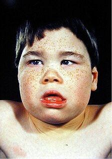
Alpha-thalassemia mental retardation syndrome (ATRX), also called alpha-thalassemia X-linked intellectual disability syndrome, nondeletion type or ATR-X syndrome, is an X-linked recessive condition associated with a mutation in the ATRX gene. Males with this condition tend to be moderately intellectually disabled and have physical characteristics including coarse facial features, microcephaly, hypertelorism, a depressed nasal bridge, a tented upper lip and an everted lower lip. Mild or moderate anemia, associated with alpha-thalassemia, is part of the condition. Females with this mutated gene have no specific signs or features, but if they do, they may demonstrate skewed X chromosome inactivation.

Keutel syndrome Medical condition

Keutel syndrome (KS) is a rare autosomal recessive genetic disorder characterized by abnormal diffuse cartilage calcification, hypoplasia of the mid-face, peripheral pulmonary stenosis, hearing loss, short distal phalanges (tips) of the fingers and mild mental retardation. Individuals with KS often present with peripheral pulmonary stenosis, brachytelephalangism, sloping forehead, midface hypoplasia, and receding chin. It is associated with abnormalities in the gene coding for matrix gla protein (MGP). Being an autosomal recessive disorder, it may be inherited from two unaffected, abnormal MGP-carrying parents. Thus, people who inherit two affected MGP genes will likely inherit KS.

Woodhouse–Sakati syndrome Medical condition

Woodhouse–Sakati syndrome, is a rare autosomal recessive multisystem disorder which causes malformations throughout the body, and deficiencies affecting the endocrine system.

FG syndrome (FGS) is a rare genetic syndrome caused by one or more recessive genes located on the X chromosome and causing physical anomalies and developmental delays. FG syndrome was named after the first letters of the surnames of the first patients noted with the disease. First reported by American geneticists John M. Opitz and Elisabeth G. Kaveggia in 1974, its major clinical features include intellectual disability, hyperactivity, hypotonia, and a characteristic facial appearance including macrocephaly.

Acropectoral syndrome Medical condition

Acropectoral syndrome is an autosomal dominant skeletal dysplasia syndrome affecting the hands, feet, sternum, and lumbosacral spine. A recently proposed candidate gene for preaxial polydactyly is LMBR1, encoding a novel transmembrane receptor, which may be an upstream regulator of SHH. The LMBR1 gene is on human chromosome 7q36.

Gillespie syndrome Medical condition

Gillespie syndrome, also called aniridia, cerebellar ataxia and mental deficiency, is a rare genetic disorder. The disorder is characterized by partial aniridia, ataxia, and, in most cases, intellectual disability. It is heterogeneous, inherited in either an autosomal dominant or autosomal recessive manner. Gillespie syndrome was first described by American ophthalmologist Fredrick Gillespie in 1965.

Cooks syndrome Medical condition

Cooks syndrome is a hereditary disorder which is characterized in the hands by bilateral nail hypoplasia on the thumb, index finger, and middle finger, absence of fingernails (anonychia) on the ring finger and little finger, lengthening of the thumbs, and bulbousness of the fingers. In the feet, it is characterized by absence of toenails and absence/hypoplasia of the distal phalanges. In the second study of this disorder, it was found that the intermediate phalanges, proximal phalanges, and metacarpals were unaffected.

Lujan–Fryns syndrome Medical condition

Lujan–Fryns syndrome (LFS) is an X-linked genetic disorder that causes mild to moderate intellectual disability and features described as Marfanoid habitus, referring to a group of physical characteristics similar to those found in Marfan syndrome. These features include a tall, thin stature and long, slender limbs. LFS is also associated with psychopathology and behavioral abnormalities, and it exhibits a number of malformations affecting the brain and heart. The disorder is inherited in an X-linked dominant manner, and is attributed to a missense mutation in the MED12 gene. There is currently no treatment or therapy for the underlying MED12 malfunction, and the exact cause of the disorder remains unclear.

Mohr–Tranebjærg syndrome Medical condition

Mohr–Tranebjærg syndrome (MTS) is a rare X-linked recessive syndrome also known as deafness–dystonia syndrome and caused by mutation in the TIMM8A gene. It is characterized by clinical manifestations commencing with early childhood onset hearing loss, followed by adolescent onset progressive dystonia or ataxia, visual impairment from early adulthood onwards and dementia from the 4th decade onwards. The severity of the symptoms may vary, but they progress usually to severe deafness and dystonia and sometimes are accompanied by cortical deterioration of vision and mental deterioration.

Malpuech facial clefting syndrome, also called Malpuech syndrome or Gypsy type facial clefting syndrome, is a rare congenital syndrome. It is characterized by facial clefting, a caudal appendage, growth deficiency, intellectual and developmental disability, and abnormalities of the renal system (kidneys) and the male genitalia. Abnormalities of the heart, and other skeletal malformations may also be present. The syndrome was initially described by Georges Malpuech and associates in 1983. It is thought to be genetically related to Juberg-Hayward syndrome. Malpuech syndrome has also been considered as part of a spectrum of congenital genetic disorders associated with similar facial, urogenital and skeletal anomalies. Termed "3MC syndrome", this proposed spectrum includes Malpuech, Michels and Mingarelli-Carnevale (OSA) syndromes. Mutations in the COLLEC11 and MASP1 genes are believed to be a cause of these syndromes. The incidence of Malpuech syndrome is unknown. The pattern of inheritance is autosomal recessive, which means a defective (mutated) gene associated with the syndrome is located on an autosome, and the syndrome occurs when two copies of this defective gene are inherited.

Ectrodactyly Medical condition

Ectrodactyly, split hand, or cleft hand involves the deficiency or absence of one or more central digits of the hand or foot and is also known as split hand/split foot malformation (SHFM). The hands and feet of people with ectrodactyly (ectrodactyls) are often described as "claw-like" and may include only the thumb and one finger with similar abnormalities of the feet.

Brachydactyly type D Abnormal shortening of the distal part of the thumb

Brachydactyly type D, also known as short thumb or stub thumb and inaccurately referred to as clubbed thumb, is a condition clinically recognised by a thumb being relatively short and round with an accompanying wider nail bed. The distal phalanx of affected thumbs is approximately two-thirds the length of full-length thumbs. It is the most common type of brachydactyly, or shortness of digits, affecting approximately 2-3% of the population, and is associated with the HOXD13 gene, located on chromosome 2q31.1

White–Sutton syndrome (WHSUS) is a rare neurodevelopmental disorder that affects different systems of the human body. It is mainly characterized by developmental delay, intellectual disability, craniofacial abnormalities and commonly features of autism spectrum disorder (ASD).

References

  1. Online Mendelian Inheritance in Man (OMIM): Nasodigitoacoustic syndrome - 301026
  2. 1 2 3 4 5 6 Keipert, JA; Fitzgerald, MG; Danks, DM (Feb 1973). "A new syndrome of broad terminal phalanges and facial abnormalities". Australian Paediatric Journal. 9 (1): 10–13. doi:10.1111/j.1440-1754.1973.tb02215.x. PMID   4708024.
  3. 1 2 3 4 5 Amor, D. J.; Dahl, H-H.; Bahlo, M.; Bankier, A. (Oct 2007). "Keipert syndrome (Nasodigitoacoustic syndrome) is X-linked and maps to Xq22.2–Xq28". American Journal of Medical Genetics Part A. 143A (19): 2236–2241. doi:10.1002/ajmg.a.31917. PMID   17726694. S2CID   34320632.
  4. 1 2 3 4 5 6 7 8 Cappon, SM; Khalifa, MM (Jul–Aug 2000). "Additional case of Keipert syndrome and review of the literature" (Free full text). Medical Science Monitor. 6 (4): 776–778. PMID   11208408.
  5. 1 2 "Prevalence and Incidence of Nasodigitoacoustic syndrome". WrongDiagnosis.com. Retrieved April 7, 2011.
  6. 1 2 Balci, S; Dagli, S (Oct 1996). "Keipert syndrome in two brothers from Turkey". Clinical Genetics. 50 (4): 223–228. doi:10.1111/j.1399-0004.1996.tb02631.x. PMID   9001804.
  7. 1 2 3 4 5 6 Nik-Zainal, S.; Holder, S. E.; Cruwys, M.; Hall, C. M.; Shaw-Smith, C. (Jul 2008). "Keipert syndrome: two further cases and review of the literature". Clinical Dysmorphology. 17 (3): 169–175. doi:10.1097/MCD.0b013e3282f4afc3. PMID   18541962.
  8. 1 2 Gorlin (1995). illustration, p. 209.
  9. 1 2 Gorlin (1995). p. 208
  10. 1 2 3 Dumic, M.; Kokic, D. D.; Matic, T.; Potocki, K. (Nov 2006). "Daughter and her mildly affected father with Keipert syndrome". American Journal of Medical Genetics Part A. 140A (22): 2488–2492. doi:10.1002/ajmg.a.31489. PMID   17036315. S2CID   2978286.
  11. Reardon, W.; Hall, C. M. (Apr 2003). "Broad thumbs and halluces with deafness: A patient with Keipert syndrome". American Journal of Medical Genetics. 118A (1): 86–89. doi:10.1002/ajmg.a.10063. PMID   12605449. S2CID   27419345.
  12. Amor DJ, Stephenson SEM, Mustapha M, Mensah MA, Ockeloen CW, Lee WS, Tankard RM, Phelan DG, Shinawi M, de Brouwer APM, Pfundt R, Dowling C, Toler TL, Sutton VR, Agolini E, Rinelli M, Capolino R, Martinelli D, Zampino G, Dumić M, Reardon W, Shaw-Smith C, Leventer RJ, Delatycki MB, Kleefstra T7, Mundlos S, Mortier G, Bahlo M, Allen NJ, Lockhart P (2019) Pathogenic variants in GPC4 cause Keipert syndrome. Am J Hum Genet
  13. Online Mendelian Inheritance in Man (OMIM): Keutel syndrome - 245150
  14. Online Mendelian Inheritance in Man (OMIM): Meunke syndrome - 602849
  15. Online Mendelian Inheritance in Man (OMIM): Teunissen-Cremers syndrome - 184460
  16. Online Mendelian Inheritance in Man (OMIM): Rubinstein-Taybi syndrome - 180849
  17. Feghali, J.; Lefebvre, P.; Staecker, H.; Kopke, R.; Frenz, D.; Malgrange, B.; Liu, W.; Moonen, G.; Ruben, R.; Van De Water, T. R. (Apr 1998). "Mammalian auditory hair cell regeneration/repair and protection: A review and future directions". Ear, Nose, & Throat Journal. 77 (4): 276, 280, 282–285. doi:10.1177/014556139807700409. PMID   9581394. S2CID   46494159.
  18. Smith, R.; Hildebrand, M.; Van Camp, G.; Pagon, R.; Bird, T.; Dolan, C.; Stephens, K. (1993). "Deafness and Hereditary Hearing Loss Overview". PMID   20301607.{{cite journal}}: Cite journal requires |journal= (help)
  19. Kikidis, D.; Nikolopoulos, T. P.; Kampessis, G.; Stamatiou, G.; Chrysovergis, A. (2011). "Sudden Sensorineural Hearing Loss: Subclinical Viral and Toxoplasmosis Infections as Aetiology and How They Alter the Clinical Course". ORL. 73 (2): 110–115. doi:10.1159/000324210. PMID   21389742. S2CID   25767318.
  20. Ali Khan, M.; Al-Yousef, S.; Huhta, J.; Bricker, J.; Mullins, C.; Sawyer, W. (May 1989). "Critical pulmonary valve stenosis in patients less than 1 year of age: Treatment with percutaneous gradational balloon pulmonary valvuloplasty". American Heart Journal. 117 (5): 1008–1014. doi:10.1016/0002-8703(89)90854-5. PMID   2711961.

Publications