Aicardi syndrome is a rare genetic malformation syndrome characterized by the partial or complete absence of a key structure in the brain called the corpus callosum, the presence of retinal lacunes, and epileptic seizures in the form of infantile spasms.[2] Other malformations of the brain and skeleton may also occur. The syndrome includes intellectual disability that is usually severe or moderate. So far, the syndrome has only been diagnosed in girls and in boys with two X chromosomes (Klinefelter syndrome).[3]
Those with Aicardi syndrome are in need of various specialist and habilitation instances. Epilepsy is treated with medication, but additional treatment may also be needed. In order to utilize the girls' eyesight and investigate the need for visual aids, examination by ophthalmologist is indicated early in life. Problems from the gastrointestinal tract are frequent. In adulthood, continued habilitation efforts and support in daily life are needed.[3]
The syndrome is named after the French child neurologist Jean Dennis Aicardi, who in 1965 described it in eight girls.[3] A causative gene has not been identified. Symptoms typically appear before a baby reaches about 5 months of age.[citation needed]
Signs and symptoms
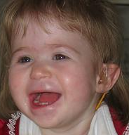
Those with Aicardi syndrome develop normally during the first months, but later various symptoms appear due to the syndrome's characteristic malformations in the brain. It is common for girls with Aicardi syndrome to have a small head (microcephaly).[3]
At three to six months of age, the girls have epileptic seizures, often of the infantile spasm type caused by changes in the brain's gray matter, the cerebral cortex. The seizures occur either as so-called flexor spasms, when the child's neck suddenly bends forward while the arms make a clasping movement, or as other types of epileptic seizures. Seizures come in series at short intervals and may increase in number from day to day until they are broken with medication. Epilepsy usually persists for life.[3]
Most have a severe intellectual disability with a major impact on language, communication and motor skills. A few have a moderate intellectual disability. Mild intellectual disability also occurs but is very rare.[3]
The eyes are always affected, and most people have impaired vision. During an eye examination, areas with less pigment (retinal lacunae) appear as white spots in the fundus, which is due to the absence of retinal pigment cells and other structures in these areas. If the lacunae are located in the macula, they affect acuity. Other types of eye abnormalities are also common, such as one eye being smaller than normal (microphthalmia), changes in the optic nerve, and incomplete closure/slitting of the membranes of the eye (coloboma). Rapid, involuntary eye movements (nystagmus) are common. Since girls with Aicardi syndrome have an intellectual disability that makes it difficult to participate in an eye examination, it is difficult to measure vision accurately.[3]
Problems from the gastrointestinal tract are common, for example constipation, diarrhea and that the normal valve mechanism between the stomach and the esophagus (upper mouth of the stomach) does not work normally and the stomach contents therefore leak up into the esophagus (gastroesophageal reflux). Some may also have difficulty eating.[3]
Extra ribs or lack of ribs and vertebral deformities often occur. While growing up, several of the girls get a crooked back (scoliosis). There are reports of isolated cases of tumors, especially brain tumors.[3]
Aicardi syndrome is a non-progressive condition and in itself does not lead to any deterioration, but various complications mean that there is an increased mortality among girls with the syndrome. Very little is known about the long-term prognosis, but there are occasional reports that the epilepsy may become milder with increasing age. The oldest women with the syndrome described so far are in their 40s.[3]
The syndrome is probably caused by a change (mutation) in one or more genes on the short arm of the X chromosome (Xp22), but which gene or genes are mutated is not yet (2015) known.[3]
Male fetuses with this change are unlikely to survive, which is because they only have one X chromosome. The individual boys with the syndrome described have also had the sex chromosome abnormality XXY syndrome (Klinefelter syndrome). Girls, who have two X chromosomes, can be born with the syndrome, because their second (normal) X chromosome compensates to some extent for the mutated gene.[3]
The mutation leads to a characteristic malformation of the brain stem with a complete absence the corpus callosum. As a rule, there are also signs that groups of brain cells have migrated incorrectly and placed themselves in the wrong place in the brain (heterotopias), an incorrect folding of the cerebral cortex (gyration abnormalities) or that the brain hemispheres are of different size.[3]
Heredity
Aicardi syndrome is an autosomal dominant X-linked disease and arises as a new mutation. The mutation has then usually occurred in one of the parents' germ cells (eggs or sperm). The probability that they will again have a child with the disease is then estimated at less than 1 percent. However, the new mutation in the child becomes hereditary and can theoretically be passed on to the next generation.[3] All cases of Aicardi syndrome are thought to be due to new mutations. No person with Aicardi syndrome is known to have transmitted the X-linked gene responsible for the syndrome to the next generation.[5]
Diagnosis
Aicardi syndrome is typically characterized by the following triad of features - however, one of the "classic" features being missing does not preclude a diagnosis of Aicardi Syndrome, if other supporting features are present.[6]
Suspicion of infantile spasms or other epileptic seizures during the first months of life should always be urgently investigated. There can be many different causes besides Aicardi syndrome. The investigation includes EEG (electroencephalogram), which in case of infantile spasms shows a characteristic pattern (hypsarrhythmia), magnetic resonance imaging (MRI) of the brain, blood and urine samples and examination of the spinal fluid (cerebrospinal fluid).[3]
In Aicardi syndrome, MRI of the brain shows that the cerebral cortex is completely or partially missing. Sometimes it is possible to see that the cerebral cortex is thin and underdeveloped. Other changes can occur at the same time, for example fluid bubbles (cysts) in the brain's fluid-producing structures (plexus choriodeus), different sized brain hemispheres and islands of nerve cells that did not migrate to the right place in the brain during fetal development. It is also possible to see that the fold pattern on the surface of the cerebrum has a different appearance (polygyry, microgyry). Absence of the cerebral cortex and other malformations of the brain also occur in conditions other than Aicardi syndrome.[3]
On eye examination, the retinal lacunae appear as white spots in the fundus, where the retina is missing. Sometimes there are slits in the eye (coloboma), retinal detachment and abnormally small or differently sized eyes.[3]
When X-raying the skeleton, it is sometimes possible to see that there are vertebral changes and extra ribs or that ribs are missing.[3]
Treatment
Treatment of Aicardi syndrome primarily involves management of seizures and early/continuing intervention programs for developmental delays.[citation needed]Additional comorbidities and complications sometimes seen with Aicardi syndrome include porencephalic cysts and hydrocephalus, and gastro-intestinal problems. Treatment for porencephalic cysts and/or hydrocephalus is often via a shunt or endoscopicfenestration of the cysts, though some require no treatment. Placement of a feeding tube, fundoplication, and surgeries to correct hernias or other gastrointestinal structural problems are sometimes used to treat gastro-intestinal issues.[citation needed]
Girls with Aicardi syndrome come into contact with many different specialists in healthcare early on. It is therefore important that efforts are coordinated.[3]
The drug treatment given for infantile spasms and other types of epilepsy is also given to girls with Aicardi syndrome. Epilepsy is often difficult to treat.[3]
If medication does not help, after an examination at a regional hospital, a decision can be made as to whether another treatment may be appropriate. Since the cause of the epileptic seizures is found in many different places in the brain, however, epilepsy surgery is rarely an option in Aicardi syndrome.[3]
Treatment with a ketogenic diet may be considered. It involves a carefully calculated diet that is rich in fat, contains a minimum of carbohydrates and provides the daily need for protein. The excess of fat forms starvation bodies (ketones) which can be used instead of glucose as a fuel source for the metabolism in the brain. For some children, this leads to fewer seizures. Treatment is started at regional hospitals by special teams with doctors, nurses and dieticians.[3]
Another treatment option when the drug treatment of the epilepsy does not help is a so-called vagus nerve stimulator (VNS). The vagus nerve is one of twelve nerves that originate directly from the brain (cranial nerves). A small battery-powered box (generator) is operated under the skin under the left collarbone and a thin wire (electrode) from the generator is operated around the left vagus nerve. The generator is then set so that it sends electrical impulses to the brain via the vagus nerve at fixed intervals and fixed strength, which can be gradually increased if necessary. This can lead to a reduction in the number and strength of the seizures but almost never results in the seizures disappearing completely. This treatment is also started and followed up at the regional hospitals.[3]
It is important that the girls' eyesight is used in the best possible way. They should therefore be examined by a pediatric ophthalmologist at an early stage to investigate visual function and the need for visual aids.[3]
Problems from the gastrointestinal tract need to be investigated and can be treated with medication. The girls who have difficulty eating may need to receive nutrition via a nasal tube or a so-called button (PEG, percutaneous endoscopic gastrostomy), an operatively created connection to the stomach via the abdominal wall. It is important to closely monitor girls' growth.[3]
Preventive dental care and contact with a children's dental care specialist (pedodontist) is needed, as the girls may find it difficult to participate in tooth brushing and dental treatments.[3]
Due to the risk of developing scoliosis, the back should be examined regularly. Scoliosis is primarily treated with a brace but may sometimes require surgery.[3]
Girls with Aicardi syndrome need rehabilitation interventions that also include vision rehabilitation. A habilitation team includes professional categories with special knowledge of disabilities and their effects on everyday life, health and development. The interventions take place in the medical, educational, psychological, social and technical fields. They consist, among other things, of investigation, treatment, testing of assistive devices, information about the disability and conversational support. Information about society's support and advice on adapting the home and other environments in which the child lives is also given. Parents, siblings and other relatives also receive support. The family may need help with the coordination of various efforts.[3]
The interventions are planned based on the needs of the child and the family, vary over time and always take place in close collaboration with people in the child's network. In order to develop the ability to communicate, it is important to work early on with educational efforts as well as alternative and supplementary communication routes (AKK).[3]
A close collaboration takes place with the municipality, which can offer various forms of interventions to facilitate the family's everyday life. Personal assistance can be given to those who, due to severe and permanent disabilities, need help with basic needs, but also to expand the possibility of an active life despite extensive disabilities. Respite care, a contact family or short-term accommodation are other examples of support measures.[3]
Adults
Adult women with Aicardi syndrome need continued habilitation efforts and support in daily life. This could be, for example, support and care in a home with special services and daily activities.[3]
Prognosis
The prognosis varies widely from case to case, depending on the severity of the symptoms. However, almost all people reported with Aicardi syndrome to date have experienced developmental delay of a significant degree, typically resulting in mild to moderate to profound intellectual disability. The age range of the individuals reported with Aicardi syndrome is from birth to the mid-40s.[citation needed]
Worldwide prevalence of Aicardi syndrome is estimated at several thousand, with approximately 900 cases reported in the United States.[8] There is no definite information on how common Aicardi syndrome is, but the incidence is estimated to be around one in 100,000 newborns. There may be people who do not have the fully developed syndrome and who have not been diagnosed.[3]
History
This disorder was first recognized as a distinct syndrome in 1965 by Jean Aicardi, a French pediatric neurologist and epileptologist.[9][6]
Related Research Articles
Porencephaly is an extremely rare cephalic disorder involving encephalomalacia. It is a neurological disorder of the central nervous system characterized by cysts or cavities within the cerebral hemisphere. Porencephaly was termed by Heschl in 1859 to describe a cavity in the human brain. Derived from Greek roots, the word porencephaly means 'holes in the brain'. The cysts and cavities are more likely to be the result of destructive (encephaloclastic) cause, but can also be from abnormal development (malformative), direct damage, inflammation, or hemorrhage. The cysts and cavities cause a wide range of physiological, physical, and neurological symptoms. Depending on the patient, this disorder may cause only minor neurological problems, without any disruption of intelligence, while others may be severely disabled or die before the second decade of their lives. However, this disorder is far more common within infants, and porencephaly can occur both before or after birth.
Septo-optic dysplasia (SOD), known also as de Morsier syndrome, is a rare congenital malformation syndrome that features a combination of the underdevelopment of the optic nerve, pituitary gland dysfunction, and absence of the septum pellucidum . Two or more of these features need to be present for a clinical diagnosis—only 30% of patients have all three. French-Swiss doctor Georges de Morsier first recognized the relation of a rudimentary or absent septum pellucidum with hypoplasia of the optic nerves and chiasm in 1956.
Polymicrogyria (PMG) is a condition that affects the development of the human brain by multiple small gyri (microgyri) creating excessive folding of the brain leading to an abnormally thick cortex. This abnormality can affect either one region of the brain or multiple regions.
Bilateral frontoparietal polymicrogyria is a genetic disorder with autosomal recessive inheritance that causes a cortical malformation. Our brain has folds in the cortex to increase surface area called gyri and patients with polymicrogyria have an increase number of folds and smaller folds than usual. Polymicrogyria is defined as a cerebral malformation of cortical development in which the normal gyral pattern of the surface of the brain is replaced by an excessive number of small, fused gyri separated by shallow sulci and abnormal cortical lamination. From ongoing research, mutation in GPR56, a member of the adhesion G protein-coupled receptor (GPCR) family, results in BFPP. These mutations are located in different regions of the protein without any evidence of a relationship between the position of the mutation and phenotypic severity. It is also found that GPR56 plays a role in cortical pattering.
Corpus callosotomy is a palliative surgical procedure for the treatment of medically refractory epilepsy. In this procedure the corpus callosum is cut through, in an effort to limit the spread of epileptic activity between the two halves of the brain.
Lennox–Gastaut syndrome (LGS) is a complex, rare, and severe childhood-onset epilepsy syndrome. It is characterized by multiple and concurrent seizure types including tonic seizure, cognitive dysfunction, and slow spike waves on electroencephalogram (EEG), which are very abnormal. Typically, it presents in children aged 3–5 years and most of the time persists into adulthood with slight changes in the electroclinical phenotype. It has been associated with perinatal injuries, congenital infections, brain malformations, brain tumors, genetic disorders such as tuberous sclerosis and numerous gene mutations. Sometimes LGS is observed after infantile epileptic spasm syndrome. The prognosis for LGS is marked by a 5% mortality in childhood and persistent seizures into adulthood.
Agenesis of the corpus callosum (ACC) is a rare birth defect in which there is a complete or partial absence of the corpus callosum. It occurs when the development of the corpus callosum, the band of white matter connecting the two hemispheres in the brain, in the embryo is disrupted. The result of this is that the fibers that would otherwise form the corpus callosum are instead longitudinally oriented along the ipsilateral ventricular wall and form structures called Probst bundles.
Sturge–Weber syndrome, sometimes referred to as encephalotrigeminal angiomatosis, is a rare congenital neurological and skin disorder. It is one of the phakomatoses and is often associated with port-wine stains of the face, glaucoma, seizures, intellectual disability, and ipsilateral leptomeningeal angioma. Sturge–Weber syndrome can be classified into three different types. Type 1 includes facial and leptomeningeal angiomas as well as the possibility of glaucoma or choroidal lesions. Normally, only one side of the brain is affected. This type is the most common. Type 2 involvement includes a facial angioma with a possibility of glaucoma developing. There is no evidence of brain involvement. Symptoms can show at any time beyond the initial diagnosis of the facial angioma. The symptoms can include glaucoma, cerebral blood flow abnormalities and headaches. More research is needed on this type of Sturge–Weber syndrome. Type 3 has leptomeningeal angioma involvement exclusively. The facial angioma is absent and glaucoma rarely occurs. This type is only diagnosed via brain scan.
Pachygyria is a congenital malformation of the cerebral hemisphere. It results in unusually thick convolutions of the cerebral cortex. Typically, children have developmental delay and seizures, the onset and severity depending on the severity of the cortical malformation. Infantile spasms are common in affected children, as is intractable epilepsy.
Epileptic spasms is an uncommon-to-rare epileptic disorder in infants, children and adults. One of the other names of the disorder, West syndrome, is in memory of the English physician, William James West (1793–1848), who first described it in an article published in The Lancet in 1841. The original case actually described his own son, James Edwin West (1840–1860). Other names for it are "generalized flexion epilepsy", "infantile epileptic encephalopathy", "infantile myoclonic encephalopathy", "jackknife convulsions", "massive myoclonia" and "Salaam spasms". The term "infantile spasms" can be used to describe the specific seizure manifestation in the syndrome, but is also used as a synonym for the syndrome itself. West syndrome in modern usage is the triad of infantile spasms, a pathognomonic EEG pattern, and developmental regression – although the international definition requires only two out of these three elements.
1p36 deletion syndrome is a congenital genetic disorder characterized by moderate to severe intellectual disability, delayed growth, hypotonia, seizures, limited speech ability, malformations, hearing and vision impairment, and distinct facial features. The symptoms may vary, depending on the exact location of the chromosomal deletion.
Ring chromosome 20, ring-shaped chromosome 20 or r(20) syndrome is a rare human chromosome abnormality where the two arms of chromosome 20 fuse to form a ring chromosome. The syndrome is associated with epileptic seizures, behaviour disorders and intellectual disability.
Papillorenal syndrome is an autosomal dominant genetic disorder marked by underdevelopment (hypoplasia) of the kidney and colobomas of the optic nerve.
Ohtahara syndrome (OS), also known as early infantile epileptic encephalopathy (EIEE) is a progressive epileptic encephalopathy. The syndrome is outwardly characterized by tonic spasms and partial seizures within the first few months of life, and receives its more elaborate name from the pattern of burst activity on an electroencephalogram (EEG). It is an extremely debilitating progressive neurological disorder, involving intractable seizures and severe intellectual disabilities. No single cause has been identified, although in many cases structural brain damage is present.
Epilepsy-intellectual disability in females also known as PCDH19 gene-related epilepsy or epileptic encephalopathy, early infantile, 9 (EIEE9), is a rare type of epilepsy that affects predominately females and is characterized by clusters of brief seizures, which start in infancy or early childhood, and is occasionally accompanied by varying degrees of cognitive impairment. The striking pattern of onset seizures at a young age, genetic testing and laboratory results, potential developmental delays or developmental regression and associated disorders, eases diagnosis.
An epilepsy syndrome is defined as "a characteristic cluster of clinical and EEG features, often supported by specific etiological findings ."
SYNGAP1-related intellectual disability is a monogenetic developmental and epileptic encephalopathy that affects the central nervous system. Symptoms include intellectual disability, epilepsy, autism, sensory processing deficits, hypotonia and unstable gait.
CDKL5 deficiency disorder (CDD) is a rare genetic disorder caused by pathogenic variants in the gene CDKL5.
Malignant migrating partial seizures of infancy (MMPSI) is a rare epileptic syndrome that onsets before 6 months of age, commonly in the first few weeks of life. Once seizures start, the site of seizure activity repeatedly migrates from one area of the brain to another, with few periods of remission in between. These seizures are 'focal' (updated term for 'partial'), meaning they do not affect both sides of the brain at the same time. These continuous seizures cause damage to the brain, hence the descriptor 'malignant.'
References
This article incorporates text that is in the public domain under the Swedish URL §9 (1960:729) as a constitution, decision or statement by Swedish authorities.
↑ RESERVED, INSERM US14-- ALL RIGHTS. "Orphanet: Aicardi syndrome". www.orpha.net. Archived from the original on 10 March 2018. Retrieved 17 June 2019.{{cite web}}: CS1 maint: numeric names: authors list (link)
↑ "Aicardi Syndrome". National Institute of Neurological Disorders and Stroke. 2022-07-25. Retrieved 2022-12-30. There is no cure for Aicardi syndrome.
↑ Kroner, Barbara L.; Preiss, Liliana R.; Ardini, Mary-Anne; Gaillard, William D. (29 January 2008). "New Incidence, Prevalence, and Survival of Aicardi Syndrome From 408 Cases". Journal of Child Neurology. 23 (5): 531–535. doi:10.1177/0883073807309782. PMID18182643. S2CID28004201.
↑ Aicardi J, Lefebvre J, Lerique-Koechlin A. A new syndrome: spasm in flexion, callosal agenesis, ocular abnormalities. Electroenceph Clin Neurophysiol 1965; 19: 609–610
This page is based on this Wikipedia article Text is available under the CC BY-SA 4.0 license; additional terms may apply. Images, videos and audio are available under their respective licenses.