Related Research Articles

Amputation is the removal of a limb by trauma, medical illness, or surgery. As a surgical measure, it is used to control pain or a disease process in the affected limb, such as malignancy or gangrene. In some cases, it is carried out on individuals as a preventive surgery for such problems. A special case is that of congenital amputation, a congenital disorder, where fetal limbs have been cut off by constrictive bands. In some countries, amputation is currently used to punish people who commit crimes. Amputation has also been used as a tactic in war and acts of terrorism; it may also occur as a war injury. In some cultures and religions, minor amputations or mutilations are considered a ritual accomplishment. When done by a person, the person executing the amputation is an amputator. The oldest evidence of this practice comes from a skeleton found buried in Liang Tebo cave, East Kalimantan, Indonesian Borneo dating back to at least 31,000 years ago, where it was done when the amputee was a young child.

In medicine, a prosthesis, or a prosthetic implant, is an artificial device that replaces a missing body part, which may be lost through trauma, disease, or a condition present at birth. Prostheses are intended to restore the normal functions of the missing body part. Amputee rehabilitation is primarily coordinated by a physiatrist as part of an inter-disciplinary team consisting of physiatrists, prosthetists, nurses, physical therapists, and occupational therapists. Prostheses can be created by hand or with computer-aided design (CAD), a software interface that helps creators design and analyze the creation with computer-generated 2-D and 3-D graphics as well as analysis and optimization tools.

In humans and other primates, the knee joins the thigh with the leg and consists of two joints: one between the femur and tibia, and one between the femur and patella. It is the largest joint in the human body. The knee is a modified hinge joint, which permits flexion and extension as well as slight internal and external rotation. The knee is vulnerable to injury and to the development of osteoarthritis.

A bone tumor is an abnormal growth of tissue in bone, traditionally classified as noncancerous (benign) or cancerous (malignant). Cancerous bone tumors usually originate from a cancer in another part of the body such as from lung, breast, thyroid, kidney and prostate. There may be a lump, pain, or neurological signs from pressure. A bone tumor might present with a pathologic fracture. Other symptoms may include fatigue, fever, weight loss, anemia and nausea. Sometimes there are no symptoms and the tumour is found when investigating another problem.

An osteosarcoma (OS) or osteogenic sarcoma (OGS) is a cancerous tumor in a bone. Specifically, it is an aggressive malignant neoplasm that arises from primitive transformed cells of mesenchymal origin and that exhibits osteoblastic differentiation and produces malignant osteoid.

Orthopedic surgery or orthopedics is the branch of surgery concerned with conditions involving the musculoskeletal system. Orthopedic surgeons use both surgical and nonsurgical means to treat musculoskeletal trauma, spine diseases, sports injuries, degenerative diseases, infections, tumors, and congenital disorders.
An osteotomy is a surgical operation whereby a bone is cut to shorten or lengthen it or to change its alignment. It is sometimes performed to correct a hallux valgus, or to straighten a bone that has healed crookedly following a fracture. It is also used to correct a coxa vara, genu valgum, and genu varum. The operation is done under a general anaesthetic.

Coxa vara is a deformity of the hip, whereby the angle between the head and the shaft of the femur is reduced to less than 120 degrees. This results in the leg being shortened and the development of a limp. It may be congenital and is commonly caused by injury, such as a fracture. It can also occur when the bone tissue in the neck of the femur is softer than normal, causing it to bend under the weight of the body. This may either be congenital or the result of a bone disorder. The most common cause of coxa vara is either congenital or developmental. Other common causes include metabolic bone diseases, post-Perthes deformity, osteomyelitis, and post traumatic. Shepherd's Crook deformity is a severe form of coxa vara where the proximal femur is severely deformed with a reduction in the neck shaft angle beyond 90 degrees. It is most commonly a sequela of osteogenesis imperfecta, Paget's disease, osteomyelitis, tumour and tumour-like conditions.
Hemicorporectomy is a radical surgery in which the body below the waist is amputated, transecting the lumbar spine. This removes the legs, the genitalia, urinary system, pelvic bones, anus, and rectum. It is a major procedure recommended only as a last resort for people with severe and potentially fatal illnesses such as osteomyelitis, tumors, severe traumas and intractable decubiti in, or around, the pelvis. By 2009, 66 cases had been reported in medical literature.

Genu valgum, commonly called "knock-knee", is a condition in which the knees angle in and touch each other when the legs are straightened. Individuals with severe valgus deformities are typically unable to touch their feet together while simultaneously straightening the legs. The term originates from the Latin genu, 'knee', and valgus which means "bent outwards", but is also used to describe the distal portion of the knee joint which bends outwards and thus the proximal portion seems to be bent inwards.

Genu varum is a varus deformity marked by (outward) bowing at the knee, which means that the lower leg is angled inward (medially) in relation to the thigh's axis, giving the limb overall the appearance of an archer's bow. Usually medial angulation of both lower limb bones is involved.

Hip replacement is a surgical procedure in which the hip joint is replaced by a prosthetic implant, that is, a hip prosthesis. Hip replacement surgery can be performed as a total replacement or a hemi/semi(half) replacement. Such joint replacement orthopaedic surgery is generally conducted to relieve arthritis pain or in some hip fractures. A total hip replacement consists of replacing both the acetabulum and the femoral head while hemiarthroplasty generally only replaces the femoral head. Hip replacement is one of the most common orthopaedic operations, though patient satisfaction varies widely. Approximately 58% of total hip replacements are estimated to last 25 years. The average cost of a total hip replacement in 2012 was $40,364 in the United States, and about $7,700 to $12,000 in most European countries.

Hemipelvectomy, also known as a pelvic resection, is a surgical procedure that involves the removal of part of the pelvic girdle. This procedure is most commonly performed to treat oncologic conditions of the pelvis. Hemipelvectomy can be further classified as internal and external hemipelvectomy. An internal hemipelvectomy is a limb-sparing procedure where the innominate bone is resected while preserving the ipsilateral limb. An external hemipelvectomy involves the resection of the innominate bone plus amputation of the ipsilateral limb.

Proximal femoral focal deficiency (PFFD), also known as Congenital Femoral Deficiency (CFD), is a rare, non-hereditary birth defect that affects the pelvis, particularly the hip bone, and the proximal femur. The disorder may affect one side or both, with the hip being deformed and the leg shortened.

A hip dislocation is when the thighbone (femur) separates from the hip bone (pelvis). Specifically it is when the ball–shaped head of the femur separates from its cup–shaped socket in the hip bone, known as the acetabulum. The joint of the femur and pelvis is very stable, secured by both bony and soft-tissue constraints. With that, dislocation would require significant force which typically results from significant trauma such as from a motor vehicle collision or from a fall from elevation. Hip dislocations can also occur following a hip replacement or from a developmental abnormality known as hip dysplasia.

Pigeon toe, also known as in-toeing, is a condition which causes the toes to point inward when walking. It is most common in infants and children under two years of age and, when not the result of simple muscle weakness, normally arises from underlying conditions, such as a twisted shin bone or an excessive anteversion resulting in the twisting of the thigh bone when the front part of a person's foot is turned in.
Limb-sparing techniques, also known as limb-saving or limb-salvage techniques, are performed in order to preserve the look and function of limbs. Limb-sparing techniques are used to preserve limbs affected by trauma, arthritis, cancers such as high-grade bone sarcomas, and vascular conditions such as diabetic foot ulcers. As the techniques for chemotherapy, radiation, and diagnostic modalities improve, there has been a trend toward limb-sparing procedures to avoid amputation, which has been associated with a lower 5-year survival rate and cost-effectiveness compared to limb salvage in the long-run. There are many different types of limb-sparing techniques, including arthrodesis, arthroplasty, endoprosthetic reconstruction, various types of implants, rotationplasty, osseointegration limb replacement, fasciotomy, and revascularization.

Gait deviations are nominally referred to as any variation of standard human gait, typically manifesting as a coping mechanism in response to an anatomical impairment. Lower-limb amputees are unable to maintain the characteristic walking patterns of an able-bodied individual due to the removal of some portion of the impaired leg. Without the anatomical structure and neuromechanical control of the removed leg segment, amputees must use alternative compensatory strategies to walk efficiently. Prosthetic limbs provide support to the user and more advanced models attempt to mimic the function of the missing anatomy, including biomechanically controlled ankle and knee joints. However, amputees still display quantifiable differences in many measures of ambulation when compared to able-bodied individuals. Several common observations are whole-body movements, slower and wider steps, shorter strides, and increased sway.
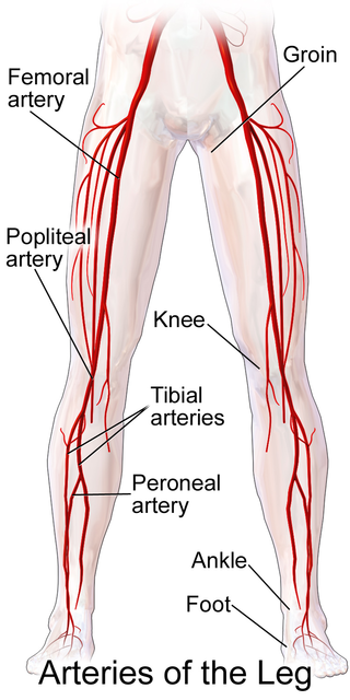
Popliteal bypass surgery, more commonly known as femoropopliteal bypass or more generally as lower extremity bypass surgery, is a surgical procedure used to treat diseased leg arteries above or below the knee. It is used as a medical intervention to salvage limbs that are at risk of amputation and to improve walking ability in people with severe intermittent claudication and ischemic rest pain.
Bone malrotation refers to the situation that results when a bone heals out of rotational alignment from another bone, or part of bone. It often occurs as the result of a surgical complication after a fracture where intramedullary nailing (IMN) occurs, especially in the femur and tibial bones, but can also occur genetically at birth. The severity of this complication is often neglected due to its complexity to detect and treat, yet if left untreated, bone malrotation can significantly impact regular bodily functioning, and even lead to severe arthritis. Detection throughout history has become more advanced and accurate, ranging from clinical assessment to ultrasounds to CT scans. Treatment can include an osteotomy, a major surgical procedure where bones are cut and realigned correctly, or compensatory methods, where individuals learn to externally or internally rotate their limb to compensate for the rotation. Further research is currently being examined in this area to reduce occurrences of malrotation, including detailed computer navigation to improve visual accuracy during surgery.
References
- ↑ Agarwal M, Puri A, Anchan C, Shah M, Jambhekar N (2007). "Rotationplasty for bone tumors: is there still a role?". Clin. Orthop. Relat. Res. 459: 76–81. doi:10.1097/BLO.0b013e31805470f0. PMID 17414168. S2CID 31227954.
- ↑ Kotz, R. "Rotationplasty." Seminars in surgical oncology 13.1 (1997): 34-40. Print. [ verification needed ]
- ↑ Rotation-plasty for congenital defects of the femur, by C. P. van Nes, in the Journal of Bone and Joint Surgery; Volume 32-B, Issue 1 / February 1950
- ↑ Ramseier, Leonhard E.; Dumont, Charles E.; Ulrich Exner, G. (January 2008). "Rotationplasty (Borggreve/Van Nes and modifications) as an alternative to amputation in failed reconstructions after resection of tumours around the knee joint". Scandinavian Journal of Plastic and Reconstructive Surgery and Hand Surgery. 42 (4): 199–201. doi:10.1080/02844310802069434. ISSN 0284-4311. PMID 18763196. S2CID 10486883.
- ↑ "RotationPlasty : A Surgeon's Approach - The Dynamics of Rotationplasty". www.rotationplasty.org. Retrieved 2020-04-27.
- ↑ Brown, Kenneth L.B. (January 2001). "Resection, Rotationplasty, and Femoropelvic Arthrodesis in Severe Congenital Femoral Deficiency". The Journal of Bone and Joint Surgery, American Volume. 83 (1): 78–85. doi:10.2106/00004623-200101000-00011. ISSN 0021-9355. PMID 11205862. S2CID 23701234.
- ↑ Kotz, Rainer (January 1997). "Rotationplasty". Seminars in Surgical Oncology. 13 (1): 34–40. doi:10.1002/(sici)1098-2388(199701/02)13:1<34::aid-ssu6>3.0.co;2-5. ISSN 8756-0437. PMID 9025180.
- ↑ Kotz, R. "Rotationplasty." Seminars in surgical oncology 13.1 (1997): 34-40. Print. [ verification needed ]
- ↑ "RotationPlasty : A Surgeon's Approach - The Dynamics of Rotationplasty". www.rotationplasty.org. Retrieved 2020-04-27.
- ↑ Hillmann, A.; Gosheger, G.; Hoffmann, C.; Ozaki, T.; Winkelmann, W. (2000-10-17). "Rotationplasty - surgical treatment modality after failed limb salvage procedure". Archives of Orthopaedic and Trauma Surgery. 120 (10): 555–558. doi:10.1007/s004020000175. ISSN 0936-8051. PMID 11110135. S2CID 6829184.
- ↑ Rödl, Robert W; Pohlmann, Ursula; Gosheger, Georg; Lindner, Norbert J; Winkelmann, Winfried (January 2002). "Rotationplasty--quality of life after 10 years in 22 patients". Acta Orthopaedica Scandinavica. 73 (1): 85–88. doi: 10.1080/000164702317281468 . ISSN 0001-6470. PMID 11928918. S2CID 30887593.