S1PR1

Last updated

S1PR1
3v2y.png
Available structures
PDB Ortholog search: PDBe RCSB
Identifiers
Aliases S1PR1 , CD363, CHEDG1, D1S3362, ECGF1, EDG-1, EDG1, S1P1, sphingosine-1-phosphate receptor 1
External IDs OMIM: 601974; MGI: 1096355; HomoloGene: 1071; GeneCards: S1PR1; OMA:S1PR1 - orthologs
Orthologs
SpeciesHumanMouse
Entrez
Ensembl
UniProt
RefSeq (mRNA)

NM_001400
NM_001320730

NM_007901

RefSeq (protein)

NP_001307659
NP_001391

NP_031927

Location (UCSC) Chr 1: 101.24 – 101.24 Mb Chr 3: 115.5 – 115.51 Mb
PubMed search [3] [4]
Wikidata
View/Edit Human View/Edit Mouse

Sphingosine-1-phosphate receptor 1 (S1P receptor 1 or S1PR1), also known as endothelial differentiation gene 1 (EDG1) is a protein that in humans is encoded by the S1PR1 gene. S1PR1 is a G-protein-coupled receptor which binds the bioactive signaling molecule sphingosine 1-phosphate (S1P). S1PR1 belongs to a sphingosine-1-phosphate receptor subfamily comprising five members (S1PR1-5). [5] S1PR1 was originally identified as an abundant transcript in endothelial cells [6] and it has an important role in regulating endothelial cell cytoskeletal structure, migration, capillary-like network formation and vascular maturation. [7] [8] In addition, S1PR1 signaling is important in the regulation of lymphocyte maturation, migration and trafficking. [9] [10]

Contents

Structure

S1PR1 like the other members of the GPCR family is composed of seven-transmembrane helices arranged in a structurally conserved bundle. [5] Like other GPCRs, in the extracellular region S1PR1 is composed of three loops: ECL1 between helices II and III, ECL2 between helices IV and V and ECL3 between helices VI and VII. Compared to the other members of the family, S1PR1 has some specific features. The N terminus of the protein folds as a helical cap above the top of the receptor and therefore it limits the access of the ligands to the amphipathic binding pocket. This marked amphipathicity is indeed in agreement with the zwitterionic nature of S1P. In addition, helices ECL1 and ECL2 pack tightly against the N-terminal helix, further occluding the access of the ligand from the extracellular space. S1P or S1P analogs are likely to reach the binding pocket from within the cell membrane and not from the extracellular space, may be through an opening between helices I and VII. Compared to the other GPCRs, this region is more open due to a different positioning of helices I and II toward helix III. [5] This occlusion of the ligand access space from the extracellular space could also explain the slow saturation of receptor binding in the presence of excess ligand. [11]

Function

Like the other members of the GPCR family, S1PR1 senses its ligand from outside the cell and activates intracellular signal pathways that at last lead to cellular responses. The signal is transduced through the association of the receptor with different G proteins, which recruits a series of systems for downstream amplification of the signal. [12]

Immune system

Immune cell trafficking

S1P and its receptors play a key role in regulating immune cell trafficking by forming gradients that guide immune cells between tissues and vascular compartments. S1PR1 is pivotal in promoting T-cell egress from lymphoid organs, while changes in S1P levels can influence immune cell migration and positioning in lymphoid and non-lymphoid tissues during inflammation or immune surveillance. [13]

S1PR1, primarily located on the cell membrane of most lymphocytes, binds to the abundant ligand S1P in the bloodstream to promote lymphocyte egress from lymphoid organs, allowing them to travel to affected tissues. S1PR1 is responsive to the S1P gradient between the lymphoid tissues (low S1P) and the lymph (high S1P), facilitating T cell movement through the endothelial barrier. [14] However, upon T cell activation in lymphoid organs via cytokine and T-cell receptor signaling, the protein Cluster of Differentiation 69 (CD69) is expressed and forms a complex with S1PR1. This interaction, involving CD69 transmembrane domain and the helix-4 of S1PR1, leads to S1PR1 internalization and degradation, preventing S1P binding and downstream signaling. [15] This mechanism results in the temporary retention of lymphocytes within the lymph organs, enhancing the chances of successful lymphocyte activation, especially if the initial activation signal was weak. Upon antigen encounter or type I interferon stimulation in lymphoid organs, S1PR1 expression is decreased through CD69 interaction and downregulation of the transcription factor Kruppel‑like factor 2. [16] Effector T cells eventually re-express S1PR1 to exit the lymph node and enter peripheral tissues. However, increased S1P levels in lymphoid tissues, due to inhibition of S1P lyase, inflammation, or synthetic S1PR1 ligands like FTY720, can block T cell egress by dissipating the S1P gradient, inducing S1PR1 internalization, and enhancing endothelial junctional contacts to close egress ports. [16]

Immune cell regulation

S1PR1 activation is heavily involved in immune cell regulation and development. Sphingosine-1-phosphate receptor 1 is also involved in immune-modulation and directly involved in suppression of innate immune responses from T cells. [17] Depending on the G protein coupled with the S1PR1, diverse cellular effects are achieved: Gαi and Gαo modulate cellular survival, proliferation and motility; Gα12 and Gα13 modulate cytoskeletal remodeling and cell-shape changes and Gαq modulates several cellular effector functions. [12] All the intracellular functions occur via the interaction with Gαi and Gαo: these two proteins recruit other proteins for downstream amplification of the signal. [12] The main downstream effector functions of S1P-S1PR1 system are as follows:

  1. The phosphatidylinositol 3-kinase (PI3K) and the lipid dependent protein kinase B (PKB) signaling pathway increases the survival of lymphocytes and other immune cells by inhibiting apoptosis.
  2. Phosphoinositide 3-kinase (PI3K) and the GTPase RAC are responsible of the lymphocytes migration and their interactions with other cells or with connective-tissue surfaces. [12] S1PR1-deficient thymocytes do not emigrate from the thymus, resulting in an increased numbers of mature thymocytes in the thymus and in medullary hyperplasia, and few S1PR1-deficient T cells can be detected in the blood, lymph nodes, spleen or non-lymphoid organs in these mouse models. [9] [10] The proliferation of immune cells is due to S1P-mediated signals via the GTPase RAS and extracellular-signal regulated kinase (ERK). IV) The Phospholipase C (PLC)-induced increases in intracellular calcium levels allow the secretion of cytokines and other immune mediators. [12]

Vasculogenesis

S1PR1 is one of the main receptors responsible for vascular growth and development, at least during embryogenesis. [18] In vascular endothelial cells the binding of S1P to S1PR1 induces migration, proliferation, cell survival and morphogenesis into capillary-like structures. [19] Moreover, the binding of S1P to S1PR1 is implicated in the formation of cell-cell adherens junctions, therefore inhibiting paracellular permeability of solutes and macromolecules. [20] [21] It was also shown in vivo that S1P synergizes with angiogenic factors such as FGF-2 and VEGF in inducing angiogenesis and vascular maturation through S1PR1. [21] [22] showed that S1PR1-KO mice died during development due to a defect in vascular stabilization, suggesting that this receptor is essential for vascular development. In conclusion, several evidences confirm that S1P via S1PR1 is a potent regulator of vascular growth and development, at least during embryogenesis. [18]

Clinical significance

Cancer

S1PR1 is involved in the motility of cancer cells upon stimulation by S1P. The signal pathway involves RAC-CDC42 and correlates with ERK1 and ERK2 activation. The RAC-CDC42 pathway leads to cell migration, whereas the ERK pathway leads to proliferation and neovascularization [23] [24] demonstrated that S1PR1 is strongly induced in endothelial cells during tumor angiogenesis and a siRNA against S1PR1 was able to inhibit angiogenesis and tumor growth. S1PR1 is also involved in other types of cancer: fibrosarcoma cells migrate upon activation of S1PR1 by S1P via RAC1–CDC42 dependent pathway) [25] [26] and ovarian cancer cell invasion involves S1PR1 or S1PR3 and calcium mobilization. [27]

Multiple sclerosis

S1PR1 is involved in multiple sclerosis. Fingolimod, a drug which internalizes the receptor, is approved as a disease modifying agent in Multiple sclerosis. There are other Sphingosine-1-phosphate receptor modulators. Van Doorn et al. (2010) [28] observed a strong increase in S1PR1 (and S1PR3) expression in hypertrophic astrocytes both in the active and inactive Multiple sclerosis lesions from patients compared to the unaffected patients.

Evolution

Paralogues for S1PR1 Gene [29]

Interactions

S1PR1 has been shown to interact with 5-HT1A receptor, [30] GNAI1, [31] and GNAI3. [31]

See also

Related Research Articles

<span class="mw-page-title-main">Natural killer cell</span> Type of cytotoxic lymphocyte

Natural killer cells, also known as NK cells or large granular lymphocytes (LGL), are a type of cytotoxic lymphocyte critical to the innate immune system. They belong to the rapidly expanding family of known innate lymphoid cells (ILC) and represent 5–20% of all circulating lymphocytes in humans. The role of NK cells is analogous to that of cytotoxic T cells in the vertebrate adaptive immune response. NK cells provide rapid responses to virus-infected cells, stressed cells, tumor cells, and other intracellular pathogens based on signals from several activating and inhibitory receptors. Most immune cells detect the antigen presented on major histocompatibility complex I (MHC-I) on infected cell surfaces, but NK cells can recognize and kill stressed cells in the absence of antibodies and MHC, allowing for a much faster immune reaction. They were named "natural killers" because of the notion that they do not require activation to kill cells that are missing "self" markers of MHC class I. This role is especially important because harmful cells that are missing MHC I markers cannot be detected and destroyed by other immune cells, such as T lymphocyte cells.

<span class="mw-page-title-main">Fas ligand</span> Protein-coding gene in the species Homo sapiens

Fas ligand is a type-II transmembrane protein expressed on various types of cells, including cytotoxic T lymphocytes, monocytes, neutrophils, breast epithelial cells, vascular endothelial cells and natural killer (NK) cells. It binds with its receptor, called FAS receptor and plays a crucial role in the regulation of the immune system and in induction of apoptosis, a programmed cell death.

<span class="mw-page-title-main">Lipid signaling</span> Biological signaling using lipid molecules

Lipid signaling, broadly defined, refers to any biological cell signaling event involving a lipid messenger that binds a protein target, such as a receptor, kinase or phosphatase, which in turn mediate the effects of these lipids on specific cellular responses. Lipid signaling is thought to be qualitatively different from other classical signaling paradigms because lipids can freely diffuse through membranes. One consequence of this is that lipid messengers cannot be stored in vesicles prior to release and so are often biosynthesized "on demand" at their intended site of action. As such, many lipid signaling molecules cannot circulate freely in solution but, rather, exist bound to special carrier proteins in serum.

<span class="mw-page-title-main">L-selectin</span> Mammalian protein found in Homo sapiens

L-selectin, also known as CD62L, is a cell adhesion molecule found on the cell surface of leukocytes, and the blastocyst. It is coded for in the human by the SELL gene. L-selectin belongs to the selectin family of proteins, which recognize sialylated carbohydrate groups containing a Sialyl LewisX (sLeX) determinant. L-selectin plays an important role in both the innate and adaptive immune responses by facilitating leukocyte-endothelial cell adhesion events. These tethering interactions are essential for the trafficking of monocytes and neutrophils into inflamed tissue as well as the homing of lymphocytes to secondary lymphoid organs. L-selectin is also expressed by lymphoid primed hematopoietic stem cells and may participate in the migration of these stem cells to the primary lymphoid organs. In addition to its function in the immune response, L-selectin is expressed on embryonic cells and facilitates the attachment of the blastocyst to the endometrial endothelium during human embryo implantation.

<span class="mw-page-title-main">CCL21</span> Mammalian protein found in Homo sapiens

Chemokine ligand 21 (CCL21) is a small cytokine belonging to the CC chemokine family. This chemokine is also known as 6Ckine, exodus-2, and secondary lymphoid-tissue chemokine (SLC). CCL21 elicits its effects by binding to a cell surface chemokine receptor known as CCR7. The main function of CCL21 is to guide CCR7 expressing leukocytes to the secondary lymphoid organs, such as lymph nodes and Peyer´s patches.

Sphingosine-1-phosphate (S1P) is a signaling sphingolipid, also known as lysosphingolipid. It is also referred to as a bioactive lipid mediator. Sphingolipids at large form a class of lipids characterized by a particular aliphatic aminoalcohol, which is sphingosine.

<span class="mw-page-title-main">CD137</span> Member of the tumor necrosis factor (TNF) receptor family

CD137, a member of the tumor necrosis factor (TNF) receptor family, is a type 1 transmembrane protein, expressed on surfaces of leukocytes and non-immune cells. Its alternative names are tumor necrosis factor receptor superfamily member 9 (TNFRSF9), 4-1BB, and induced by lymphocyte activation (ILA). It is of interest to immunologists as a co-stimulatory immune checkpoint molecule, and as a potential target in cancer immunotherapy.

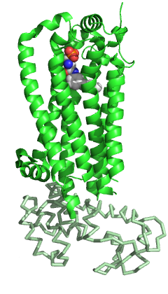
The lysophospholipid receptor (LPL-R) group are members of the G protein-coupled receptor family of integral membrane proteins that are important for lipid signaling. In humans, there are eleven LPL receptors, each encoded by a separate gene. These LPL receptor genes are also sometimes referred to as "Edg".

<span class="mw-page-title-main">CD69</span> Human lectin protein

CD69 is a human transmembrane C-Type lectin protein encoded by the CD69 gene. It is an early activation marker that is expressed in hematopoietic stem cells, T cells, and many other cell types in the immune system. It is also implicated in T cell differentiation as well as lymphocyte retention in lymphoid organs.

<span class="mw-page-title-main">LPAR1</span> Protein

Lysophosphatidic acid receptor 1 also known as LPA1 is a protein that in humans is encoded by the LPAR1 gene. LPA1 is a G protein-coupled receptor that binds the lipid signaling molecule lysophosphatidic acid (LPA).

<span class="mw-page-title-main">S1PR3</span> Protein and coding gene in humans

Sphingosine-1-phosphate receptor 3 also known as S1PR3 is a human gene which encodes a G protein-coupled receptor which binds the lipid signaling molecule sphingosine 1-phosphate (S1P). Hence this receptor is also known as S1P3.

<span class="mw-page-title-main">GPR6</span> Protein-coding gene in the species Homo sapiens

G protein-coupled receptor 6, also known as GPR6, is a protein which in humans is encoded by the GPR6 gene.

<span class="mw-page-title-main">S1PR4</span> Protein-coding gene in the species Homo sapiens

Sphingosine-1-phosphate receptor 4 also known as S1PR4 is a human gene which encodes a G protein-coupled receptor which binds the lipid signaling molecule sphingosine 1-phosphate (S1P). Hence this receptor is also known as S1P4.

<span class="mw-page-title-main">LPAR2</span> Protein-coding gene in the species Homo sapiens

Lysophosphatidic acid receptor 2 also known as LPA2 is a protein that in humans is encoded by the LPAR2 gene. LPA2 is a G protein-coupled receptor that binds the lipid signaling molecule lysophosphatidic acid (LPA).

<span class="mw-page-title-main">S1PR2</span> Protein and coding gene in humans

Sphingosine-1-phosphate receptor 2, also known as S1PR2 or S1P2, is a human gene which encodes a G protein-coupled receptor which binds the lipid signaling molecule sphingosine 1-phosphate (S1P).

<span class="mw-page-title-main">S1PR5</span> Protein-coding gene in the species Homo sapiens

Sphingosine-1-phosphate receptor 5 also known as S1PR5 is a human gene which encodes a G protein-coupled receptor which binds the lipid signaling molecule sphingosine 1-phosphate (S1P). Hence this receptor is also known as S1P5.

<span class="mw-page-title-main">GPR3</span> Protein

G-protein coupled receptor 3 is a protein that in humans is encoded by the GPR3 gene. The protein encoded by this gene is a member of the G protein-coupled receptor family of transmembrane receptors and is involved in signal transduction.

Lymph node stromal cells are essential to the structure and function of the lymph node whose functions include: creating an internal tissue scaffold for the support of hematopoietic cells; the release of small molecule chemical messengers that facilitate interactions between hematopoietic cells; the facilitation of the migration of hematopoietic cells; the presentation of antigens to immune cells at the initiation of the adaptive immune system; and the homeostasis of lymphocyte numbers. Stromal cells originate from multipotent mesenchymal stem cells.

<span class="mw-page-title-main">Sphingosine-1-phosphate receptor</span>

The sphingosine-1-phosphate receptors are a class of G protein-coupled receptors that are targets of the lipid signalling molecule Sphingosine-1-phosphate (S1P). They are divided into five subtypes: S1PR1, S1PR2, S1PR3, S1PR4 and S1PR5.

Sarah Spiegel is professor and chair of the Department of Biochemistry and Molecular Biology at Virginia Commonwealth University (VCU). In the mid-1990s she discovered the sphingosine-1-phosphate (S1P) molecule, a lipid which has been identified as a signaler for the spread of cancer, inflammation, and cardiovascular disease. Her research continues to focus on S1P.

References

  1. 1 2 3 GRCh38: Ensembl release 89: ENSG00000170989 Ensembl, May 2017
  2. 1 2 3 GRCm38: Ensembl release 89: ENSMUSG00000045092 Ensembl, May 2017
  3. "Human PubMed Reference:". National Center for Biotechnology Information, U.S. National Library of Medicine.
  4. "Mouse PubMed Reference:". National Center for Biotechnology Information, U.S. National Library of Medicine.
  5. 1 2 3 Hanson MA, Roth CB, Jo E, Griffith MT, Scott FL, Reinhart G, et al. (February 2012). "Crystal structure of a lipid G protein-coupled receptor". Science. 335 (6070): 851–855. Bibcode:2012Sci...335..851H. doi:10.1126/science.1215904. PMC   3338336 . PMID   22344443.
  6. Hla T, Maciag T (June 1990). "An abundant transcript induced in differentiating human endothelial cells encodes a polypeptide with structural similarities to G-protein-coupled receptors". The Journal of Biological Chemistry. 265 (16): 9308–9313. doi: 10.1016/S0021-9258(19)38849-0 . PMID   2160972.
  7. Lee MJ, Van Brocklyn JR, Thangada S, Liu CH, Hand AR, Menzeleev R, et al. (March 1998). "Sphingosine-1-phosphate as a ligand for the G protein-coupled receptor EDG-1". Science. 279 (5356): 1552–1555. Bibcode:1998Sci...279.1552L. doi:10.1126/science.279.5356.1552. PMID   9488656.
  8. Liu CH, Thangada S, Lee MJ, Van Brocklyn JR, Spiegel S, Hla T (April 1999). "Ligand-induced trafficking of the sphingosine-1-phosphate receptor EDG-1". Molecular Biology of the Cell. 10 (4): 1179–1190. doi:10.1091/mbc.10.4.1179. PMC   25247 . PMID   10198065.
  9. 1 2 Allende ML, Dreier JL, Mandala S, Proia RL (April 2004). "Expression of the sphingosine 1-phosphate receptor, S1P1, on T-cells controls thymic emigration". The Journal of Biological Chemistry. 279 (15): 15396–15401. doi: 10.1074/jbc.M314291200 . PMID   14732704.
  10. 1 2 Matloubian M, Lo CG, Cinamon G, Lesneski MJ, Xu Y, Brinkmann V, et al. (January 2004). "Lymphocyte egress from thymus and peripheral lymphoid organs is dependent on S1P receptor 1". Nature. 427 (6972): 355–360. Bibcode:2004Natur.427..355M. doi:10.1038/nature02284. PMID   14737169. S2CID   4371877.
  11. Rosen H, Gonzalez-Cabrera PJ, Sanna MG, Brown S (2009). "Sphingosine 1-phosphate receptor signaling". Annual Review of Biochemistry. 78: 743–768. doi:10.1146/annurev.biochem.78.072407.103733. PMID   19231986.
  12. 1 2 3 4 5 Rosen H (September 2005). "Chemical approaches to the lysophospholipid receptors". Prostaglandins & Other Lipid Mediators. 77 (1–4): 179–184. doi:10.1016/j.prostaglandins.2004.09.011. PMID   16099402.
  13. Garris CS, Blaho VA, Hla T, Han MH (July 2014). "Sphingosine-1-phosphate receptor 1 signalling in T cells: trafficking and beyond". Immunology. 142 (3): 347–353. doi:10.1111/imm.12272. PMC   4080950 . PMID   24597601.
  14. Chi H (January 2011). "Sphingosine-1-phosphate and immune regulation: trafficking and beyond". Trends in Pharmacological Sciences. 32 (1): 16–24. doi:10.1016/j.tips.2010.11.002. PMC   3017656 . PMID   21159389.
  15. Cibrián D, Sánchez-Madrid F (June 2017). "CD69: from activation marker to metabolic gatekeeper". European Journal of Immunology. 47 (6): 946–953. doi:10.1002/eji.201646837. PMC   6485631 . PMID   28475283.
  16. 1 2 Rivera J, Proia RL, Olivera A (October 2008). "The alliance of sphingosine-1-phosphate and its receptors in immunity". Nature Reviews. Immunology. 8 (10): 753–763. doi:10.1038/nri2400. PMC   2600775 . PMID   18787560.
  17. Sharma N, Akhade AS, Qadri A (April 2013). "Sphingosine-1-phosphate suppresses TLR-induced CXCL8 secretion from human T cells". Journal of Leukocyte Biology. 93 (4): 521–528. doi:10.1189/jlb.0712328. PMID   23345392. S2CID   21897008.
  18. 1 2 Chae SS, Paik JH, Allende ML, Proia RL, Hla T (April 2004). "Regulation of limb development by the sphingosine 1-phosphate receptor S1p1/EDG-1 occurs via the hypoxia/VEGF axis". Developmental Biology. 268 (2): 441–447. doi: 10.1016/j.ydbio.2004.01.001 . PMID   15063179.
  19. Lee MJ, Thangada S, Claffey KP, Ancellin N, Liu CH, Kluk M, et al. (October 1999). "Vascular endothelial cell adherens junction assembly and morphogenesis induced by sphingosine-1-phosphate". Cell. 99 (3): 301–312. doi: 10.1016/S0092-8674(00)81661-X . PMID   10555146. S2CID   1126846.
  20. Sanchez T, Estrada-Hernandez T, Paik JH, Wu MT, Venkataraman K, Brinkmann V, et al. (November 2003). "Phosphorylation and action of the immunomodulator FTY720 inhibits vascular endothelial cell growth factor-induced vascular permeability". The Journal of Biological Chemistry. 278 (47): 47281–47290. doi: 10.1074/jbc.M306896200 . PMID   12954648.
  21. 1 2 Garcia JG, Liu F, Verin AD, Birukova A, Dechert MA, Gerthoffer WT, et al. (September 2001). "Sphingosine 1-phosphate promotes endothelial cell barrier integrity by Edg-dependent cytoskeletal rearrangement". The Journal of Clinical Investigation. 108 (5): 689–701. doi:10.1172/JCI12450. PMC   209379 . PMID   11544274.
  22. Liu Y, Wada R, Yamashita T, Mi Y, Deng CX, Hobson JP, et al. (October 2000). "Edg-1, the G protein-coupled receptor for sphingosine-1-phosphate, is essential for vascular maturation". The Journal of Clinical Investigation. 106 (8): 951–961. doi:10.1172/JCI10905. PMC   314347 . PMID   11032855.
  23. Pyne NJ, Pyne S (July 2010). "Sphingosine 1-phosphate and cancer" (PDF). Nature Reviews. Cancer. 10 (7): 489–503. doi:10.1038/nrc2875. PMID   20555359. S2CID   32955497.
  24. Chae SS, Paik JH, Furneaux H, Hla T (October 2004). "Requirement for sphingosine 1-phosphate receptor-1 in tumor angiogenesis demonstrated by in vivo RNA interference". The Journal of Clinical Investigation. 114 (8): 1082–1089. doi:10.1172/JCI22716. PMC   522258 . PMID   15489955.
  25. Fisher KE, Pop A, Koh W, Anthis NJ, Saunders WB, Davis GE (December 2006). "Tumor cell invasion of collagen matrices requires coordinate lipid agonist-induced G-protein and membrane-type matrix metalloproteinase-1-dependent signaling". Molecular Cancer. 5: 69. doi: 10.1186/1476-4598-5-69 . PMC   1762019 . PMID   17156449.
  26. Nyalendo C, Michaud M, Beaulieu E, Roghi C, Murphy G, Gingras D, et al. (May 2007). "Src-dependent phosphorylation of membrane type I matrix metalloproteinase on cytoplasmic tyrosine 573: role in endothelial and tumor cell migration". The Journal of Biological Chemistry. 282 (21): 15690–15699. doi: 10.1074/jbc.M608045200 . PMID   17389600.
  27. Park KS, Kim MK, Lee HY, Kim SD, Lee SY, Kim JM, et al. (April 2007). "S1P stimulates chemotactic migration and invasion in OVCAR3 ovarian cancer cells". Biochemical and Biophysical Research Communications. 356 (1): 239–244. doi:10.1016/j.bbrc.2007.02.112. PMID   17349972.
  28. Van Doorn R, Van Horssen J, Verzijl D, Witte M, Ronken E, Van Het Hof B, et al. (September 2010). "Sphingosine 1-phosphate receptor 1 and 3 are upregulated in multiple sclerosis lesions". Glia. 58 (12): 1465–1476. doi:10.1002/glia.21021. PMID   20648639. S2CID   26000783.
  29. "GeneCards®: The Human Gene Database".
  30. Salim K, Fenton T, Bacha J, Urien-Rodriguez H, Bonnert T, Skynner HA, et al. (May 2002). "Oligomerization of G-protein-coupled receptors shown by selective co-immunoprecipitation". The Journal of Biological Chemistry. 277 (18): 15482–15485. doi: 10.1074/jbc.M201539200 . PMID   11854302.
  31. 1 2 Lee MJ, Evans M, Hla T (May 1996). "The inducible G protein-coupled receptor edg-1 signals via the G(i)/mitogen-activated protein kinase pathway". The Journal of Biological Chemistry. 271 (19): 11272–11279. doi: 10.1074/jbc.271.19.11272 . PMID   8626678.

This article incorporates text from the United States National Library of Medicine, which is in the public domain.