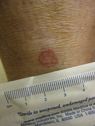
A skin condition, also known as cutaneous condition, is any medical condition that affects the integumentary system—the organ system that encloses the body and includes skin, nails, and related muscle and glands. The major function of this system is as a barrier against the external environment.

Keratin 16 is a protein that in humans is encoded by the KRT16 gene.

Darier's disease (DAR) is a rare, inherited skin disorder that presents with multiple greasy, crusting, thick brown bumps that merge into patches. It is an autosomal dominant disorder discovered by French dermatologist Ferdinand-Jean Darier.

A seborrheic keratosis is a non-cancerous (benign) skin tumour that originates from cells, namely keratinocytes, in the outer layer of the skin called the epidermis. Like liver spots, seborrheic keratoses are seen more often as people age.

Actinic keratosis (AK), sometimes called solar keratosis or senile keratosis, is a pre-cancerous area of thick, scaly, or crusty skin. Actinic keratosis is a disorder of epidermal keratinocytes that is induced by ultraviolet (UV) light exposure. These growths are more common in fair-skinned people and those who are frequently in the sun. They are believed to form when skin gets damaged by UV radiation from the sun or indoor tanning beds, usually over the course of decades. Given their pre-cancerous nature, if left untreated, they may turn into a type of skin cancer called squamous cell carcinoma. Untreated lesions have up to a 20% risk of progression to squamous cell carcinoma, so treatment by a dermatologist is recommended.

Palmoplantar keratodermas are a heterogeneous group of disorders characterized by abnormal thickening of the stratum corneum of the palms and soles.

Meleda disease (MDM) or "mal de Meleda", also called Mljet disease, keratosis palmoplantaris and transgradiens of Siemens, is an extremely rare autosomal recessive congenital skin disorder in which dry, thick patches of skin develop on the soles of the hands and feet, a condition known as palmoplantar hyperkeratosis. Meleda Disease is a skin condition which usually can be identified not long after birth. This is a genetic condition but it is very rare. The hands and feet usually are the first to show signs of the disease but the disease can advance to other parts of the body. Signs of the disease include thickening of the skin, on hands and soles of feet, which can turn red in color. There currently is no cure and treatment is limited, but Acitretin can be used in severe cases.

Epidermodysplasia verruciformis (EV) is a skin condition characterised by warty skin lesions. It results from an abnormal susceptibility to HPV infection (HPV) and is associated with a high lifetime risk of squamous cell carcinomas in skin. It generally presents with scaly spots and small bumps particularly on the hands, feet, face and neck; typically beginning in childhood or in a young adult. The bumps tend to be flat, grow in number and then merge to form plaques. On the trunk, it typically appears like pityriasis versicolor; lesions there being slightly scaly and tan, brown, red or looking pale. On the elbows, it may appear like psoriasis. On the forehead, neck and trunk, the lesions may appear like seborrheic keratosis.
ATP2A2 also known as sarcoplasmic/endoplasmic reticulum calcium ATPase 2 (SERCA2) is an ATPase associated with Darier's disease and Acrokeratosis verruciformis.
Florid cutaneous papillomatosis (FCP), is an obligate paraneoplastic syndrome.

Urbach–Wiethe disease is a very rare recessive genetic disorder, with approximately 400 reported cases since its discovery. It was first officially reported in 1929 by Erich Urbach and Camillo Wiethe, although cases may be recognized dating back as early as 1908.

Acrokeratoelastoidosis of Costa or Acrokeratoelastoidosis is a hereditary form of marginal keratoderma, and can be defined as a palmoplantar keratoderma. It is distinguished by tiny, firm pearly or warty papules on the sides of the hands and, occasionally, the feet. It is less common than the hereditary type of marginal keratoderma, keratoelastoidosis marginalis.

Porokeratosis is a specific disorder of keratinization that is characterized histologically by the presence of a cornoid lamella, a thin column of closely stacked, parakeratotic cells extending through the stratum corneum with a thin or absent granular layer.

Inflammatory Linear Verrucous Epidermal Nevus is a rare disease of the skin that presents as multiple, discrete, red papules that tend to coalesce into linear plaques that follow the Lines of Blaschko. The plaques can be slightly warty (psoriaform) or scaly (eczema-like). ILVEN is caused by somatic mutations that result in genetic mosaicism. There is no cure, but different medical treatments can alleviate the symptoms.
Actinic granuloma (AG) was first described by O'Brien in 1975 as a rare granulomatous disease. Lesions appear on sun-exposed areas, usually on the face, neck, and scalp, with a slight preference for middle-aged women. They are typically asymptomatic, single or multiple, annular or polycyclic lesions measuring up to 6 cm in diameter, with slow centrifugal expansion, an erythematous elevated edge, and a hypopigmented, atrophic center.
Granuloma multiforme is a cutaneous condition most commonly seen in central Africa, and rarely elsewhere, characterized by skin lesions that are on the upper trunk and arms in sun-exposed areas. It may be confused with tuberculoid leprosy, with which it has clinical similarities. The condition was first noted by Gosset in the 1940s, but it was not until 1964 that Leiker coined the term to describe "a disease resembling leprosy" in his study in Nigeria.
Progressive nodular histiocytosis is a cutaneous condition clinically characterized by the development of two types of skin lesions: superficial papules and deeper larger subcutaneous nodules. Progressive nodular histiocytosis was first reported in 1978 by Taunton et al. It is a subclass of non-Langerhans cell histiocytosis and a subgroup of xanthogranuloma.

Bowenoid papulosis is a cutaneous condition characterized by the presence of pigmented verrucous papules on the body of the penis. They are associated with human papillomavirus, the causative agent of genital warts. The lesions have a typical dysplastic histology and are generally considered benign, although a small percentage will develop malignant characteristics.
Howel–Evans syndrome is an extremely rare condition involving thickening of the skin in the palms of the hands and the soles of the feet (hyperkeratosis). This familial disease is associated with a high lifetime risk of esophageal cancer. For this reason, it is sometimes known as tylosis with oesophageal cancer (TOC).


