Anti-centromere antibodies

Last updated
Immunofluorescence staining pattern of anti-centromere antibodies on HEp-20-10 cells CENTROMERE.jpg
Immunofluorescence staining pattern of anti-centromere antibodies on HEp-20-10 cells

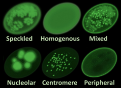
Anti-centromere antibodies (ACAs; often styled solid, anticentromere) are autoantibodies specific to centromere and kinetochore function. They occur in some autoimmune diseases, frequently in limited systemic scleroderma (formerly called CREST syndrome), and occasionally in the diffuse form of scleroderma. They are rare in other rheumatic diseases and in healthy persons.

Anti-centromere antibodies are found in approximately 60% of patients with limited systemic scleroderma and in 15% of those with the diffuse form of scleroderma. The specificity of this test is >98%. Thus, a positive anti-centromere antibody finding is strongly suggestive of limited systemic scleroderma. Anti-centromere antibodies present early in the course of disease and are notably predictive of limited cutaneous involvement and a decreased likelihood of aggressive internal organ involvement, such as fibrosis in the lungs. [1]

When present in primary biliary cirrhosis, ACAs are prognostic of portal hypertension such that serum ACA levels correlate with the severity of portal hypertension. [2]

Related Research Articles

<span class="mw-page-title-main">Systemic scleroderma</span> Medical condition

Systemic scleroderma, or systemic sclerosis, is an autoimmune rheumatic disease characterised by excessive production and accumulation of collagen, called fibrosis, in the skin and internal organs and by injuries to small arteries. There are two major subgroups of systemic sclerosis based on the extent of skin involvement: limited and diffuse. The limited form affects areas below, but not above, the elbows and knees with or without involvement of the face. The diffuse form also affects the skin above the elbows and knees and can also spread to the torso. Visceral organs, including the kidneys, heart, lungs, and gastrointestinal tract can also be affected by the fibrotic process. Prognosis is determined by the form of the disease and the extent of visceral involvement. Patients with limited systemic sclerosis have a better prognosis than those with the diffuse form. Death is most often caused by lung, heart, and kidney involvement. The risk of cancer is increased slightly.

<span class="mw-page-title-main">Caroli disease</span> Medical condition

Caroli disease is a rare inherited disorder characterized by cystic dilatation of the bile ducts within the liver. There are two patterns of Caroli disease: focal or simple Caroli disease consists of abnormally widened bile ducts affecting an isolated portion of liver. The second form is more diffuse, and when associated with portal hypertension and congenital hepatic fibrosis, is often referred to as "Caroli syndrome". The underlying differences between the two types are not well understood. Caroli disease is also associated with liver failure and polycystic kidney disease. The disease affects about one in 1,000,000 people, with more reported cases of Caroli syndrome than of Caroli disease.

<span class="mw-page-title-main">Antinuclear antibody</span> Autoantibody that binds to contents of the cell nucleus

Antinuclear antibodies are autoantibodies that bind to contents of the cell nucleus. In normal individuals, the immune system produces antibodies to foreign proteins (antigens) but not to human proteins (autoantigens). In some cases, antibodies to human antigens are produced.

<span class="mw-page-title-main">Autoimmune hepatitis</span> Chronic, autoimmune disease of the liver

Autoimmune hepatitis, formerly known as lupoid hepatitis, plasma cell hepatitis, or autoimmune chronic active hepatitis, is a chronic, autoimmune disease of the liver that occurs when the body's immune system attacks liver cells, causing the liver to be inflamed. Common initial symptoms may include fatigue, nausea, muscle aches, or weight loss or signs of acute liver inflammation including fever, jaundice, and right upper quadrant abdominal pain. Individuals with autoimmune hepatitis often have no initial symptoms and the disease may be detected by abnormal liver function tests and increased protein levels during routine bloodwork or the observation of an abnormal-looking liver during abdominal surgery.

<span class="mw-page-title-main">Primary biliary cholangitis</span> Autoimmune disease of the liver

Primary biliary cholangitis (PBC), previously known as primary biliary cirrhosis, is an autoimmune disease of the liver. It results from a slow, progressive destruction of the small bile ducts of the liver, causing bile and other toxins to build up in the liver, a condition called cholestasis. Further slow damage to the liver tissue can lead to scarring, fibrosis, and eventually cirrhosis.

<span class="mw-page-title-main">Portal hypertension</span> Abnormally increased portal venous pressure

Portal hypertension is defined as increased portal venous pressure, with a hepatic venous pressure gradient greater than 5 mmHg. Normal portal pressure is 1–4 mmHg; clinically insignificant portal hypertension is present at portal pressures 5–9 mmHg; clinically significant portal hypertension is present at portal pressures greater than 10 mmHg. The portal vein and its branches supply most of the blood and nutrients from the intestine to the liver.

<span class="mw-page-title-main">Primary sclerosing cholangitis</span> Medical condition

Primary sclerosing cholangitis (PSC) is a long-term progressive disease of the liver and gallbladder characterized by inflammation and scarring of the bile ducts, which normally allow bile to drain from the gallbladder. Affected individuals may have no symptoms or may experience signs and symptoms of liver disease, such as yellow discoloration of the skin and eyes, itching, and abdominal pain.

<span class="mw-page-title-main">CREST syndrome</span> Medical condition

CREST syndrome, also known as the limited cutaneous form of systemic sclerosis (lcSSc), is a multisystem connective tissue disorder. The acronym "CREST" refers to the five main features: calcinosis, Raynaud's phenomenon, esophageal dysmotility, sclerodactyly, and telangiectasia.

An autoantibody is an antibody produced by the immune system that is directed against one or more of the individual's own proteins. Many autoimmune diseases are associated with such antibodies.

<span class="mw-page-title-main">Morphea</span> Form of scleroderma involving isolated patches of hardened skin

Morphea is a form of scleroderma that mainly involves isolated patches of hardened skin on the face, hands, and feet, or anywhere else on the body, usually with no internal organ involvement. However, in Deep Morphea inflammation and sclerosis can be found in the deep dermis, panniculus, fascia, superficial muscle and bone.

<span class="mw-page-title-main">Gastric antral vascular ectasia</span> Medical condition of the stomach

Gastric antral vascular ectasia (GAVE) is an uncommon cause of chronic gastrointestinal bleeding or iron deficiency anemia. The condition is associated with dilated small blood vessels in the pyloric antrum, which is a distal part of the stomach. The dilated vessels result in intestinal bleeding. It is also called watermelon stomach because streaky long red areas that are present in the stomach may resemble the markings on watermelon.

<span class="mw-page-title-main">Anti-mitochondrial antibody</span> Autoantibodies against liver mitochondria, indicating primary biliary cholangitis

Anti-mitochondrial antibodies (AMA) are autoantibodies, consisting of immunoglobulins formed against mitochondria, primarily the mitochondria in cells of the liver.

Mixed connective tissue disease, commonly abbreviated as MCTD, is an autoimmune disease characterized by the presence of elevated blood levels of a specific autoantibody, now called anti-U1 ribonucleoprotein (RNP) together with a mix of symptoms of systemic lupus erythematosus (SLE), scleroderma, and polymyositis. The idea behind the "mixed" disease is that this specific autoantibody is also present in other autoimmune diseases such as systemic lupus erythematosus, polymyositis, scleroderma, etc. MCTD was characterized as an individual disease in 1972 by Sharp et al., and the term was introduced by Leroy in 1980.

Anti-glycoprotein-210 antibodies are directed at gp210 and are found within primary biliary cirrhosis (PBC) patients in high frequency. AGPA recognize the cytoplasmic-oriented carboxyl terminus (tail) of the protein. While AGPA is found as a prognostic marker in only a minority of PBC patients, those that did had higher mortality and were predicted a poor outcome. In addition, patients that responded to ursodeoxycholic acid (UDCA) therapy and, therefore, had AGPA reductions failed to develop end-stage liver disease relative to untreated cohort with anti-gp210 Ab. PBC patients with potentially destructive AGPA have increased expression of Nup210 in the bile duct, a potential immune tolerance-escaping factor.

<span class="mw-page-title-main">Portal hypertensive gastropathy</span> Changes in the mucosa of the stomach in patients with portal hypertension

Portal hypertensive gastropathy refers to changes in the mucosa of the stomach in patients with portal hypertension; by far the most common cause of this is cirrhosis of the liver. These changes in the mucosa include friability of the mucosa and the presence of ectatic blood vessels at the surface. Patients with portal hypertensive gastropathy may experience bleeding from the stomach, which may uncommonly manifest itself in vomiting blood or melena; however, portal hypertension may cause several other more common sources of upper gastrointestinal bleeding, such as esophageal varices and gastric varices. On endoscopic evaluation of the stomach, this condition shows a characteristic mosaic or "snake-skin" appearance to the mucosa of the stomach.

Anti-topoisomerase antibodies (ATA) are autoantibodies directed against topoisomerase and found in several diseases, most importantly scleroderma. Diseases with ATA are autoimmune disease because they react with self-proteins. They are also referred to as anti-DNA topoisomerase I antibody.

<span class="mw-page-title-main">Cirrhosis</span> Chronic disease of the liver, characterized by fibrosis

Cirrhosis, also known as liver cirrhosis or hepatic cirrhosis, and end-stage liver disease, is the impaired liver function caused by the formation of scar tissue known as fibrosis due to damage caused by liver disease. Damage to the liver leads to repair of liver tissue and subsequent formation of scar tissue. Over time, scar tissue can replace normal functioning tissue, leading to the impaired liver function of cirrhosis. The disease typically develops slowly over months or years. Early symptoms may include tiredness, weakness, loss of appetite, unexplained weight loss, nausea and vomiting, and discomfort in the right upper quadrant of the abdomen. As the disease worsens, symptoms may include itchiness, swelling in the lower legs, fluid build-up in the abdomen, jaundice, bruising easily, and the development of spider-like blood vessels in the skin. The fluid build-up in the abdomen may develop into spontaneous infections. More serious complications include hepatic encephalopathy, bleeding from dilated veins in the esophagus, stomach, or intestines, and liver cancer.

<span class="mw-page-title-main">Scleroderma</span> Group of autoimmune diseases resulting in abnormal growth of connective tissue

Scleroderma is a group of autoimmune diseases that may result in changes to the skin, blood vessels, muscles, and internal organs. The disease can be either localized to the skin or involve other organs, as well. Symptoms may include areas of thickened skin, stiffness, feeling tired, and poor blood flow to the fingers or toes with cold exposure. One form of the condition, known as CREST syndrome, classically results in calcium deposits, Raynaud's syndrome, esophageal problems, thickening of the skin of the fingers and toes, and areas of small, dilated blood vessels.

Anti-histone antibodies are autoantibodies that are a subset of the anti-nuclear antibody family, which specifically target histone protein subunits or histone complexes. They were first reported by Henry Kunkel, H.R. Holman, and H.R.G. Dreicher in their studies of cellular causes of lupus erythematosus in 1959–60. Today, anti-histone antibodies are still used as a marker for systemic lupus erythematosus, but are also implicated in other autoimmune diseases like Sjögren syndrome, dermatomyositis, or rheumatoid arthritis. Anti-histone antibodies can be used as a marker for drug-induced lupus.

Anti-Scl-70 is an anti-topoisomerase antibody-type of anti-nuclear autoantibodies, seen mainly in diffuse systemic scleroderma, but is also seen in 10–18% of cases of the more limited form of systemic scleroderma called CREST syndrome. Anti Scl-70 antibodies are associated with more severe scleroderma disease.

References

  1. JB Imboden, DB Hellmann, JH Stone. Current Rheumatology Diagnosis & Treatment, Second Edition. McGraw-Hill, 2007.
  2. Nakamura M, Kondo H, Mori T, et al. (2007). "Anti-gp210 and anti-centromere antibodies are different risk factors for the progression of primary biliary cirrhosis". Hepatology. 45 (1): 118–27. doi: 10.1002/hep.21472 . PMID   17187436.
  3. Al-Mughales JA (2022). "Anti-Nuclear Antibodies Patterns in Patients With Systemic Lupus Erythematosus and Their Correlation With Other Diagnostic Immunological Parameters". Front Immunol. 13: 850759. doi: 10.3389/fimmu.2022.850759 . PMC   8964090 . PMID   35359932.
    Minor edits by Mikael Häggström, MD
    - Attribution 4.0 International (CC BY 4.0) license