
In bacteriology, gram-positive bacteria are bacteria that give a positive result in the Gram stain test, which is traditionally used to quickly classify bacteria into two broad categories according to their type of cell wall.

Microtubules are polymers of tubulin that form part of the cytoskeleton and provide structure and shape to eukaryotic cells. Microtubules can grow as long as 50 micrometres and are highly dynamic. The outer diameter of a microtubule is between 23 and 27 nm while the inner diameter is between 11 and 15 nm. They are formed by the polymerization of a dimer of two globular proteins, alpha and beta tubulin into protofilaments that can then associate laterally to form a hollow tube, the microtubule. The most common form of a microtubule consists of 13 protofilaments in the tubular arrangement.
In cell biology, an organelle is a specialized subunit, usually within a cell, that has a specific function. The name organelle comes from the idea that these structures are parts of cells, as organs are to the body, hence organelle, the suffix -elle being a diminutive. Organelles are either separately enclosed within their own lipid bilayers or are spatially distinct functional units without a surrounding lipid bilayer. Although most organelles are functional units within cells, some functional units that extend outside of cells are often termed organelles, such as cilia, the flagellum and archaellum, and the trichocyst.

A flagellum is a hairlike appendage that protrudes from certain plant and mammalian sperm cells, and from a wide range of microorganisms to provide motility. Many protists with flagella are termed as flagellates.

The cilium, plural cilia is a membrane-bound organelle found on most types of cell, and certain microorganisms known as ciliates. Cilia are absent in bacteria and archaea. The cilium has the shape of a slender threadlike projection that extends from the surface of the much larger cell body. Eukaryotic flagella found on sperm cells and many protozoans have a similar structure to motile cilia that enables swimming through liquids; they are longer than cilia and have a different undulating motion.

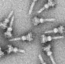
The evolution of flagella is of great interest to biologists because the three known varieties of flagella – each represent a sophisticated cellular structure that requires the interaction of many different systems.
The microtubule-organizing center (MTOC) is a structure found in eukaryotic cells from which microtubules emerge. MTOCs have two main functions: the organization of eukaryotic flagella and cilia and the organization of the mitotic and meiotic spindle apparatus, which separate the chromosomes during cell division. The MTOC is a major site of microtubule nucleation and can be visualized in cells by immunohistochemical detection of γ-tubulin. The morphological characteristics of MTOCs vary between the different phyla and kingdoms. In animals, the two most important types of MTOCs are 1) the basal bodies associated with cilia and flagella and 2) the centrosome associated with spindle formation.

Rod cells are photoreceptor cells in the retina of the eye that can function in lower light better than the other type of visual photoreceptor, cone cells. Rods are usually found concentrated at the outer edges of the retina and are used in peripheral vision. On average, there are approximately 92 million rod cells in the human retina. Rod cells are more sensitive than cone cells and are almost entirely responsible for night vision. However, rods have little role in color vision, which is the main reason why colors are much less apparent in dim light.

An axoneme, also called an axial filament is the microtubule-based cytoskeletal structure that forms the core of a cilium or flagellum. Cilia and flagella are found on many cells, organisms, and microorganisms, to provide motility. The axoneme serves as the "skeleton" of these organelles, both giving support to the structure and, in some cases, the ability to bend. Though distinctions of function and length may be made between cilia and flagella, the internal structure of the axoneme is common to both.
The bacterium, despite its simplicity, contains a well-developed cell structure which is responsible for some of its unique biological structures and pathogenicity. Many structural features are unique to bacteria and are not found among archaea or eukaryotes. Because of the simplicity of bacteria relative to larger organisms and the ease with which they can be manipulated experimentally, the cell structure of bacteria has been well studied, revealing many biochemical principles that have been subsequently applied to other organisms.
The radial spoke is a multi-unit protein structure found in the axonemes of eukaryotic cilia and flagella. Although experiments have determined the importance of the radial spoke in the proper function of these organelles, its structure and mode of action remain poorly understood.

Tetraselmis is a genus of phytoplankton. Tetraselmis is a green algal genus within the order Chlorodendrales, and they are characterized by their intensely-colored green chloroplast, their flagellated cell bodies, the presence of a pyrenoid within the chloroplast, and a scale-produced thecal-wall. Species within this genus are found in both marine and freshwater ecosystems across the globe; their habitat range is mainly limited by water depth due to their photosynthetic nature. Thus, they live in diverse water environments if enough nutrients and light are available for net photosynthetic activity. Tetraselmis species have proven to be useful for both research and industry. Tetraselmis species have been studied for understanding plankton growth rates, and recently a colonial species is being used to gain an understanding of multicellularity evolution. Additionally, many species are currently being examined for their use as biofuels due to their high lipid content.
An undulipodium or a 9+2 organelle is a motile filamentous intracellular projection of eukaryotic cells. It is basically synonymous to flagella and cilia which are differing terms for similar molecular structures used on different types of cells, and usually correspond to different waveforms.
The spindle pole body (SPB) is the microtubule organizing center in yeast cells, functionally equivalent to the centrosome. Unlike the centrosome the SPB does not contain centrioles. The SPB organises the microtubule cytoskeleton which plays many roles in the cell. It is important for organising the spindle and thus in cell division.

Mastigonemes are lateral "hairs" that attach to protistan flagella. Flimsy hairs attach to the flagella of euglenid flagellates, while stiff hairs occur in stramenopile and cryptophyte protists. Stramenopile hairs are approximately 15 nm in diameter, and usually consist of flexible basal part that inserts into the cell membrane, a tubular shaft that itself terminates in smaller "hairs". They reverse the thrust caused when a flagellum beats. The consequence is that the cell is drawn into the water and particles of food are drawn to the surface of heterotrophic species.
Type three secretion system is a protein appendage found in several Gram-negative bacteria.

Sperm motility describes the ability of sperm to move properly through the female reproductive tract or through water to reach the egg. Sperm motility can also be thought of as the quality, which is a factor in successful conception; sperm that do not "swim" properly will not reach the egg in order to fertilize it. Sperm motility in mammals also facilitates the passage of the sperm through the cumulus oophorus and the zona pellucida, which surround the mammalian oocyte.
The archaellum is a unique structure on the cell surface of many archaea, that allows for swimming motility. The archaellum consists of a rigid helical filament that is attached to the cell membrane by a molecular motor. This molecular motor – composed of cytosolic, membrane, and pseudo-periplasmic proteins – is responsible for the assembly of the filament and, once assembled, for its rotation. The rotation of the filament propels archaeal cells in liquid medium, in a manner similar to the propeller of a boat. The bacterial analog of the archaellum is the flagellum, which is also responsible for their swimming motility and can also be compared to a rotating corkscrew. Although the movement of archaella and flagella is sometimes described as "whip-like", this is incorrect, as only cilia from Eukaryotes move in this manner. Indeed, even "flagellum" is a misnomer, as bacterial flagella work are also propeller-like structures
Aquaspirillum (ak'wă-spī-ril'ŭm) is a genus of helical aerobic bacteria in the family Neisseriaceae that lives in freshwater.
Stygiella /ˌstɪ.d͡ʒiˈɛ.lə/ is a genus of free-living marine flagellates belonging to the family Stygiellidae in the jakobids (excavata).
