Arthur M. Lesk

Last updated
Arthur Lesk

Arthur M. Lesk ISMB 2015.jpg
Arthur M. Lesk speaking at the ISMB conference in 2015.
Born
Arthur Mallay Lesk
Alma mater
Known forIntroduction to Bioinformatics [1] and other textbooks
Scientific career
Institutions
Thesis Valence Bond Configuration Interaction Studies on the Chemical Bonding of the Noble Gases  (1966)
Website bmb.psu.edu/directory/aml25

Arthur Mallay Lesk, is a protein science researcher, who is a professor of biochemistry and molecular biology at the Pennsylvania State University in University Park.

Contents

Education

Lesk received a bachelor's degree, magna cum laude, from Harvard University in 1961. He received his doctoral degree from Princeton University in 1966. He also received a master's degree from the University of Cambridge in the United Kingdom in 1999. [2] [3]

Research

Lesk has made significant contributions to the study of protein evolution. [4] He and Cyrus Chothia, working at the Medical Research Council (UK) Laboratory of Molecular Biology in Cambridge, United Kingdom, discovered the relationship between changes in amino-acid sequence and changes in protein structure by analyzing the mechanism of evolution in protein families. [5] [6] This discovery has provided the quantitative basis for the most successful and widely used method of structure prediction, known as homology modelling.

Lesk and Chothia also studied the conformations of antigen-binding sites of immunoglobulins. They discovered the “canonical-structure model” for the conformation of the complementarity-determining regions of antibodies, and they applied this model to the analysis of antibody-germ-line genes, including the prediction of the structure of the corresponding proteins. This work has supported the “humanization” of antibodies for therapy in the treatment of cancer. “This approach to cancer therapy is based on the observation of H. Waldmann that rats can raise antibodies against human cancers, but that the rat antibodies lead to immune responses, similar to allergies, in human patients,” Lesk explains. “Humanization of these antibodies is the formation of hybrid molecules that are more human than rat, but that retain the therapeutic activity while reducing the patient’s immune response.”

Lesk’s work also involves the detailed comparison of proteins in different structural states as a means for understanding the mechanisms that enable the proteins to change conformation, both as part of their normal activity and in disease. The discovery and analysis of these mechanisms was the key to understanding conformation changes in serine protease inhibitors, also known as serpins, mutations of which are an important cause of several diseases, including emphysema and certain types of inherited mental illness.

Lesk used a systematic analysis of protein-folding patterns to develop a mathematical representation that aids in the recognition and classification of these patterns. He also wrote the first computer program to generate schematic diagrams of proteins using molecular graphics, and he developed many algorithms now used by other researchers to analyze the structures of proteins.

Lesk has served as chair of the Task Group on Biological Macromolecules for the Committee on Data for Science and Technology (CODATA), which aimed to foster worldwide coordination of databases in molecular biology to enhance their quality and utility. He has given invited lectures and presentations related to his research at universities and professional conferences worldwide.

Lesk is a member of the American Physical Society. He has published 189 scientific articles and 10 books related to his research. [1] [7] [8] [9] [10] [11] [12] [13] [14] [15] [16]

Prior to joining Penn State during the fall semester of 2003, Lesk was on the faculty of the clinical school at the University of Cambridge from 1990 to 2003. He was a group leader in the biocomputing program at the European Molecular Biology Laboratory in Heidelberg, Germany, from 1987 to 1990; a visiting scientist at MRC Laboratory of Molecular Biology in Cambridge, United Kingdom, between 1977 and 1990; and a professor of chemistry at Fairleigh Dickinson University in New Jersey from 1971 to 1987. He has held visiting fellowships at the University of Otago in New Zealand and Monash University in Australia. He also is a Life Member of Clare Hall, Cambridge in the United Kingdom.

In 2023 Lesk was awarded the Carl Brändén Award. [17] The award honors an outstanding protein scientist who has also made exceptional contributions in the areas of education and/or service to the field. [18]

Schematic diagrams of protein structures

Lesk, along with Karl D. Hardman, [19] wrote one of the first computer programs for generating the schematic diagram of protein structure. It is known to produce one of the best representations of the protein structures and employs the classification scheme for Ribbon Diagrams created by Jane Richardson. Most of the protein structure illustrations in Lesk's book (see reference list below) are generated using this program. Although these schematic diagrams are less detailed compared to other representations, such as pictures simulating wire models or space-filling models, the simplifications make them more effective in presenting the topological relationships among elements of secondary structure of the proteins. [20] This was then further improved by creating a program to produce stereoscopic pairs of diagrams. As a result, the viewer’s ability to perceive spatial relationship in complex molecules was enhanced. [20]

The basic operation of the program begins with the execution of line drawing. There are four phases involved in this program: [19]

  1. The input phase – Program reads the input files. There are two input files. They are the coordinates and the details of the contents and appearance of the picture.
  2. Picture generation – Geometric transformation of coordinates are generated by the program into picture elements. For example, a cylinder of appropriate size and orientation about the z-axis represents α-helix; each peptide plane is determined for Ribbon Diagrams and β-sheets; and spline fit is used for curved sheets.
  3. Hidden-line removal – This step is only required by the cylinders of α-helices and the arrows of β-sheets, not skeletal models. Picture of these structures are classified by three levels of “optical density” – transparent, translucent, or opaque. If lines are passing behind the transparent object, it is not changed. If it passes behind a translucent object, it is altered into dashed lines. If it is opaque, the lines passing through the object are removed completely. This step can be replaced with an alternative step to create a Colour-Raster Output. The lines are ignored and the windows are painted according to the user.
  4. Output – Character strings are extended to sets of line segments through a set of stroke tables. Line segments are placed into the two-dimensional space.

Personal life

Arthur Lesk's son, Victor Lesk, followed his father into the field of structural biology and bioinformatics, and has held a post-doctoral research position with Michael Sternberg at Imperial College London. [21] His daughter, Valerie Lesk, is an academic in the Division of Psychology at the University of Bradford, UK.

Related Research Articles

<span class="mw-page-title-main">Antibody</span> Protein(s) forming a major part of an organisms immune system

An antibody (Ab), also known as an immunoglobulin (Ig), is a large, Y-shaped protein used by the immune system to identify and neutralize foreign objects such as pathogenic bacteria and viruses. The antibody recognizes a unique molecule of the pathogen, called an antigen. Each tip of the "Y" of an antibody contains a paratope that is specific for one particular epitope on an antigen, allowing these two structures to bind together with precision. Using this binding mechanism, an antibody can tag a microbe or an infected cell for attack by other parts of the immune system, or can neutralize it directly.

<span class="mw-page-title-main">Protein</span> Biomolecule consisting of chains of amino acid residues

Proteins are large biomolecules and macromolecules that comprise one or more long chains of amino acid residues. Proteins perform a vast array of functions within organisms, including catalysing metabolic reactions, DNA replication, responding to stimuli, providing structure to cells and organisms, and transporting molecules from one location to another. Proteins differ from one another primarily in their sequence of amino acids, which is dictated by the nucleotide sequence of their genes, and which usually results in protein folding into a specific 3D structure that determines its activity.

<span class="mw-page-title-main">Protein tertiary structure</span> Three dimensional shape of a protein

Protein tertiary structure is the three-dimensional shape of a protein. The tertiary structure will have a single polypeptide chain "backbone" with one or more protein secondary structures, the protein domains. Amino acid side chains may interact and bond in a number of ways. The interactions and bonds of side chains within a particular protein determine its tertiary structure. The protein tertiary structure is defined by its atomic coordinates. These coordinates may refer either to a protein domain or to the entire tertiary structure. A number of tertiary structures may fold into a quaternary structure.

<span class="mw-page-title-main">Structural bioinformatics</span> Bioinformatics subfield

Structural bioinformatics is the branch of bioinformatics that is related to the analysis and prediction of the three-dimensional structure of biological macromolecules such as proteins, RNA, and DNA. It deals with generalizations about macromolecular 3D structures such as comparisons of overall folds and local motifs, principles of molecular folding, evolution, binding interactions, and structure/function relationships, working both from experimentally solved structures and from computational models. The term structural has the same meaning as in structural biology, and structural bioinformatics can be seen as a part of computational structural biology. The main objective of structural bioinformatics is the creation of new methods of analysing and manipulating biological macromolecular data in order to solve problems in biology and generate new knowledge.

An epitope, also known as antigenic determinant, is the part of an antigen that is recognized by the immune system, specifically by antibodies, B cells, or T cells. The part of an antibody that binds to the epitope is called a paratope. Although epitopes are usually non-self proteins, sequences derived from the host that can be recognized are also epitopes.

<span class="mw-page-title-main">Protein structure</span> Three-dimensional arrangement of atoms in an amino acid-chain molecule

Protein structure is the three-dimensional arrangement of atoms in an amino acid-chain molecule. Proteins are polymers – specifically polypeptides – formed from sequences of amino acids, which are the monomers of the polymer. A single amino acid monomer may also be called a residue, which indicates a repeating unit of a polymer. Proteins form by amino acids undergoing condensation reactions, in which the amino acids lose one water molecule per reaction in order to attach to one another with a peptide bond. By convention, a chain under 30 amino acids is often identified as a peptide, rather than a protein. To be able to perform their biological function, proteins fold into one or more specific spatial conformations driven by a number of non-covalent interactions, such as hydrogen bonding, ionic interactions, Van der Waals forces, and hydrophobic packing. To understand the functions of proteins at a molecular level, it is often necessary to determine their three-dimensional structure. This is the topic of the scientific field of structural biology, which employs techniques such as X-ray crystallography, NMR spectroscopy, cryo-electron microscopy (cryo-EM) and dual polarisation interferometry, to determine the structure of proteins.

<span class="mw-page-title-main">Epitope mapping</span> Identifying the binding site of an antibody on its target antigen

In immunology, epitope mapping is the process of experimentally identifying the binding site, or epitope, of an antibody on its target antigen. Identification and characterization of antibody binding sites aid in the discovery and development of new therapeutics, vaccines, and diagnostics. Epitope characterization can also help elucidate the binding mechanism of an antibody and can strengthen intellectual property (patent) protection. Experimental epitope mapping data can be incorporated into robust algorithms to facilitate in silico prediction of B-cell epitopes based on sequence and/or structural data.

<span class="mw-page-title-main">Michael Levitt</span> Nobel laureate, biophysicist, and professor of structural biology (born 1947)

Michael Levitt, is a South African-born biophysicist and a professor of structural biology at Stanford University, a position he has held since 1987. Levitt received the 2013 Nobel Prize in Chemistry, together with Martin Karplus and Arieh Warshel, for "the development of multiscale models for complex chemical systems". In 2018, Levitt was a founding co-editor of the Annual Review of Biomedical Data Science.

Molecular graphics is the discipline and philosophy of studying molecules and their properties through graphical representation. IUPAC limits the definition to representations on a "graphical display device". Ever since Dalton's atoms and Kekulé's benzene, there has been a rich history of hand-drawn atoms and molecules, and these representations have had an important influence on modern molecular graphics.

<span class="mw-page-title-main">Beta barrel</span>

In protein structures, a beta barrel is a beta sheet composed of tandem repeats that twists and coils to form a closed toroidal structure in which the first strand is bonded to the last strand. Beta-strands in many beta-barrels are arranged in an antiparallel fashion. Beta barrel structures are named for resemblance to the barrels used to contain liquids. Most of them are water-soluble proteins and frequently bind hydrophobic ligands in the barrel center, as in lipocalins. Others span cell membranes and are commonly found in porins. Porin-like barrel structures are encoded by as many as 2–3% of the genes in Gram-negative bacteria. It has been shown that more than 600 proteins with various function contain the beta barrel structure.

<span class="mw-page-title-main">MRC Laboratory of Molecular Biology</span> Research institute in Cambridge, England

The Medical Research Council (MRC) Laboratory of Molecular Biology (LMB) is a research institute in Cambridge, England, involved in the revolution in molecular biology which occurred in the 1950–60s. Since then it has remained a major medical research laboratory at the forefront of scientific discovery, dedicated to improving the understanding of key biological processes at atomic, molecular and cellular levels using multidisciplinary methods, with a focus on using this knowledge to address key issues in human health.

<span class="mw-page-title-main">Complementarity-determining region</span> Part of the variable chains in immunoglobulins and T cell receptors

Complementarity-determining regions (CDRs) are part of the variable chains in immunoglobulins (antibodies) and T cell receptors, generated by B-cells and T-cells respectively, where these molecules bind to their specific antigen. A set of CDRs constitutes a paratope. As the most variable parts of the molecules, CDRs are crucial to the diversity of antigen specificities generated by lymphocytes.

<span class="mw-page-title-main">Protein domain</span> Self-stable region of a proteins chain that folds independently from the rest

In molecular biology, a protein domain is a region of a protein's polypeptide chain that is self-stabilizing and that folds independently from the rest. Each domain forms a compact folded three-dimensional structure. Many proteins consist of several domains, and a domain may appear in a variety of different proteins. Molecular evolution uses domains as building blocks and these may be recombined in different arrangements to create proteins with different functions. In general, domains vary in length from between about 50 amino acids up to 250 amino acids in length. The shortest domains, such as zinc fingers, are stabilized by metal ions or disulfide bridges. Domains often form functional units, such as the calcium-binding EF hand domain of calmodulin. Because they are independently stable, domains can be "swapped" by genetic engineering between one protein and another to make chimeric proteins.
<span class="mw-page-title-main">Ribbon diagram</span> 3D schematic representation of protein structure

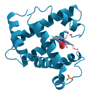
Ribbon diagrams, also known as Richardson diagrams, are 3D schematic representations of protein structure and are one of the most common methods of protein depiction used today. The ribbon depicts the general course and organisation of the protein backbone in 3D and serves as a visual framework for hanging details of the entire atomic structure, such as the balls for the oxygen atoms attached to myoglobin's active site in the adjacent figure. Ribbon diagrams are generated by interpolating a smooth curve through the polypeptide backbone. α-helices are shown as coiled ribbons or thick tubes, β-sheets as arrows, and non-repetitive coils or loops as lines or thin tubes. The direction of the polypeptide chain is shown locally by the arrows, and may be indicated overall by a colour ramp along the length of the ribbon.

<span class="mw-page-title-main">Janet Thornton</span> British bioinformatician and academic

Dame Janet Maureen Thornton, is a senior scientist and director emeritus at the European Bioinformatics Institute (EBI), part of the European Molecular Biology Laboratory (EMBL). She is one of the world's leading researchers in structural bioinformatics, using computational methods to understand protein structure and function. She served as director of the EBI from October 2001 to June 2015, and played a key role in ELIXIR.

<span class="mw-page-title-main">Protein dynamics</span>

Proteins are generally thought to adopt unique structures determined by their amino acid sequences. However, proteins are not strictly static objects, but rather populate ensembles of conformations. Transitions between these states occur on a variety of length scales and time scales , and have been linked to functionally relevant phenomena such as allosteric signaling and enzyme catalysis.

<span class="mw-page-title-main">Cyrus Chothia</span> English biochemist (1942–2019)

Cyrus Homi Chothia was an English biochemist who was an emeritus scientist at the Medical Research Council (MRC) Laboratory of Molecular Biology (LMB) at the University of Cambridge and emeritus fellow of Wolfson College, Cambridge.

Michael Joseph Ezra Sternberg is a professor at Imperial College London, where he is director of the Centre for Integrative Systems Biology and Bioinformatics and Head of the Structural bioinformatics Group.

<span class="mw-page-title-main">Peter Colman</span>

Peter Malcolm Colman is the head of the structural biology division at the Walter and Eliza Hall Institute of Medical Research in Melbourne, Australia.

<span class="mw-page-title-main">Anna Tramontano</span> Italian computational biologist

Anna Tramontano was an Italian computational biologist and chair professor of biochemistry at the Sapienza University of Rome. From 2011 to 2014 she was a member of the Scientific Council of the European Research Council (ERC). She was an associate editor for the journal Bioinformatics from 2005 until 2016 editing papers in the area of structural bioinformatics.

References

  1. 1 2 Lesk, Arthur M. (2002). Introduction to bioinformatics. Oxford [Oxfordshire]: Oxford University Press. ISBN   0-19-925196-7.
  2. "Academy of Europe: Lesk Arthur". www.ae-info.org. Retrieved 21 December 2022.
  3. "Reporter 28/4/99: Graces to be submitted to the Regent House at a Congregation on 8 May 1999". www.admin.cam.ac.uk. Retrieved 21 December 2022.
  4. Chothia, C.; Lesk, A. M.; Tramontano, A.; Levitt, M.; Smith-Gill, S. J.; Air, G.; Sheriff, S.; Padlan, E. A.; Davies, D.; Tulip, W. R.; Colman, P. M.; Spinelli, S.; Alzari, P. M.; Poljak, R. J. (1989). "Conformations of immunoglobulin hypervariable regions". Nature. 342 (6252): 877–883. Bibcode:1989Natur.342..877C. doi:10.1038/342877a0. PMID   2687698. S2CID   4241051.
  5. Lesk, A.; Chothia, C. (1980). "Solvent accessibility, protein surfaces, and protein folding". Biophysical Journal. 32 (1): 35–47. Bibcode:1980BpJ....32...35L. doi:10.1016/S0006-3495(80)84914-9. PMC   1327253 . PMID   7248454.
  6. Chothia, C.; Lesk, A. (1986). "The relation between the divergence of sequence and structure in proteins". The EMBO Journal. 5 (4): 823–826. doi:10.1002/j.1460-2075.1986.tb04288.x. PMC   1166865 . PMID   3709526.
  7. Lesk, Arthur M. (1982). Introduction to physical chemistry . Englewood Cliffs, NJ: Prentice-Hall. ISBN   0-13-492710-9.
  8. Lesk, Arthur M. (1988). Computational molecular biology: sources and methods for sequence analysis . Oxford [Oxfordshire]: Oxford University Press. ISBN   0-19-854218-6.
  9. Lesk, Arthur M. (1991). Protein architecture: a practical approach. Ithaca, N.Y: IRL Press. ISBN   0-19-963055-0.
  10. Lesk, Arthur M. (2001). Introduction to protein architecture: the structural biology of proteins. Oxford [Oxfordshire]: Oxford University Press. ISBN   0-19-850474-8.
  11. Lesk, Arthur M. (2004). Introduction to symmetry and group theory for chemists. Boston: Kluwer Academic Publishers. ISBN   1-4020-2150-X.
  12. Lesk, Arthur M. (2004). Introduction to protein science: architecture, function and genomics. Oxford [Oxfordshire]: Oxford University Press. ISBN   0-19-926511-9.
  13. Lesk, Arthur M. (2005). Database annotation in molecular biology. New York: John Wiley. ISBN   0-470-85681-5.
  14. Lesk, Arthur M.; Anna Tramontano (2006). Protein Structure Prediction: Concepts and Applications. Weinheim: Wiley-VCH. ISBN   3-527-31167-X.
  15. Lesk, Arthur M. (2007). Introduction to genomics . Oxford [Oxfordshire]: Oxford University Press. ISBN   978-0-19-929695-8.
  16. Lesk, Arthur M. (2010). Introduction to Protein Science: Architecture, Function, and Genomics. Oxford University Press, USA. ISBN   978-0-19-954130-0.
  17. "The Protein Society announces its 2023 award winners". EurekAlert!. Retrieved 2024-01-07.
  18. "Protein Society Awards Nominations Form". www.proteinsociety.org. Retrieved 2024-01-07.
  19. 1 2 Lesk, A.; Hardman, K. (1985). Computer-generated pictures of proteins. Methods in Enzymology. Vol. 115. pp.  381–390. doi:10.1016/0076-6879(85)15027-5. ISBN   9780121820152. PMID   2934605.
  20. 1 2 Lesk, A.; Hardman, K. (1982). "Computer-generated schematic diagrams of protein structures". Science. 216 (4545): 539–540. Bibcode:1982Sci...216..539L. doi:10.1126/science.7071602. PMID   7071602.
  21. Dobbins, S. E.; Lesk, V. I.; Sternberg, M. J. E. (2008). "Insights into protein flexibility: The relationship between normal modes and conformational change upon protein-protein docking". Proceedings of the National Academy of Sciences. 105 (30): 10390–10395. Bibcode:2008PNAS..10510390D. doi: 10.1073/pnas.0802496105 . PMC   2475499 . PMID   18641126.