Balanol

Last updated
Balanol
Balanol.svg
Identifiers
  • 2-{[2,6-dihydroxy-4-({[(3S,4R)-3-[(4-hydroxybenzene)amido]azepan-4-yl]oxy}carbonyl)phenyl]carbonyl}-3-hydroxybenzoic acid
CAS Number
PubChem CID
IUPHAR/BPS
DrugBank
ChemSpider
UNII
Chemical and physical data
Formula C28H26N2O10
Molar mass 550.520 g·mol−1
 X mark.svgNYes check.svgY  (what is this?)    (verify)

Balanol is a fungal metabolite produced by the fungus Verticillium balanoides . [1] It is a potent inhibitor of the serine/threonine kinases protein kinase A (PKA) and protein kinase C (PKC), binding in a similar manner with that of ATP. [2] Balanol was discovered in 1993 in the search for novel inhibitors of PKC, a member of a family of serine/threonine kinases whose overactivation is associated with numerous human diseases of signal transduction including cancer. However, much of the research on balanol focuses on how chemical modifications of the molecular structure affect binding to PKA. [3] Indeed, balanol, its chemically altered analogs, and their interactions with PKA in particular are used to illuminate the roles of selectivity and protein flexibility in the inhibition of kinases. For instance, the X-ray crystal structure of balanol in complex with PKA was used in order to confer selectivity and to improve pharmacological efficacy of inhibitors of the H. sapiens Akt (PKB), another serine/threonine protein kinase implicated in the proper functioning of many cellular processes. [4]

Contents

Structure

The chemical structure was initially characterized through a combination of IR spectroscopy, hydrogen-1 NMR, carbon-13 NMR, and 2D NMR spectra data, and the crystal structure of balanol in complex with PKA was solved in 1999. [5] The balanol molecule consists of three regions: a benzophenone, hexahydroazepane, and 4-hydroxy benzoyl moiety. The benzophenone and hexahydroazepane moieties are connected via an ester linkage, and the azepane and benzoyl moieties are connected through an amide linkage. [6] Furthermore, balanol is sometimes referred to as ophiocordin, an antifungal agent produced by the fungus Cordyceps ophioglossoides whose structure is regioisometric to that of balanol; that is, in ophiocordin, the benzophenone is linked to the hexahydroazepane instead via an amide; the 4-hydroxy benzoyl group and hexahydroazepane are connected with an ester linkage. [7]

Balanol is a congener of ATP, with distinct regions of its molecular structure able to form bonds like the adenine ring, ribose, and phosphate groups of the ATP molecule. Specifically, balanol's 4-hydroxybenzamide moiety including the amide linker corresponds to the adenine of ATP, the hexahydroazepane moiety to the ribose region, and the benzophenone rings to ATP's triphosphates. [5]

Numerous balanol congeners have been synthesized in order to study the effect of chemical modifications on kinase binding and specificity. For example, modifications of the benzophenone rings, analogs of the phosphate groups of ATP, produce unusually potent and specific protein kinase inhibitors. [6] Additionally, many of these balanol congeners show substantial specificity toward PKA over PKC. Elimination of the hydroxyl group from the benzophenone ring (producing 10"-deoxybalanol), for instance, elicits selectivity two orders of magnitude greater for PKA over PKC. [8] Potent inhibitory activity was also shown to be dependent on the carboxylic acid of the benzophenone ring and the presence of a five- or seven-membered azepane ring. [3]

Balanol's flexible structure also plays an important role in its selectivity. Specifically, balanol's distal benzophenone ring is able to rotate. In fact, in complex with PKA, balanol's distal benzophenone ring was observed to be nearly orthogonal with the neighboring ring. [5] This flexibility could allow balanol to adapt to numerous protein microenvironments in order to exert its inhibitory properties on various protein kinases. Balanol's selectivity for some kinases and not others, in turn, could represent the varying degrees of flexibilities allowed by these kinases' catalytic ATP-binding sites. [6]

Biological activities

Balanol is one of the most potent, naturally occurring inhibitors of protein kinases PKC and PKA. [5] Balanol was originally discovered to inhibit PKC and many of its isoforms in humans (α, β-Ι, β-ΙΙ, γ, δ, ε, η), with an inhibition profile similar to that of staurosporine. [1]

Balanol prevents functioning by binding competitively to the catalytic domain of PKC and PKA with an affinity (Ki   4 nM) three orders of magnitude greater than for ATP. [6] Without ATP bound, these kinases are unable to catalyze the transfer of the γ-phosphate from ATP to the kinase's target substrate, and thus function is hindered.

Binding

Bound balanol extends 17.2 Å from the hydroxyl oxygen atom of its 4-hydroxybenzamide to the most distal carboxyl oxygen atom of its benzophenone moiety, fitting between the large and small catalytic lobes of PKA and extending from the inner edge to the outer mouth of the active-site ATP-binding cleft of the enzyme. The cleft's glycine-rich loop and small lobe allow for tight, induced-fit binding. [5]

Each subsite of the ATP-binding pocket in PKA is occupied by a substituent of the balanol molecule as observed in studies of recombinant mouse catalytic subunit of PKA. [5] The adenine subsite of PKA has hydrophobic components and has the potential to donate and accept electrons to make hydrogen bonds with one or two planar cyclic rings. Specifically, the backbone nitrogen of Val123 and the carbonyl oxygen atom of Glu121 can form hydrogen bonds with balanol's single hydroxyl group of the 4-hydroxyl benzoyl moiety. In the ATP molecule, the Val123 amide hydrogen atom accepts electrons from ATP's purine ring N1 atom while the Glu121 backbone carbonyl oxygen atom donates electrons to the ATP purine ring N6 atom. Mimetics of ATP like balanol can be anchored within the hydrophobic adenine-binding pocket because their planar substituents can make favorable nonpolar interactions. The ribose subsite is occupied by balanol's axepane ring. Specifically, the N1 atom of the azepane ring hydrogen bonds with the backbone carbonyl oxygen atom of Glu170 of the catalytic loop. Atoms within the azepane ring also can make favorable nonpolar contacts with residues Gly50, Flu127, and Glu170. While other interactions also occur, most of the polar and nonpolar interactions involve the benzophenone substituent of balanol, which interacts with residues comprising the phosphate-binding subsite. For instance, balanol's benzophenone ring interacts with numerous highly conserved residues in PKA such as Gly52, Phe54, Asp184 among others in addition to various nonconserved residues such as Leu74 and Gln84. Most of the conserved residues in PKA interact with ATP whereas interactions of ATP with the nonconserved residues do not occur. [5]

Six ordered water molecules also play critical roles in balanol binding to PKA. Several PKA residues such as Leu49 and Tyr330 hydrogen bond with conserved water molecules. These water molecules also mediate interactions between balanol and the residues of the catalytic cleft of PKA; for example, two water molecules form a bridge to allow balanol's N1’ atom to interact with the hydroxyl group of Tyr330. [5]

However, the tight interaction of balanol with the ATP-binding site that leads to balanol's potency as an inhibitor is not due to hydrogen bonding but rather because of the predominant nonpolar interactions the molecule makes. For instance, balanol's benzophenone rings are positioned beside the glycine-rich loop and further from the more polar catalytic loop. Compared to ATP's triphosphates, accommodating balanol's benzophenone rings induces the rearrangement of catalytic side-chains Phe54 and Ser53, enabling favorable polar and nonpolar interactions and likely contributing to balanol's potent inhibitory effect on these kinases. [5]

Action against other protein kinases

As balanol acts specifically at the ATP-binding site in the catalytic core of PKA and PKC, it was initially hypothesized that balanol could inhibit all serine/threonine kinases sharing a conserved ATP-binding site. However, balanol does not inhibit all serine/threonine kinases uniformly. Affinity to balanol varies greatly (Ki = 1.6–742 nM), with several members of the serine/threonine protein kinase family not being affected at all, even though affinity of these kinases to ATP varies little (13–60 μM). [6] Balanol exhibits the most potent inhibitory effect (Ki = 1.6–6.4  nM) on cGMP-dependent protein kinase (PKG), PKA, and PKC, including isoforms. [6] It exerts much less of an effect (Ki = 30 – 742 nM), if any, on Ca2+-calmodulin-regulated kinases, mitogen-activating protein kinase (MAPK/Erk1), and certain cyclin-dependent kinases. [6] Furthermore, balanol does not inhibit two tyrosine protein kinases, the SC kinase, nor the epidermal growth factor receptor kinase. [2]

Related Research Articles

Chymotrypsin Digestive enzyme

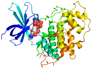
Chymotrypsin (EC 3.4.21.1, chymotrypsins A and B, alpha-chymar ophth, avazyme, chymar, chymotest, enzeon, quimar, quimotrase, alpha-chymar, alpha-chymotrypsin A, alpha-chymotrypsin) is a digestive enzyme component of pancreatic juice acting in the duodenum, where it performs proteolysis, the breakdown of proteins and polypeptides. Chymotrypsin preferentially cleaves peptide amide bonds where the side chain of the amino acid N-terminal to the scissile amide bond (the P1 position) is a large hydrophobic amino acid (tyrosine, tryptophan, and phenylalanine). These amino acids contain an aromatic ring in their side chain that fits into a hydrophobic pocket (the S1 position) of the enzyme. It is activated in the presence of trypsin. The hydrophobic and shape complementarity between the peptide substrate P1 side chain and the enzyme S1 binding cavity accounts for the substrate specificity of this enzyme. Chymotrypsin also hydrolyzes other amide bonds in peptides at slower rates, particularly those containing leucine and methionine at the P1 position.

Protein kinase enzyme that adds phosphate groups to other proteins

A protein kinase is a kinase which selectively modifies other proteins by covalently adding phosphates to them (phosphorylation) as opposed to kinases which modify lipids, carbohydrates, or other molecules. Phosphorylation usually results in a functional change of the target protein (substrate) by changing enzyme activity, cellular location, or association with other proteins. The human genome contains about 500 protein kinase genes and they constitute about 2% of all human genes. There are two main types of protein kinase. The great majority are serine/threonine kinases, which phosphorylate the hydroxyl groups of serines and threonines in their targets and the other are tyrosine kinases, although additional types exist. Protein kinases are also found in bacteria and plants. Up to 30% of all human proteins may be modified by kinase activity, and kinases are known to regulate the majority of cellular pathways, especially those involved in signal transduction.

Kinase Enzyme catalyzing transfer of phosphate groups onto specific substrates

In biochemistry, a kinase is an enzyme that catalyzes the transfer of phosphate groups from high-energy, phosphate-donating molecules to specific substrates. This process is known as phosphorylation, where the substrate gains a phosphate group and the high-energy ATP molecule donates a phosphate group. This transesterification produces a phosphorylated substrate and ADP. Conversely, it is referred to as dephosphorylation when the phosphorylated substrate donates a phosphate group and ADP gains a phosphate group. These two processes, phosphorylation and dephosphorylation, occur four times during glycolysis.

Active site Active region of an enzyme

In biology, the active site is the region of an enzyme where substrate molecules bind and undergo a chemical reaction. The active site consists of amino acid residues that form temporary bonds with the substrate and residues that catalyse a reaction of that substrate. Although the active site occupies only ~10–20% of the volume of an enzyme, it is the most important part as it directly catalyzes the chemical reaction. It usually consists of three to four amino acids, while other amino acids within the protein are required to maintain the tertiary structure of the enzymes.

In cell biology, protein kinase A (PKA) is a family of enzymes whose activity is dependent on cellular levels of cyclic AMP (cAMP). PKA is also known as cAMP-dependent protein kinase. PKA has several functions in the cell, including regulation of glycogen, sugar, and lipid metabolism.

GSK-3

Glycogen synthase kinase 3 (GSK-3) is a serine/threonine protein kinase that mediates the addition of phosphate molecules onto serine and threonine amino acid residues. First discovered in 1980 as a regulatory kinase for its namesake, glycogen synthase (GS), GSK-3 has since been identified as a protein kinase for over 100 different proteins in a variety of different pathways. In mammals, including humans, GSK-3 exists in two isoforms encoded by two paralogous genes GSK-3α (GSK3A) and GSK-3β (GSK3B). GSK-3 has been the subject of much research since it has been implicated in a number of diseases, including type 2 diabetes, Alzheimer's disease, inflammation, cancer, and bipolar disorder.

Protein kinase C, commonly abbreviated to PKC (EC 2.7.11.13), is a family of protein kinase enzymes that are involved in controlling the function of other proteins through the phosphorylation of hydroxyl groups of serine and threonine amino acid residues on these proteins, or a member of this family. PKC enzymes in turn are activated by signals such as increases in the concentration of diacylglycerol (DAG) or calcium ions (Ca2+). Hence PKC enzymes play important roles in several signal transduction cascades.

CAMK, also written as CaMK, is an abbreviation for the Ca2+/calmodulin-dependent protein kinase class of enzymes. CAMKs are activated by increases in the concentration of intracellular calcium ions (Ca2+) and calmodulin. When activated, the enzymes transfer phosphates from ATP to defined serine or threonine residues in other proteins, so they are serine/threonine-specific protein kinases. Activated CAMK is involved in the phosphorylation of transcription factors and therefore, in the regulation of expression of responding genes. CAMK also works to regulate the cell life cycle (i.e. programmed cell death), rearrangement of the cell's cytoskeletal network, and mechanisms involved in the learning and memory of an organism.

Serine/threonine-specific protein kinase

A serine/threonine protein kinase is a kinase enzyme that phosphorylates the OH group of serine or threonine. At least 125 of the 500+ human protein kinases are serine/threonine kinases (STK).

Phosphodiesterase 3

PDE3 is a phosphodiesterase. The PDEs belong to at least eleven related gene families, which are different in their primary structure, substrate affinity, responses to effectors, and regulation mechanism. Most of the PDE families are composed of more than one gene. PDE3 is clinically significant because of its role in regulating heart muscle, vascular smooth muscle and platelet aggregation. PDE3 inhibitors have been developed as pharmaceuticals, but their use is limited by arrhythmic effects and they can increase mortality in some applications.

Phosphodiesterase 2

The PDE2 enzyme is one of 21 different phosphodiesterases (PDE) found in mammals. These different PDEs can be subdivided to 11 families. The different PDEs of the same family are functionally related despite the fact that their amino acid sequences show considerable divergence. The PDEs have different substrate specificities. Some are cAMP selective hydrolases, others are cGMP selective hydrolases and the rest can hydrolyse both cAMP and cGMP.

Aurora inhibitor

Aurora kinases regulate cell cycle transit from G2 through cytokinesis and, thus, are targets in cancer therapy. There are three mammalian aurora kinase genes, encoding aurora A, B and C. Intense investigation has focused on aurora A and B as they appear to play a role in oncogenesis with aurora A identified as a low penetrance tumor susceptibility gene in mice and humans. Aurora kinases could be potential targets for novel small-molecule enzyme inhibitors.

Carboxypeptidase A

Carboxypeptidase A usually refers to the pancreatic exopeptidase that hydrolyzes peptide bonds of C-terminal residues with aromatic or aliphatic side-chains. Most scientists in the field now refer to this enzyme as CPA1, and to a related pancreatic carboxypeptidase as CPA2.

The IκB kinase (IKK) is an enzyme complex that is involved in propagating the cellular response to inflammation.

BRAF (gene)

BRAF is a human gene that encodes a protein called B-Raf. The gene is also referred to as proto-oncogene B-Raf and v-Raf murine sarcoma viral oncogene homolog B, while the protein is more formally known as serine/threonine-protein kinase B-Raf.

Bcr-Abl tyrosine-kinase inhibitors (TKI) are the first-line therapy for most patients with chronic myelogenous leukemia (CML). More than 90% of CML cases are caused by a chromosomal abnormality that results in the formation of a so-called Philadelphia chromosome. This abnormality was discovered by Peter Nowell in 1960 and is a consequence of fusion between the Abelson (Abl) tyrosine kinase gene at chromosome 9 and the break point cluster (Bcr) gene at chromosome 22, resulting in a chimeric oncogene (Bcr-Abl) and a constitutively active Bcr-Abl tyrosine kinase that has been implicated in the pathogenesis of CML. Compounds have been developed to selectively inhibit the tyrosine kinase.

BIM-1 Biological protein kinase C inhibitor

BIM-1 and the related compounds BIM-2, BIM-3, and BIM-8 are bisindolylmaleimide-based protein kinase C (PKC) inhibitors. These inhibitors also inhibit PDK1 explaining the higher inhibitory potential of LY33331 compared to the other BIM compounds a bisindolylmaleimide inhibitor toward PDK1.

c-Met inhibitors are a class of small molecules that inhibit the enzymatic activity of the c-Met tyrosine kinase, the receptor of hepatocyte growth factor/scatter factor (HGF/SF). These inhibitors may have therapeutic application in the treatment of various types of cancers.

Autophosphorylation

Autophosphorylation is a type of post-translational modification of proteins. It is generally defined as the phosphorylation of the kinase by itself. In eukaryotes, this process occurs by the addition of a phosphate group to serine, threonine or tyrosine residues within protein kinases, normally to regulate the catalytic activity. Autophosphorylation may occur when a kinases' own active site catalyzes the phosphorylation reaction, or when another kinase of the same type provides the active site that carries out the chemistry. The latter often occurs when kinase molecules dimerize. In general, the phosphate groups introduced are gamma phosphates from nucleoside triphosphates, most commonly ATP.

Four drugs from the class of direct Xa inhibitors are marketed worldwide. Rivaroxaban (Xarelto) was the first approved FXa inhibitor to become commercially available in Europe and Canada in 2008. The second one was apixaban (Eliquis), approved in Europe in 2011 and in the United States in 2012. The third one edoxaban was approved in Japan in 2011 and in Europe and the US in 2015. Betrixaban (Bevyxxa) was approved in the US in 2017.

References

  1. 1 2 Kulanthaivel P, Hallock YF, Boros C, Hamilton SM, Janzen WP, Ballas LM, Loomis CR, Jiang JB, Katz B (1993). "Balanol: A novel and potent inhibitor of protein kinase C from the fungus Verticillium balanoides". J. Am. Chem. Soc. 115 (14): 6452–6453. doi:10.1021/ja00067a087.
  2. 1 2 Koide K, Bunnage ME, Gomez Paloma L, Kanter JR, Taylor SS, Brunton LL, Nicolaou KC (September 1995). "Molecular design and biological activity of potent and selective protein kinase inhibitors related to balanol". Chemistry & Biology. 2 (9): 601–8. doi: 10.1016/1074-5521(95)90124-8 . PMID   9383464.
  3. 1 2 Pande V, Ramos MJ, Gago F (August 2008). "The Protein kinase inhibitor balanol: structure-activity relationships and structure-based computational studies". Anti-Cancer Agents in Medicinal Chemistry. 8 (6): 638–45. doi:10.2174/187152008785133056. PMID   18690827.
  4. Gassel M, Breitenlechner CB, Rüger P, Jucknischke U, Schneider T, Huber R, et al. (June 2003). "Mutants of protein kinase A that mimic the ATP-binding site of protein kinase B (AKT)". Journal of Molecular Biology. 329 (5): 1021–34. doi:10.1016/S0022-2836(03)00518-7. PMID   12798691.
  5. 1 2 3 4 5 6 7 8 9 Narayana N, Diller TC, Koide K, Bunnage ME, Nicolaou KC, Brunton LL, et al. (February 1999). "Crystal structure of the potent natural product inhibitor balanol in complex with the catalytic subunit of cAMP-dependent protein kinase". Biochemistry. 38 (8): 2367–76. doi:10.1021/bi9820659. PMID   10029530.
  6. 1 2 3 4 5 6 7 Setyawan J, Koide K, Diller TC, Bunnage ME, Taylor SS, Nicolaou KC, Brunton LL (August 1999). "Inhibition of protein kinases by balanol: specificity within the serine/threonine protein kinase subfamily". Molecular Pharmacology. 56 (2): 370–6. doi:10.1124/mol.56.2.370. PMID   10419556.
  7. Saha T, Maitra R, Chattopadhyay SK (December 2013). "A unified approach to the important protein kinase inhibitor balanol and a proposed analogue". Beilstein Journal of Organic Chemistry. 9: 2910–5. doi:10.3762/bjoc.9.327. PMC   3896276 . PMID   24454570.
  8. Gustafsson AB, Brunton LL (August 1999). "Differential and selective inhibition of protein kinase A and protein kinase C in intact cells by balanol congeners". Molecular Pharmacology. 56 (2): 377–82. doi:10.1124/mol.56.2.377. PMID   10419557.