Related Research Articles

A nerve is an enclosed, cable-like bundle of nerve fibers in the peripheral nervous system.

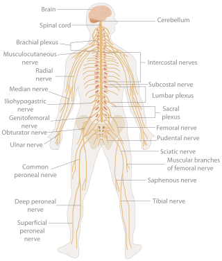
In biology, the nervous system is the highly complex part of an animal that coordinates its actions and sensory information by transmitting signals to and from different parts of its body. The nervous system detects environmental changes that impact the body, then works in tandem with the endocrine system to respond to such events. Nervous tissue first arose in wormlike organisms about 550 to 600 million years ago. In vertebrates, it consists of two main parts, the central nervous system (CNS) and the peripheral nervous system (PNS). The CNS consists of the brain and spinal cord. The PNS consists mainly of nerves, which are enclosed bundles of the long fibers, or axons, that connect the CNS to every other part of the body. Nerves that transmit signals from the brain are called motor nerves (efferent), while those nerves that transmit information from the body to the CNS are called sensory nerves (afferent). The PNS is divided into two separate subsystems, the somatic and autonomic, nervous systems. The autonomic nervous system is further subdivided into the sympathetic, parasympathetic and enteric nervous systems. The sympathetic nervous system is activated in cases of emergencies to mobilize energy, while the parasympathetic nervous system is activated when organisms are in a relaxed state. The enteric nervous system functions to control the gastrointestinal system. Nerves that exit from the brain are called cranial nerves while those exiting from the spinal cord are called spinal nerves.

The peripheral nervous system (PNS) is one of two components that make up the nervous system of bilateral animals, with the other part being the central nervous system (CNS). The PNS consists of nerves and ganglia, which lie outside the brain and the spinal cord. The main function of the PNS is to connect the CNS to the limbs and organs, essentially serving as a relay between the brain and spinal cord and the rest of the body. Unlike the CNS, the PNS is not protected by the vertebral column and skull, or by the blood–brain barrier, which leaves it exposed to toxins.

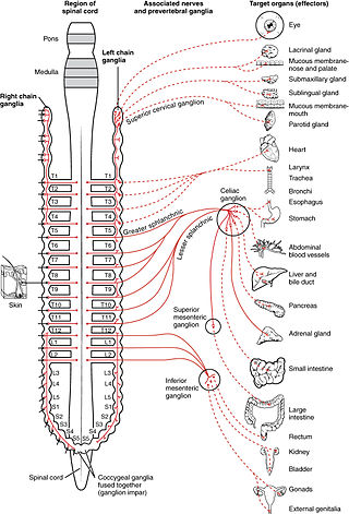
The autonomic nervous system (ANS), formerly referred to as the vegetative nervous system, is a division of the nervous system that operates internal organs, smooth muscle and glands. The autonomic nervous system is a control system that acts largely unconsciously and regulates bodily functions, such as the heart rate, its force of contraction, digestion, respiratory rate, pupillary response, urination, and sexual arousal. This system is the primary mechanism in control of the fight-or-flight response.

The parasympathetic nervous system (PSNS) is one of the three divisions of the autonomic nervous system, the others being the sympathetic nervous system and the enteric nervous system. The enteric nervous system is sometimes considered part of the autonomic nervous system, and sometimes considered an independent system.

The sympathetic nervous system (SNS) is one of the three divisions of the autonomic nervous system, the others being the parasympathetic nervous system and the enteric nervous system. The enteric nervous system is sometimes considered part of the autonomic nervous system, and sometimes considered an independent system.

The enteric nervous system (ENS) or intrinsic nervous system is one of the three main divisions of the autonomic nervous system (ANS), the other being the sympathetic (SNS) and parasympathetic nervous system (PSNS), and consists of a mesh-like system of neurons that governs the function of the gastrointestinal tract. It is capable of acting independently of the SNS and PSNS, although it may be influenced by them. The ENS is nicknamed the "second brain". It is derived from neural crest cells.

A spinal nerve is a mixed nerve, which carries motor, sensory, and autonomic signals between the spinal cord and the body. In the human body there are 31 pairs of spinal nerves, one on each side of the vertebral column. These are grouped into the corresponding cervical, thoracic, lumbar, sacral and coccygeal regions of the spine. There are eight pairs of cervical nerves, twelve pairs of thoracic nerves, five pairs of lumbar nerves, five pairs of sacral nerves, and one pair of coccygeal nerves. The spinal nerves are part of the peripheral nervous system.

The somatic nervous system (SNS) is made up of nerves that link the brain and spinal cord to voluntary or skeletal muscles that are under conscious control as well as to skin sensory receptors. Specialized nerve fiber ends called sensory receptors are responsible for detecting information within and outside of the body.

The fight-or-flight or the fight-flight-freeze-or-fawn is a physiological reaction that occurs in response to a perceived harmful event, attack, or threat to survival. It was first described by Walter Bradford Cannon. His theory states that animals react to threats with a general discharge of the sympathetic nervous system, preparing the animal for fighting or fleeing. More specifically, the adrenal medulla produces a hormonal cascade that results in the secretion of catecholamines, especially norepinephrine and epinephrine. The hormones estrogen, testosterone, and cortisol, as well as the neurotransmitters dopamine and serotonin, also affect how organisms react to stress. The hormone osteocalcin might also play a part.
Physiological psychology is a subdivision of behavioral neuroscience that studies the neural mechanisms of perception and behavior through direct manipulation of the brains of nonhuman animal subjects in controlled experiments. This field of psychology takes an empirical and practical approach when studying the brain and human behavior. Most scientists in this field believe that the mind is a phenomenon that stems from the nervous system. By studying and gaining knowledge about the mechanisms of the nervous system, physiological psychologists can uncover many truths about human behavior. Unlike other subdivisions within biological psychology, the main focus of psychological research is the development of theories that describe brain-behavior relationships.

A reflex arc is a neural pathway that controls a reflex. In vertebrates, most sensory neurons do not pass directly into the brain, but synapse in the spinal cord. This allows for faster reflex actions to occur by activating spinal motor neurons without the delay of routing signals through the brain. The brain will receive the input while the reflex is being carried out and the analysis of the signal takes place after the reflex action.

The cardiac conduction system transmits the signals generated by the sinoatrial node – the heart's pacemaker, to cause the heart muscle to contract, and pump blood through the body's circulatory system. The pacemaking signal travels through the right atrium to the atrioventricular node, along the bundle of His, and through the bundle branches to Purkinje fibers in the walls of the ventricles. The Purkinje fibers transmit the signals more rapidly to stimulate contraction of the ventricles.

The neuroimmune system is a system of structures and processes involving the biochemical and electrophysiological interactions between the nervous system and immune system which protect neurons from pathogens. It serves to protect neurons against disease by maintaining selectively permeable barriers, mediating neuroinflammation and wound healing in damaged neurons, and mobilizing host defenses against pathogens.

The lateral grey column is one of the three grey columns of the spinal cord ; the others being the anterior and posterior grey columns. The lateral grey column is primarily involved with activity in the sympathetic division of the autonomic motor system. It projects to the side as a triangular field in the thoracic and upper lumbar regions of the postero-lateral part of the anterior grey column.

The axon reflex is the response stimulated by peripheral nerves of the body that travels away from the nerve cell body and branches to stimulate target organs. Reflexes are single reactions that respond to a stimulus making up the building blocks of the overall signaling in the body's nervous system. Neurons are the excitable cells that process and transmit these reflex signals through their axons, dendrites, and cell bodies. Axons directly facilitate intercellular communication projecting from the neuronal cell body to other neurons, local muscle tissue, glands and arterioles. In the axon reflex, signaling starts in the middle of the axon at the stimulation site and transmits signals directly to the effector organ skipping both an integration center and a chemical synapse present in the spinal cord reflex. The impulse is limited to a single bifurcated axon, or a neuron whose axon branches into two divisions and does not cause a general response to surrounding tissue.
Cardiac physiology or heart function is the study of healthy, unimpaired function of the heart: involving blood flow; myocardium structure; the electrical conduction system of the heart; the cardiac cycle and cardiac output and how these interact and depend on one another.

The following diagram is provided as an overview of and topical guide to the human nervous system:
Neurocardiology is the study of the neurophysiological, neurological and neuroanatomical aspects of cardiology, including especially the neurological origins of cardiac disorders. The effects of stress on the heart are studied in terms of the heart's interactions with both the peripheral nervous system and the central nervous system.

The classification of peripheral nerves in the peripheral nervous system (PNS) groups the nerves into two main groups, the somatic and the autonomic nervous systems. Together, these two systems provide information regarding the location and status of the limbs, organs, and the remainder of the body to the central nervous system (CNS) via nerves and ganglia present outside of the spinal cord and brain. The somatic nervous system directs all voluntary movements of the skeletal muscles, and can be sub-divided into afferent and efferent neuronal flow. The autonomic nervous system is divided primarily into the sympathetic and parasympathetic nervous systems with a third system, the enteric nervous system, receiving less recognition.
References
- ↑ Carlson, N. R. (2013). Structure and Functions of Cells of the Nervous System. Physiology of Behavior (Eleventh ed., p. 29). Upper Saddle River: Pearson Education, Inc.
- ↑ Tortora, G. J., & Derrickson, B. (2012). The Spinal Cord and Spinal Nerves. Principles of Anatomy (p. 492). Hobobken: John Wiley and Sons Inc.
- ↑ The Central Nervous System. (n.d.). Molecular and Cell Biology Home. Retrieved May 2, 2013, from http://mcb.berkeley.edu/courses/mcb135e/central.html
- ↑ Tortora, G. J., & Derrickson, B. (2012). Nervous Tissue. Principles of Anatomy and Physiology (pp. 448,449). Hobobken: John Wiley and Sons Inc.
- 1 2 3 Chudler, E. (n.d.). Neuroscience For Kids - Explore the nervous system . UW Faculty Web Server. Retrieved May 2, 2013, from http://faculty.washington.edu/chudler/nsdivide.html#cns
- 1 2 3 4 5 6 7 8 9 Kremer, J. M., & McMullen, W. (2010). Biopac Student Lab. Goleta: Biopac Systems, Inc.
- ↑ Phelps, Brady I. (2011). "Orienting Response". Encyclopedia of Child Behavior and Development. p. 1044. doi:10.1007/978-0-387-79061-9_2037. ISBN 978-0-387-77579-1.
- ↑ Associate Degree Nursing Physiology Review. (2008, August 1). Austin Community College - Start Here. Get There.. Retrieved May 2, 2013, from http://www.austincc.edu/apreview/PhysText/Cardiac.html
This article needs additional or more specific categories .(November 2021) |