Bovine leukaemia virus RNA packaging signal

Last updated
Bovine leukaemia virus RNA packaging signal
RF00192.jpg
Predicted secondary structure and sequence conservation of BLV_package
Identifiers
SymbolBLV_package
Rfam RF00192
Other data
RNA type Cis-reg
Domain(s) Eukaryota; Viruses
SO SO:0000233
PDB structures PDBe

This family represents the bovine leukaemia virus RNA encapsidation (packaging) signal, which is essential for efficient viral replication. [1]

Contents

Discovery

Before its discovery, the location of the encapsidation (packaging) signal within the bovine leukaemia virus (BLV) was suggested by Manksy et al. to either be “between the major splice donor site (in the r region of the viral RNA) and the gag start codon or between the primer binding site (located just downstream of the 5’ LTR) and the gag start codon.” [2] In 1994, researchers from the University of Wisconsin Medical School made mutations in various locations of BLV through the process of deletion analysis to determine if viral replication efficiency would be affected. They found mutants made with deletions in the 5’ end of the gag gene and in the 5’ untranslated leader region had a 7-fold and a 50-fold decrease in replication respectively. In contrast, they did not find a significant decrease in viral replication in mutants made with deletions in the 3’ end of the gag gene. These results suggest there are two regions within BLV necessary for successful RNA encapsidation. [2]

Function

The RNA encapsidation (packaging) signal participates in the process of RNA packaging and aids in making viral packaging and encapsidation more efficient. [3] RNA packaging is characterized by an initial recognition event between the Gag polyprotein and the RNA encapsidation (packaging) signal. Research suggests this specifically takes place at the 5’ end of the viral genome and involves stable RNA secondary structures. These structures interact with amino acids located in the nucleocapsid, or NC domain, of the Gag protein. [1] Once the encapsidation region and the Gag protein interact, the NC domain recruits the genome of the virus to be packaged into the retrovirus capsid. [3] Recently, it has also been proposed that the matrix (MA) domain of Gag plays an important role in recognizing genomic RNA early on in this process. Furthermore, it has been found that this MA domain is involved in gRNA packaging due to the presence of charged residues in this region, which are vital for successful packaging to take place. [4]

Necessity in viral replication

It is unknown if viral replication can occur at all without the presence of this RNA encapsidaiton (packaging) signal. Mutation or deletion of the RNA encapsidation (packaging) signal reduces RNA viral packaging, but still allows for the viral particle to be made. Mutation of the NC domain in Gag, similarly, halts RNA packaging, but also prevents the assembly of retroviral particles. If the encapsidation (packaging) signal is not essential for this particle assembly, then the protein interactions among Gag molecules are likely solely responsible for viral replication. However, further research of the NC domain involving recombinant Gag proteins show the encapsidation signal is necessary for viral particle assembly. Two hypotheses currently stand to explain these contradictory findings. First, small cellular RNAs, specifically tRNAs, in the retroviruses could be utilized during particle production. Second, viral particles without genomic RNA could use other cellular RNAs as structural support during replication. [3]

As retroviral particles consistently have similar amounts of RNA, lack of genomic RNA in some viral particles must be supplemented by cellular RNA. When treated with RNase, retoviral cores are disrupted. Therefore, regardless of whether the NC domain of Gag needs assistance in assembling retroviruses, the RNA encapsidation (packaging) signal does ensure the presence of RNA material in the viral particles, which serve as important structural elements. [3] [ non-primary source needed ]

Structure

Regions

The encapsidation (packaging) signal of BLV is made up of two regions. One of the regions (nucleotides 551 to 698) includes 147 base pairs beginning after the primer binding site and ending just after the start of the gag gene (nucleotide 628). This region is called the primary signal and is essential for viral replication to occur. [2]

The other region, composed of 132 nucleotide base pairs, is located within the gag gene between nucleotides 1015 and 1147. This region, also known as the secondary signal, will facilitate RNA encapsidation if the primary region is present and fully functional. [2]

The encapsidation (packaging) signal of BLV, then, is considered to be discontinuous since its two regions are not sequential. [1]

Secondary structures

The encapsidation (packaging) signal is made up of secondary structures. There are two stable stem-loop structures in the primary region and one stem-loop structure in the secondary region. [5] The two primary stem-loops are located in the matrix (MA) domain of the gag gene, downstream of the start codon, while the secondary signal stem-loop is found in the capsid (CA) domain of gag. [6] When mutations in the primary region make one of its stem-loops inoperable, viral replication decreases by a factor of 7. Also, replication decreases 40-fold when neither primary signal stem-loop is properly functioning, creating a phenotype similar to BLV strains that do not have a primary signal region. A similar effect is found in the secondary signal stem-loop, where mutations in this region reduce viral replication 4 to 6 times. However, replication rates of mutant BLV strains can be reestablished to normal levels by implementing counter mutations. Furthermore, when the encapsidation signal regions of type 1 or type 2 human T-cell leukemia virus (HTLV), a virus within the same family as BLV, replace these mutated primary and secondary regions of mutated BLV, replication improves, implying the encapsidation regions of HTLV and BLV are homologous. Chimeric forms of BLV RNA are also not as efficient, suggesting secondary and tertiary structures of these encapsidation regions are important for replication success. [5]

SL1 and SL2

Stem-loop 1 (SL2) and stem-loop 2 (SL2) are the specific secondary structures of the primary signaling region of BLV. Both SL1 and SL2 are essential for proper RNA encapsidation and viral replication to occur. SL1 and SL2 are believed to have different functions, though, because two copies of either one of these secondary structures alone result in inefficient replication. [5]

Implications

Further research studying the RNA encapsidation (packaging) signal region and its interactions with other domains within BLV will lead to a better understanding of viral packaging and replication. It is predicted BLV will soon be studied in animal models because of its current impact on the U.S. dairy industry: BLV decreases milk production in its disease-ridden cattle hosts. This research would be beneficial for clinical application as well since BLV has been found in human breast tissue. It is currently unknown how human infection of BLV occurs. [7]

See also

Related Research Articles

Retrovirus Family of viruses

A retrovirus is a type of virus that inserts a copy of its RNA genome into the DNA of a host cell that it invades, thus changing the genome of that cell. Once inside the host cell's cytoplasm, the virus uses its own reverse transcriptase enzyme to produce DNA from its RNA genome, the reverse of the usual pattern, thus retro (backwards). The new DNA is then incorporated into the host cell genome by an integrase enzyme, at which point the retroviral DNA is referred to as a provirus. The host cell then treats the viral DNA as part of its own genome, transcribing and translating the viral genes along with the cell's own genes, producing the proteins required to assemble new copies of the virus.

Poliovirus enterovirus

Poliovirus, the causative agent of polio, is a serotype of the species Enterovirus C, in the family of Picornaviridae.

Picornavirus Family of viruses

Picornaviruses are a group of related nonenveloped RNA viruses which infect vertebrates including mammals and birds. They are viruses that represent a large family of small, positive-sense, single-stranded RNA viruses with a 30-nm icosahedral capsid. The viruses in this family can cause a range of diseases including the common cold, poliomyelitis, meningitis, hepatitis, and paralysis.

<i>Gammaretrovirus</i> Genus of viruses

Gammaretrovirus is a genus in the Retroviridae family. Example species are the murine leukemia virus and the feline leukemia virus. They cause various sarcomas, leukemias and immune deficiencies in mammals, reptiles and birds.

The genome and proteins of HIV have been the subject of extensive research since the discovery of the virus in 1983. "In the search for the causative agent, it was initially believed that the virus was a form of the Human T-cell leukemia virus (HTLV), which was known at the time to affect the human immune system and cause certain leukemias. However, researchers at the Pasteur Institute in Paris isolated a previously unknown and genetically distinct retrovirus in patients with AIDS which was later named HIV." Each virion comprises a viral envelope and associated matrix enclosing a capsid, which itself encloses two copies of the single-stranded RNA genome and several enzymes. The discovery of the virus itself occurred two years following the report of the first major cases of AIDS-associated illnesses.

The murine leukemia viruses are retroviruses named for their ability to cause cancer in murine (mouse) hosts. Some MLVs may infect other vertebrates. MLVs include both exogenous and endogenous viruses. Replicating MLVs have a positive sense, single-stranded RNA (ssRNA) genome that replicates through a DNA intermediate via the process of reverse transcription.

The Simian foamy virus (SFV) is a species of the genus Spumavirus, which belongs to the family of Retroviridae. It has been identified in a wide variety of primates, including pro-simians, New World and Old World monkeys as well as apes, and each species has been shown to harbor a unique (species-specific) strain of SFV, including African green monkeys, baboons, macaques and chimpanzees. As it is related to the more well-known retrovirus human immunodeficiency virus (HIV), its discovery in primates has led to some speculation that HIV may have been spread to the human species in Africa through contact with blood from apes, monkeys, and other primates, most likely through bushmeat hunting practices.

Long terminal repeat

A long terminal repeat (LTR) is a pair of identical sequences of DNA, several hundred base pairs long, which occur in eukaryotic genomes on either end of a series of genes or pseudogenes that form a retrotransposon or an endogenous retrovirus or a retroviral provirus. All retroviral genomes are flanked by LTRs, while there are some retrotransposons without LTRs. Typically, an element flanked by a pair of LTRs will encode a reverse transcriptase and an integrase, allowing the element to be copied and inserted at a different location of the genome. Copies of such an LTR-flanked element can often be found hundreds or thousands of times in a genome. LTR retrotransposons comprise about 8% of the human genome.

<i>Crinivirus</i> Genus of viruses

Crinivirus, formerly the lettuce infectious yellows virus group, is a genus of viruses, in the family Closteroviridae. They are linear, single-stranded positive sense RNA viruses. There are 14 species in this genus. Diseases associated with this genus include: yellowing and necrosis, particularly affecting the phloem.

Citrus tristeza virus replication signal

The Citrus tristeza virus replication signal is a regulatory element involved in a viral replication signal which is highly conserved in citrus tristeza viruses. Replication signals are required for viral replication and are usually found near the 5' and 3' termini of protein coding genes. This element is predicted to form ten stem loop structures some of which are essential for functions that provide for efficient viral replication.

Coronavirus 3′ stem-loop II-like motif (s2m)

The Coronavirus 3′ stem-loop II-like motif is a secondary structure motif identified in the 3′ untranslated region (3′UTR) of astrovirus, coronavirus and equine rhinovirus genomes. Its function is unknown, but various viral 3′ UTR regions have been found to play roles in viral replication and packaging.

Coronavirus packaging signal Regulartory element in coronaviruses

The Coronavirus packaging signal is a conserved cis-regulatory element found in Betacoronavirus. It has an important role in regulating the packaging of the viral genome into the capsid. As part of the viral life cycle, within the infected cell, the viral genome becomes associated with viral proteins and assembles into new infective progeny viruses. This process is called packaging and is vital for viral replication.

Gammaretrovirus core encapsidation signal

The Gammaretrovirus core encapsidation signal is an RNA element known to be essential for stable dimerisation and efficient genome packaging during virus assembly. Dimerisation of the viral RNA genomes is proposed to act as an RNA conformational switch which exposes conserved UCUG elements and enables efficient genome encapsidation. The structure of this element is composed of three stem-loops. Two of the stem-loops called SL-C and SL-D form a single co-axial extend helix.

Retroviral psi packaging element

The retroviral psi packaging element, also known as the Ψ RNA packaging signal, is a cis-acting RNA element identified in the genomes of the retroviruses Human immunodeficiency virus (HIV) and Simian immunodeficiency virus (SIV). It is involved in regulating the essential process of packaging the retroviral RNA genome into the viral capsid during replication. The final virion contains a dimer of two identical unspliced copies of the viral genome.

HBV RNA encapsidation signal epsilon

The HBV RNA encapsidation signal epsilon is an element essential for HBV virus replication.

Avian HBV RNA encapsidation signal epsilon

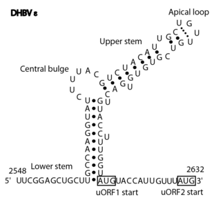
The Avian HBV RNA encapsidation signal epsilon is an RNA structure that is shown to facilitate encapsidation of the pregenomic RNA required for viral replication. There are two main classes of encapsidation signals in avian hepatitis B viruses - Duck hepatitis B virus (DHBV) and Heron hepatitis B virus (HHBV) like. DHBV is used as a model to understand human Hepatitis B virus. Although studies have shown that the HHBV epsilon has less pairing in the upper stem than DHBV, this pairing is not absolutely required for DHBV infection in ducks.

Mason-Pfizer monkey virus (M-PMV), formerly Simian retrovirus (SRV), is a species of retroviruses that usually infect and cause a fatal immune deficiency in Asian macaques. The ssRNA virus appears sporadically in mammary carcinoma of captive macaques at breeding facilities which expected as the natural host, but the prevalence of this virus in feral macaques remains unknown. M-PMV was transmitted naturally by virus-containing body fluids, via biting, scratching, grooming, and fighting. Cross contaminated instruments or equipment (fomite) can also spread this virus among animals.

Bovine foamy virus (BFV) is a ss(+)RNA retrovirus that belongs to the genus spumaviridae. Spumaviruses differ from the other six members of family retroviridae, both structurally and in pathogenic nature. Spumaviruses derive their name from spuma the latin for "foam". The 'foam' aspect of 'foamy virus' comes from syncytium formation and the rapid vacuolization of infected cells, creating a 'foamy' appearance.

Coronavirus genomes are positive-sense single-stranded RNA molecules with an untranslated region (UTR) at the 5′ end which is called the 5′ UTR. The 5′ UTR is responsible for important biological functions, such as viral replication, transcription and packaging. The 5′ UTR has a conserved RNA secondary structure but different Coronavirus genera have different structural features described below.

Flavivirus 5 UTR Untranslated regions in the genome of viruses in the genus Flavivirus

Flavivirus 5' UTR are untranslated regions in the genome of viruses in the genus Flavivirus.

References

  1. 1 2 3 Mansky LM, Gajary LC (September 2002). "The primary nucleotide sequence of the bovine leukemia virus RNA packaging signal can influence efficient RNA packaging and virus replication". Virology. 301 (2): 272–280. doi: 10.1006/viro.2002.1578 . PMID   12359429.
  2. 1 2 3 4 Mansky LM, Krueger AE, Temin HM (June 1995). "The bovine leukemia virus encapsidation signal is discontinuous and extends into the 5' end of the gag gene". Journal of Virology. 69 (6): 3282–3289. doi:10.1128/JVI.69.6.3282-3289.1995. PMC   189039 . PMID   7745675.
  3. 1 2 3 4 Muriaux D, Mirro J, Harvin D, Rein A (April 2001). "RNA is a structural element in retrovirus particles". Proceedings of the National Academy of Sciences of the United States of America. 98 (9): 5246–5251. Bibcode:2001PNAS...98.5246M. doi:10.1073/pnas.091000398. PMC   33195 . PMID   11320254.
  4. Sun M, Grigsby IF, Gorelick RJ, Mansky LM, Musier-Forsyth K (January 2014). "Retrovirus-specific differences in matrix and nucleocapsid protein-nucleic acid interactions: implications for genomic RNA packaging". Journal of Virology. 88 (2): 1271–1280. doi:10.1128/JVI.02151-13. PMC   3911680 . PMID   24227839.
  5. 1 2 3 Mansky, LM; Wisniewski, RM (April 1998). "The bovine leukemia virus encapsidation signal is composed of RNA secondary structures". Journal of Virology. 72 (4): 3196–3204. doi: 10.1128/JVI.72.4.3196-3204.1998 .
  6. Jewell NA, Mansky LM (August 2000). "In the beginning: genome recognition, RNA encapsidation and the initiation of complex retrovirus assembly". The Journal of General Virology. 81 (Pt 8): 1889–1899. doi: 10.1099/0022-1317-81-8-1889 . PMID   10900025.
  7. Qualley DF, Sokolove VL, Ross JL (March 2015). "Bovine leukemia virus nucleocapsid protein is an efficient nucleic acid chaperone". Biochemical and Biophysical Research Communications. 458 (3): 687–692. doi:10.1016/j.bbrc.2015.02.025. PMID   25686502.