
Staining is a technique used to enhance contrast in samples, generally at the microscopic level. Stains and dyes are frequently used in histology and in the medical fields of histopathology, hematology, and cytopathology that focus on the study and diagnoses of disease at a microscopic level. Stains may be used to define biological tissues, cell populations, or organelles within individual cells.
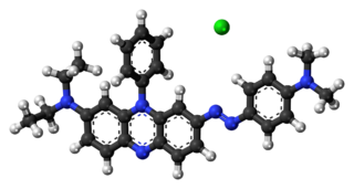
Janus Green B is a basic dye and vital stain used in histology. It is also used to stain mitochondria supravitally, as was introduced by Leonor Michaelis in 1900.
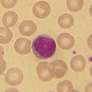
Wright's stain is a hematologic stain that facilitates the differentiation of blood cell types. It is classically a mixture of eosin (red) and methylene blue dyes. It is used primarily to stain peripheral blood smears, urine samples, and bone marrow aspirates, which are examined under a light microscope. In cytogenetics, it is used to stain chromosomes to facilitate diagnosis of syndromes and diseases.
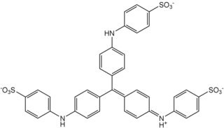
Methyl blue is a chemical compound with the molecular formula C37H27N3Na2O9S3. It is used as a stain in histology, and stains collagen blue in tissue sections. It can be used in some differential staining techniques such as Mallory's connective tissue stain and Gömöri trichrome stain, and can be used to mediate electron transfer in microbial fuel cells. Fungal cell walls are also stained by methyl blue.
Trypan blue is an azo dye. It is a direct dye for cotton textiles. In biosciences, it is used as a vital stain to selectively colour dead tissues or cells blue.
MilliporeSigma is an American chemical, life science and biotechnology company, before 2014 known as Sigma-Aldrich, owned by Merck KGaA.

Ponceau S, Acid Red 112, or C.I. 27195 is a sodium salt of a diazo dye of a light red color, that may be used to prepare a stain for rapid reversible detection of protein bands on nitrocellulose or polyvinylidene fluoride (PVDF) membranes, as well as on cellulose acetate membranes. A Ponceau S stain is useful because it does not appear to have a deleterious effect on the sequencing of blotted polypeptides and is therefore one method of choice for locating polypeptides on western blots for blot-sequencing. It is also easily reversed with water washes, facilitating subsequent immunological detection. The stain can be completely removed from the protein bands by continued washing. Common stain formulations include 0.1% (w/v) Ponceau S in 5% acetic acid or 2% (w/v) Ponceau S in 30% trichloroacetic acid and 30% sulfosalicylic acid.

Ponceau 2R, Xylidine ponceau, Ponceau G, Red R, Acid Red 26, Food Red 5, or C.I. 16150 is a red azo dye used in histology for staining. It is easily soluble in water and slightly in ethanol. It usually comes as a disodium salt.

Hematoxylin and eosin stain is one of the principal tissue stains used in histology. It is the most widely used stain in medical diagnosis and is often the gold standard. For example, when a pathologist looks at a biopsy of a suspected cancer, the histological section is likely to be stained with H&E.
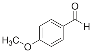
4-Anisaldehyde, or p-Anisaldehyde, is an organic compound with the formula CH3OC6H4CHO. The molecule consists of a benzene ring with an formyl and a methoxy group. It is a colorless liquid with a strong aroma. It provides sweet, floral and strong aniseed odor. Two isomers of 4-anisaldehyde are known, ortho-anisaldehyde and meta-anisaldehyde. They are less commonly encountered.

Bromocresol green (BCG) is a dye of the triphenylmethane family. It belongs to a class of dyes called sulfonephthaleins. It is used as a pH indicator in applications such as growth mediums for microorganisms and titrations. In clinical practise, it is commonly used as a diagnostic technique. The most common use of bromocresol green is to measure serum albumin concentration within mammalian blood samples in possible cases of kidney failure and liver disease.

In staining dyes, nigrosin is a mixture of black synthetic dyes made by heating a mixture of nitrobenzene, aniline, and hydrochloric acid in the presence of copper or iron. Related to induline, it is a mixture of phenazine-based compounds. Its main industrial uses are as a colorant for lacquers and varnishes and in marker pen inks. Sulfonation of nigrosin yields a water-soluble anionic dye, nigrosin WS.

DiOC6 (3,3′-dihexyloxacarbocyanine iodide) is a fluorescent dye used for the staining of a cell's endoplasmic reticulum, vesicle membranes and mitochondria. Binding to these structures occurs via the dye's hydrophilic groups. DiOC6 can be used to label living cells, however they are quickly damaged due to the dye's extreme phototoxicity, so cells stained with this dye can only be exposed to light for short periods of time. When exposed to blue light, the dye fluoresces green.

Toluidine blue, also known as TBO or tolonium chloride (INN) is a blue cationic (basic) dye used in histology and sometimes clinically.
Brilliant blue is a shade of blue, or may refer to:
Cresyl violet is an organic compound with the chemical formula C19H18ClN3O. It is a basic dye and is used as a common stain in histology.
Silver proteinate is used in electron microscopy with periodic acid and thiocarbohydrazide or thiosemicarbohydrazide as a positive stain for carbohydrates such as glycogen. It can also be used for light microscopy to stain nerve tissue. It is normally available as 8% silver in combination with albumin.

Supravital staining is a method of staining used in microscopy to examine living cells that have been removed from an organism. It differs from intravital staining, which is done by injecting or otherwise introducing the stain into the body. Thus a supravital stain may have a greater toxicity, as only a few cells need to survive it a short while. The term "vital stain" is used by some authors to refer specifically to an intravital stain, and by others interchangeably with a supravital stain, the core concept being that the cell being examined is still alive. As the cells are alive and unfixed, outside the body, supravital stains are temporary in nature.
In hemocytometry, Türk's solution is a hematological stain prepared in 99% acetic acid (glacial) and distilled water. The solution destroys the red blood cells and platelets within a blood sample, and stains the nuclei of the white blood cells, making them easier to see and count.

