Carcinocythemia

Last updated
Carcinocythemia
Other namesCarcinoma cell leukemia
Carcinocythemia - malignant tumour cells in peripheral blood (cropped v1).png
A case of carcinocythemia. The large, round cells are breast cancer cells circulating in the peripheral blood. [1]
Specialty Hematology, oncology
CausesUsually secondary to metastatic cancer in the bone marrow
Differential diagnosis Acute leukemia, lymphoma, leukemoid reaction, circulating immature cells from chemotherapy, circulating endothelial cells, megakaryocytes or osteoclasts
Prognosis Poor; 15% survival rate at 6 months

Carcinocythemia, also known as carcinoma cell leukemia, [2] is a condition in which cells from malignant tumours of non-hematopoietic origin are visible on the peripheral blood smear. [3] [4] It is an extremely rare condition, [5] with 33 cases identified in the literature from 1960 to 2018. [4] Carcinocythemia typically occurs secondary to infiltration of the bone marrow by metastatic cancer [6] and carries a very poor prognosis. [3] [4] [5]

Contents

Presentation

Carcinocythemia occurs most commonly in breast cancer, followed by small cell lung cancer, and usually appears late in the course of the disease. [4] Thrombosis and disseminated intravascular coagulation are frequently reported in association with carcinocythemia. [2] [4] The prognosis is poor: a review of 26 patients found that 85% died within 6 months of the diagnosis, with an average time of 6.1 weeks between diagnosis and death. [4]

The amount of tumour cells on the blood smear can range from 1 to 80 percent of the total white blood cell count, [4] with lower percentages being more common. [3] Carcinocythemia is distinct from the presence of circulating tumour cells (CTCs), as CTCs usually occur in such low quantities that they cannot be seen on blood smear examination, requiring special techniques for detection. [2] [7]

Mechanism

The mechanism of carcinocythemia is poorly understood. Some patients with carcinocythemia show evidence of impaired spleen function, and it has been suggested that dysfunction of the reticuloendothelial system, preventing phagocytosis of malignant cells, could contribute to the presence of tumour cells in the blood. [4] [8]

Diagnosis

Carcinocythemia can be detected on a routine blood smear examination or manual differential. [8] If the number of suspicious cells is low, a smear can be prepared from the buffy coat of the blood sample to concentrate the cells. [3]

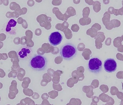
Tumour cells in peripheral blood may look similar to circulating blasts or lymphoma cells. [3] [9] Features that aid in distinguishing tumour cells from other cells include their very large size, mature nuclear chromatin pattern, vacuolated cytoplasm, and their tendency to appear in clumps or clusters, although some of these characteristics are shared by megakaryoblasts and monoblasts. Tumour cells are often found at the edge of the blood smear due to their large size, so this area should be examined thoroughly if carcinocythemia is suspected. [4]

Cytochemical staining and immunohistochemistry techniques can help determine the lineage of the cells. [4] When immunophenotyped by flow cytometry, the cells are generally CD45 negative and may express CD56, a profile that is non-specific but unusual for hematologic malignancies. [3] In some cases, flow cytometry and FISH results may be misleading, as circulating tumour cells can exhibit cell markers and chromosomal abnormalities associated with hematologic diseases. [4]

Bone marrow examination is indicated in carcinocythemia to better characterize the tumour cells. [3] [4]

Differential diagnosis

Carcinocythemia must be distinguished from the following conditions: [3] [4] [8]

History

The presence of tumour cells in the peripheral blood of a cancer patient was first described in an 1869 case report in the Medical Journal of Australia . [8] [10] The term carcinocythemia was first used in 1976 by Robert Carey. [8] [11] In 1984, a review of 10 cases was published, noting the condition's poor prognosis. [12]

Other animals

As of 2018, there were two documented cases of carcinocythemia in dogs and one case in a cat. [13]

Related Research Articles

<span class="mw-page-title-main">Lymphoma</span> Hematologic cancer that affects lymphocytes

Lymphoma is a group of blood and lymph tumors that develop from lymphocytes. The name typically refers to just the cancerous versions rather than all such tumours. Signs and symptoms may include enlarged lymph nodes, fever, drenching sweats, unintended weight loss, itching, and constantly feeling tired. The enlarged lymph nodes are usually painless. The sweats are most common at night.

<span class="mw-page-title-main">Adenocarcinoma</span> Medical condition

Adenocarcinoma is a type of cancerous tumor that can occur in several parts of the body. It is defined as neoplasia of epithelial tissue that has glandular origin, glandular characteristics, or both. Adenocarcinomas are part of the larger grouping of carcinomas, but are also sometimes called by more precise terms omitting the word, where these exist. Thus invasive ductal carcinoma, the most common form of breast cancer, is adenocarcinoma but does not use the term in its name—however, esophageal adenocarcinoma does to distinguish it from the other common type of esophageal cancer, esophageal squamous cell carcinoma. Several of the most common forms of cancer are adenocarcinomas, and the various sorts of adenocarcinoma vary greatly in all their aspects, so that few useful generalizations can be made about them.

<span class="mw-page-title-main">Chronic lymphocytic leukemia</span> Medical condition

Chronic lymphocytic leukemia (CLL) is a type of cancer in which the bone marrow makes too many lymphocytes. Early on, there are typically no symptoms. Later, non-painful lymph node swelling, feeling tired, fever, night sweats, or weight loss for no clear reason may occur. Enlargement of the spleen and low red blood cells (anemia) may also occur. It typically worsens gradually over years.

<span class="mw-page-title-main">Hybridoma technology</span> Method for producing lots of identical antibodies

Hybridoma technology is a method for producing large numbers of identical antibodies. This process starts by injecting a mouse with an antigen that provokes an immune response. A type of white blood cell, the B cell, produces antibodies that bind to the injected antigen. These antibody producing B-cells are then harvested from the mouse and, in turn, fused with immortal B cell cancer cells, a myeloma, to produce a hybrid cell line called a hybridoma, which has both the antibody-producing ability of the B-cell and the longevity and reproductivity of the myeloma. The hybridomas can be grown in culture, each culture starting with one viable hybridoma cell, producing cultures each of which consists of genetically identical hybridomas which produce one antibody per culture (monoclonal) rather than mixtures of different antibodies (polyclonal). The myeloma cell line that is used in this process is selected for its ability to grow in tissue culture and for an absence of antibody synthesis. In contrast to polyclonal antibodies, which are mixtures of many different antibody molecules, the monoclonal antibodies produced by each hybridoma line are all chemically identical.

<span class="mw-page-title-main">Adult T-cell leukemia/lymphoma</span> Human disease

Adult T-cell leukemia/lymphoma is a rare cancer of the immune system's T-cells caused by human T cell leukemia/lymphotropic virus type 1 (HTLV-1). All ATL cells contain integrated HTLV-1 provirus further supporting that causal role of the virus in the cause of the neoplasm. A small amount of HTLV-1 individuals progress to develop ATL with a long latency period between infection and ATL development. ATL is categorized into 4 subtypes: acute, smoldering, lymphoma-type, chronic. Acute and Lymphoma-type are known to particularity be aggressive with poorer prognosis.

<span class="mw-page-title-main">Myeloid sarcoma</span> Medical condition

A myeloid sarcoma is a solid tumor composed of immature white blood cells called myeloblasts. A chloroma is an extramedullary manifestation of acute myeloid leukemia; in other words, it is a solid collection of leukemic cells occurring outside of the bone marrow.

<span class="mw-page-title-main">Invasive carcinoma of no special type</span> Medical condition

Invasive carcinoma of no special type (NST) is also referred to as invasive ductal carcinoma or infiltrating ductal carcinoma(IDC) and invasive ductal carcinoma, not otherwise specified (NOS). Each of these terms represents to the same disease entity, but for international audiences this article will use invasive carcinoma NST because it is the preferred term of the World Health Organization (WHO).

Splenic marginal zone lymphoma (SMZL) is a type of cancer made up of B-cells that replace the normal architecture of the white pulp of the spleen. The neoplastic cells are both small lymphocytes and larger, transformed lymphoblasts, and they invade the mantle zone of splenic follicles and erode the marginal zone, ultimately invading the red pulp of the spleen. Frequently, the bone marrow and splenic hilar lymph nodes are involved along with the peripheral blood. The neoplastic cells circulating in the peripheral blood are termed villous lymphocytes due to their characteristic appearance.

Myelophthisic anemia is a severe type of anemia found in some people with diseases that affect the bone marrow. Myelophthisis refers to the displacement of hemopoietic bone-marrow tissue by fibrosis, tumors, or granulomas. The word comes from the roots myelo-, which refers to bone marrow, and phthysis, shrinkage or atrophy.

<span class="mw-page-title-main">Neprilysin</span> Mammalian protein found in Homo sapiens

Neprilysin, also known as membrane metallo-endopeptidase (MME), neutral endopeptidase (NEP), cluster of differentiation 10 (CD10), and common acute lymphoblastic leukemia antigen (CALLA) is an enzyme that in humans is encoded by the MME gene. Neprilysin is a zinc-dependent metalloprotease that cleaves peptides at the amino side of hydrophobic residues and inactivates several peptide hormones including glucagon, enkephalins, substance P, neurotensin, oxytocin, and bradykinin. It also degrades the amyloid beta peptide whose abnormal folding and aggregation in neural tissue has been implicated as a cause of Alzheimer's disease. Synthesized as a membrane-bound protein, the neprilysin ectodomain is released into the extracellular domain after it has been transported from the Golgi apparatus to the cell surface.

Hepatosplenic T-cell lymphoma is a rare form of lymphoma that is generally incurable, except in the case of an allogeneic stem cell transplant. It is a systemic neoplasm comprising medium-sized cytotoxic T-cells that show significant sinusoidal infiltration in the liver, spleen, and bone marrow.

<span class="mw-page-title-main">ETV6</span> Protein-coding gene in the species Homo sapiens

ETV6 protein is a transcription factor that in humans is encoded by the ETV6 gene. The ETV6 protein regulates the development and growth of diverse cell types, particularly those of hematological tissues. However, its gene, ETV6 frequently suffers various mutations that lead to an array of potentially lethal cancers, i.e., ETV6 is a clinically significant proto-oncogene in that it can fuse with other genes to drive the development and/or progression of certain cancers. However, ETV6 is also an anti-oncogene or tumor suppressor gene in that mutations in it that encode for a truncated and therefore inactive protein are also associated with certain types of cancers.

<span class="mw-page-title-main">Aggressive lymphoma</span> Medical condition

Aggressive lymphoma, also known as high-grade lymphoma, is a group of fast growing non-Hodgkin lymphoma.

<span class="mw-page-title-main">Signet ring cell carcinoma</span> Medical condition

Signet ring cell carcinoma (SRCC) is a rare form of highly malignant adenocarcinoma that produces mucin. It is an epithelial malignancy characterized by the histologic appearance of signet ring cells.

A circulating tumor cell (CTC) is a cell that has shed into the vasculature or lymphatics from a primary tumor and is carried around the body in the blood circulation. CTCs can extravasate and become seeds for the subsequent growth of additional tumors (metastases) in distant organs, a mechanism that is responsible for the vast majority of cancer-related deaths. The detection and analysis of CTCs can assist early patient prognoses and determine appropriate tailored treatments. Currently, there is one FDA-approved method for CTC detection, CellSearch, which is used to diagnose breast, colorectal and prostate cancer.

<span class="mw-page-title-main">Cancer biomarker</span> Substance or process that is indicative of the presence of cancer in the body

A cancer biomarker refers to a substance or process that is indicative of the presence of cancer in the body. A biomarker may be a molecule secreted by a tumor or a specific response of the body to the presence of cancer. Genetic, epigenetic, proteomic, glycomic, and imaging biomarkers can be used for cancer diagnosis, prognosis, and epidemiology. Ideally, such biomarkers can be assayed in non-invasively collected biofluids like blood or serum.

Thymidine kinase is an enzyme, a phosphotransferase : 2'-deoxythymidine kinase, ATP-thymidine 5'-phosphotransferase, EC 2.7.1.21 that catalyzes the reaction:

<span class="mw-page-title-main">White blood cell differential</span> Blood test

A white blood cell differential is a medical laboratory test that provides information about the types and amounts of white blood cells in a person's blood. The test, which is usually ordered as part of a complete blood count (CBC), measures the amounts of the five normal white blood cell types – neutrophils, lymphocytes, monocytes, eosinophils and basophils – as well as abnormal cell types if they are present. These results are reported as percentages and absolute values, and compared against reference ranges to determine whether the values are normal, low, or high. Changes in the amounts of white blood cells can aid in the diagnosis of many health conditions, including viral, bacterial, and parasitic infections and blood disorders such as leukemia.

<span class="mw-page-title-main">Cellular adoptive immunotherapy</span> Cellular adoptive immunotherapy

Cellular adoptive immunotherapy is a type of immunotherapy. Immune cells such as T-cells are usually isolated from patients for expansion or engineering purposes and reinfused back into patients to fight diseases using their own immune system. A major application of cellular adoptive therapy is cancer treatment, as the immune system plays a vital role in the development and growth of cancer. The primary types of cellular adoptive immunotherapies are T cell therapies. Other therapies include CAR-T therapy, CAR-NK therapy, macrophage-based immunotherapy and dendritic cell therapy.

References

  1. Ogura, Kanako; Amano, Maki; Matsumoto, Toshiharu; Sakaguchi, Asumi; Kosaka, Taijiro; Kitabatake, Toshiaki; Kojima, Kuniaki (2015). "Occult Breast Lobular Carcinoma with Numerous Circulating Tumor Cells in Peripheral Blood". Case Reports in Pathology. 2015: 1–6. doi: 10.1155/2015/135684 . ISSN   2090-6781. PMC   4496473 . PMID   26199779.
  2. 1 2 3 Johnsrud, Andrew J.; Pina-Oviedo, Sergio (2017). "Carcinocythemia (carcinoma cell leukemia)". Blood. 130 (21): 2357. doi: 10.1182/blood-2017-08-799882 . ISSN   0006-4971. PMID   29170195.
  3. 1 2 3 4 5 6 7 8 Irma Pereira; Tracy I. George; Daniel A. Arber (7 December 2011). "Chapter 20: Nonhematopoietic tumors in the blood". Atlas of Peripheral Blood: The Primary Diagnostic Tool. Lippincott Williams & Wilkins. pp. 185–7. ISBN   978-1-4511-6366-7.
  4. 1 2 3 4 5 6 7 8 9 10 11 12 13 Ronen, Shira; Kroft, Steven H.; Olteanu, Horatiu; Hosking, Paul R.; Harrington, Alexandra M. (2019). "Carcinocythemia: A rare entity becoming more common? A 3-year, single institution series of seven cases and literature review". International Journal of Laboratory Hematology. 41 (1): 69–79. doi: 10.1111/ijlh.12924 . ISSN   1751-5521. PMID   30216684. S2CID   52278041.
  5. 1 2 Noel Weidner; Richard J. Cote; Saul Suster; Lawrence M. Weiss (8 July 2009). Modern Surgical Pathology. Elsevier Health Sciences. pp. 1593–. ISBN   978-1-4377-1958-1.
  6. Michael Caligiuri; Marcel M. Levi; Kenneth Kaushansky; Marshall A. Lichtman, Josef Prchal, Linda J Burns, Oliver W Press (23 December 2015). Williams Hematology, 9E. McGraw-Hill Education. p. 658. ISBN   978-0-07-183300-4.{{cite book}}: CS1 maint: multiple names: authors list (link)
  7. Krishnamurthy, Savitri (2012). "The emerging role of circulating tumor cells in breast cancer". Cancer Cytopathology. 120 (3): 161–166. doi: 10.1002/cncy.20207 . ISSN   1934-662X. PMID   22275137. S2CID   36422597.
  8. 1 2 3 4 5 Chang, Yuan-Hsin; Hsieh, Ruey-Kuen; Chang, Ming-Chi; Chen, Gon-Shen (2007). "Breast cancer with an unusual leukemia-like presentation: case report and literature review". Medical Oncology. 25 (1): 100–103. doi:10.1007/s12032-007-0048-2. ISSN   1357-0560. PMID   18188722. S2CID   20417675.
  9. Lugassy G, Vorst EJ, Varon D, Sigler E, Shani A, Bassous-Guedj L. (1990). "Carcinocythemia. Report of two cases, one simulating a Burkitt lymphoma". Acta Cytol. 34 (2): 265–8. PMID   2157324.{{cite journal}}: CS1 maint: multiple names: authors list (link)
  10. Ashworth, T.R. (1869). "(1869) A Case of Cancer in Which Cells Similar to Those in the Tumours Were Seen in the Blood after Death". The Medical Journal of Australia. 14: 146–147.
  11. Carey, Robert W.; Taft, Priscilla D.; Bennett, John M.; Kaufman, Sheldon (1976). "Carcinocythemia (carcinoma cell leukemia)". The American Journal of Medicine. 60 (2): 273–278. doi:10.1016/0002-9343(76)90437-X. ISSN   0002-9343. PMID   1062163.
  12. Gallivan, Monica V. E.; Lokich, Jacob J. (1984). "Carcinocythemia (carcinoma cell leukemia). Report of two cases with english literature review". Cancer. 53 (5): 1100–1102. doi: 10.1002/1097-0142(19840301)53:5<1100::AID-CNCR2820530514>3.0.CO;2-K . ISSN   0008-543X. PMID   6362836.
  13. Sá e Lemos, Eva; Lima de Carvalho, Hugo; Gil da Costa, Rui M.; Pinto da Cunha, Nazaré (2018). "Carcinocythemia: First report in a cat and literature review". Veterinary Clinical Pathology. 47 (1): 142–145. doi:10.1111/vcp.12565. ISSN   0275-6382. PMID   29360147.