Cholinergic neuron

Last updated

A cholinergic neuron is a nerve cell which mainly uses the neurotransmitter acetylcholine (ACh) to send its messages. Many neurological systems are cholinergic. Cholinergic neurons provide the primary source of acetylcholine to the cerebral cortex, and promote cortical activation during both wakefulness and rapid eye movement sleep. [1] The cholinergic system of neurons has been a main focus of research in aging and neural degradation, specifically as it relates to Alzheimer's disease. [2] The dysfunction and loss of basal forebrain cholinergic neurons and their cortical projections are among the earliest pathological events in Alzheimer's disease. [3]

Contents

Neuromuscular junction using acetylcholine as the neurotransmitter Synapse diag4.png
Neuromuscular junction using acetylcholine as the neurotransmitter

Anatomy

Most research involving cholinergic neurons involves the basal forebrain cholinergic neurons. However, cholinergic neurons only represent about 5% of the total basal forebrain cell population. [1] Most of these neurons originate in different areas of the basal forebrain and have extensive projections into almost all layers of the cortex. [1] [3] Basal forebrain cholinergic neurons are homologous within a particular basal forebrain region but vary across different regions. [1] In the brainstem acetylcholine originates from the pedunculopontine nucleus and laterodorsal tegmental nucleus collectively known as the meso-pontine tegmental area or pontomesencephalotegmental complex. [4] [5]

Normal aging

Normal aging is described as aging unaccompanied by the behavioral or cognitive dysfunctions associated with the cholinergic basal forebrain system. [3] In normal aging, there are beadlike swellings within the cholinergic fibers with enlarged or thickened axons, often in grape-like clusters. [3] This fiber swelling can be induced in a laboratory setting by damaging the cell body of the cholinergic neuron, which implies there is a slow cell and fiber degeneration of affected neurons and their projecting axons. [3]

Neuroprotective effects

Nerve growth factor protects cholinergic neurons. [6] [7] The small non-toxic molecule urea has no neuroprotective effect on cholinergic neurons by itself, but when experimental brain slices were treated with nerve growth factor and urea, the number of cholinergic neurons in the brain slices was significantly enhanced when compared to slices treated with nerve growth factor only. [6] The enhancing effect of urea may be due to inhibition of the nitric oxide-system within the cholinergic neuron. [6]

Relationship to mammalian circadian system

Cholinergic neurons, along with non-cholinergic neurons, have sleep/wake regulatory functions in the basal forebrain that can be categorized based on their firing patterns in different regions. [1] The cholinergic system allows the circadian system to have the cycle of one day. The cholinergic neuron may also play a role in time memory, and the ability of an individual to form a memory around a certain time of day, which is known as "time stamping". [8] The cholinergic system is characterized by high acetylcholine release during the active phase of an individual’s circadian rhythm. [8]

Firing patterns and the circadian system

In the medial septum-diagonal band of Broca's area of the brain, cholinergic neurons have very low firing rates during both wake and non-REM sleep, and show no rhythmic bursts during hippocampal (theta) Electroencephalography activity. However, cholinergic neurons in the magnocellular preoptic nucleus and Substantia innominata have increased firing rates with fast cortical (gamma) Electroencephalography activity during wake and rapid eye movement sleep. This indicates that cholinergic neurons may be activated through α1-receptors by noradrenaline, which were released by locus coeruleus neurons during wake cycles. [1] In a basic summary, cholinergic neurons are always active during wake or rapid eye movement sleep cycles, and are more likely to activate the cerebral cortex to induce the gamma wave and Theta rhythm activities while behaviorally promoting the states of wakefulness and rapid eye movement sleep. [1]

Time memory and time stamping

The suprachiasmatic nucleus functions as the hypothalamic master clock, controlling the body's Circadian rhythm. The suprachiasmatic nucleus of mice, hamsters, and rats have a small amount of cholinergic innervation. [8] A "time memory" is the memory at a specific time of day for which an individual made an association with a certain event or location. "Time stamping" is the process by which the specific time-of-day is encoded to support the formation of a time memory. The situation must be important and specific, without unnecessary prolonging, for a time stamp to occur. Acetylcholine excites cells in the suprachiasmatic nucleus, so cholinergic transmission of more Acetylcholine into the suprachiasmatic nucleus should support the formation of a time memory. [8]

The number of free and available muscarinic acetylcholine receptors (mAChRs) is highest when acetylcholine release is at the lowest levels. When a memorable event occurs, there is a massive release of acetylcholine that will attach to mAChRs. Once too many are involved, the mAChRs will reduce or block further cholinergic input, which protects these cells and the networks from additional cholinergic input that could disrupt the signal. This allows the suprachiasmatic nucleus to perform time stamping and produce a time memory of what has just occurred to the individual. [8] If correct, this would explain the cholinergic neuron’s role in memory.

Circadian system and Alzheimer's disease

The circadian system is one of the first systems to be damaged in Alzheimer's disease. [8] Alzheimer's patients often complain of disrupted sleep, shortened rapid eye movement sleep, and increased night time awakening. These disruptions steadily worsen as the disease progresses. It is normal in aging for circadian rhythms to deteriorate as choline acetyltransferase (ChAT) fluctuations change in pattern and acetylcholine levels fluctuate more often. As Alzheimer's disease drastically changes cholinergic function, the circadian system naturally follows the changed levels. Circadian rhythmicity in acetylcholine release is critical for optimal memory processing, and a loss of this rhythmicity contributes to cognitive problems in Alzheimer's disease. [8]

Olfactory behavior

Circadian modulation of cholinergic neuronal could be important for mediating sexual behaviors in mice. Modifications of basal forebrain cholinergic neuronal activity disrupted odor discrimination of simple odors, and the investigation of social odors. [9]

Neurological disorders

Degeneration of the cholinergic neurons in the basal forebrain has been linked to progressing memory deficits related to aging, which eventually results in decreased cholinergic function. [2] The dysfunction and loss of basal forebrain cholinergic neurons has been observed in many dementias, especially Alzheimer's. [2] [3] Recent findings imply that aging-related cognitive deficits are due to impairments of cholinergic function rather than cholinergic cell loss. [2] This suggests that it will be possible to reverse cognitive declines, as the cells are not dead, but deteriorating.

Alzheimer's disease

Alzheimer's disease is the most common form of dementia, and the sixth leading cause of death in the United States. [10] [11] The proportion of deaths associated with Alzheimer's continues to grow rapidly, increasing by 66% from 2000 to 2008. [10] Alzheimer's typically involves a decline in the activity of choline acetyltransferase and acetylcholinesterase, as well as a decline in acetylcholine release. [3] [11] Cholinergic system research may provide the key to treating and reversing this devastating disease.

Histological hallmarks

Although degeneration of basal forebrain cholinergic cells has been observed in many other dementias, Alzheimer's has two distinctive histological hallmarks: Beta amyloid plaques and neurofibrillary tangles. [2] The Beta amyloid plaques are high-molecular weight fibrils and are major components of the senile Alzheimer's disease brain. [3] [12] There appears to be a vast, intrinsic microvascular pathology of the brain in these cases, which suggests a link between Beta amyloid production, impairments in cerebrovascular function, and basal forebrain cholinergic deficits in AD. [2] It appears that Beta amyloid (1-42) mediates its cytotoxic action by affecting key proteins that play a role in apoptosis induction. [2] There is also evidence that shows beta amyloid proteins actually bind to cholinergic neurons and physically inhibit ChAT activity in cultures treated with oligomers of beta amyloid. [13] The other histological hallmarks, neurofibrillary tangles, are the intracellular inclusions formed by aggregates of hyperphosphorylated tau protein. This is found only in select populations of patients with Alzheimer's. This tau protein has specific pathology, and has been found both in patients with mild cognitive impairment (a forerunner of Alzheimer's) and Alzheimer's itself. The neurofibrillary tangles seem to increase within the basal forebrain cholinergic complex with old age and at a more accelerated pace in patients with Alzheimer's. [2]

Probable cause for vulnerable cholinergic neurons

The "cholinergic hypothesis" is a well-established pathology of the involvement of cholinergic neurons on Alzheimer's disease due to their role in memory. [7] Research in 2007 determined why cholinergic neurons were becoming more vulnerable to Beta amyloid plaque formation. A pathway exists for both the maturation and degradation of Nerve Growth Factor, which causes cholinergic neurons to become vulnerable. [7] Basal forebrain cholinergic neurons are highly dependent on the constant internal supply of Nerve Growth Factor throughout life. If the supply of Nerve Growth Factor is interrupted, cholinergic atrophy could begin to occur in these neurons and change their phenotype. This supply could be interrupted if there is a failure in the protease cascade and the precursor proNGF cannot be converted to Nerve Growth Factor. This is caused by a rise in matrix metalloproteinase-9 activity, which increases the degradation of Nerve Growth Factor and reduces its production. This double failure of Nerve Growth Factor stimulation leads to the progressive atrophy of basal forebrain cholinergic neurons, which in turn contributes to Alzheimer's-related learning and memory declines. [7]

Disease model

Most studies of Alzheimer's have used mice or rat brains with Beta amyloid plaque buildup as the disease model. In 2013, Dr. Su-Chun Zhang and his research team derived cholinergic neurons from neuroepithelial stem cells in a laboratory setting, making it easier to test potential treatments without the use of live animals. [14]

Potential treatments

Potential treatments for Alzheimer's include the use of memantine, a moderate affinity uncompetitive NMDA receptor antagonist that preferentially blocks excessive N-methyl-D-aspartate (NMDA) receptor activity without disrupting normal activity. This treatment is based on the theory that degenerative neural disorders have excitotoxic processes due to the inappropriate overstimulation of the NMDA receptor. [3] In a rat model, memantine treatment given preventatively to certain rats pre-β-amyloid (1-42) lesion significantly reduced the loss of cholinergic fibers. Memantine treatment reversed attention and learning deficits in Beta amyloid (1-42) affected rats. This data indicates the ability of memantine to rescue neocortical cholinergic fibers (originating from basal forebrain cholinergic neurons) from the neurotoxic effects of Beta amyloid (1-42) oligomers. It should also be noted that memantine is able to inhibit the truncation of glycogen synthase kinase-3 (triggered by activated calpain), which is believed to play a key role in the pathogenesis of Alzheimer's, affecting tau phosphorylation (the second histological hallmark). [3]

Another treatment involves the use of exogenous choline acetyltransferase as supplementation in cholinergic neurons. Cholinergic neurons have significantly reduced choline acetyltransferase and acetylcholine activity, which is correlated to the severity of the dementia or cognitive impairments. [12] The problem with this therapy is that choline acetyltransferase is largely blocked by the blood-brain barrier. PTD-ChAT is a fusion protein made up of both protein transduction domain and choline acetyltransferase; it can pass through the blood-brain barrier and cell membranes. It regulates acetylcholine levels in the brain, curing mice treated with PTD-ChAT from their memory and cognitive deficits. [12]

Other potential diseases

Cholinergic neurons have an effect on other neurodegenerative diseases such as Parkinson's disease, Huntington's disease and Down syndrome. [2] [3] [15] As with Alzheimer's, the degeneration of basal forebrain cholinergic neurons and the decrease in the neurotransmitter acetylcholine have a drastic effect on behavioral and cognitive function. [2]

Related Research Articles

<span class="mw-page-title-main">Neurotransmitter</span> Chemical substance that enables neurotransmission

A neurotransmitter is a signaling molecule secreted by a neuron to affect another cell across a synapse. The cell receiving the signal, or target cell, may be another neuron, but could also be a gland or muscle cell.

<span class="mw-page-title-main">Acetylcholine</span> Organic chemical and neurotransmitter

Acetylcholine (ACh) is an organic compound that functions in the brain and body of many types of animals as a neurotransmitter. Its name is derived from its chemical structure: it is an ester of acetic acid and choline. Parts in the body that use or are affected by acetylcholine are referred to as cholinergic.

<span class="mw-page-title-main">Cholinergic</span> Agent which mimics choline

Cholinergic agents are compounds which mimic the action of acetylcholine and/or butyrylcholine. In general, the word "choline" describes the various quaternary ammonium salts containing the N,N,N-trimethylethanolammonium cation. Found in most animal tissues, choline is a primary component of the neurotransmitter acetylcholine and functions with inositol as a basic constituent of lecithin. Choline also prevents fat deposits in the liver and facilitates the movement of fats into cells.

<span class="mw-page-title-main">Suprachiasmatic nucleus</span> Part of the brains hypothalamus

The suprachiasmatic nucleus or nuclei (SCN) is a small region of the brain in the hypothalamus, situated directly above the optic chiasm. The SCN is the principal circadian pacemaker in mammals, responsible for generating circadian rhythms. Reception of light inputs from photosensitive retinal ganglion cells allow the SCN to coordinate the subordinate cellular clocks of the body and entrain to the environment. The neuronal and hormonal activities it generates regulate many different body functions in an approximately 24-hour cycle.

<span class="mw-page-title-main">Choline acetyltransferase</span> Protein-coding gene in the species Homo sapiens

Choline acetyltransferase is a transferase enzyme responsible for the synthesis of the neurotransmitter acetylcholine. ChAT catalyzes the transfer of an acetyl group from the coenzyme acetyl-CoA to choline, yielding acetylcholine (ACh). ChAT is found in high concentration in cholinergic neurons, both in the central nervous system (CNS) and peripheral nervous system (PNS). As with most nerve terminal proteins, ChAT is produced in the body of the neuron and is transported to the nerve terminal, where its concentration is highest. Presence of ChAT in a nerve cell classifies this cell as a "cholinergic" neuron. In humans, the choline acetyltransferase enzyme is encoded by the CHAT gene.

<span class="mw-page-title-main">Locus coeruleus</span> Stress and panic response centre

The locus coeruleus (LC), also spelled locus caeruleus or locus ceruleus, is a nucleus in the pons of the brainstem involved with physiological responses to stress and panic. It is a part of the reticular activating system.

<span class="mw-page-title-main">Coluracetam</span> Chemical compound

Coluracetam is a purported nootropic agent of the racetam family. It is contains a chemical group that is a bioisostere of the 9-amino-tetrahydroacridine family. It was initially developed and tested by the Mitsubishi Tanabe Pharma Corporation for Alzheimer's disease. After the drug failed to reach endpoints in its clinical trials it was in-licensed by BrainCells Inc for investigations into major depressive disorder (MDD), which was preceded by being awarded a "Qualifying Therapeutic Discovery Program Grant" by the state of California. Findings from phase IIa clinical trials have suggested that it would be a potential medication for comorbid MDD with generalized anxiety disorder (GAD). BrainCells Inc is currently out-licensing the drug for this purpose. It may also have potential use in prevention and treatment of ischemic retinopathy and retinal and optic nerve injury.

<span class="mw-page-title-main">End-plate potential</span>

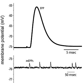
End plate potentials (EPPs) are the voltages which cause depolarization of skeletal muscle fibers caused by neurotransmitters binding to the postsynaptic membrane in the neuromuscular junction. They are called "end plates" because the postsynaptic terminals of muscle fibers have a large, saucer-like appearance. When an action potential reaches the axon terminal of a motor neuron, vesicles carrying neurotransmitters are exocytosed and the contents are released into the neuromuscular junction. These neurotransmitters bind to receptors on the postsynaptic membrane and lead to its depolarization. In the absence of an action potential, acetylcholine vesicles spontaneously leak into the neuromuscular junction and cause very small depolarizations in the postsynaptic membrane. This small response (~0.4mV) is called a miniature end plate potential (MEPP) and is generated by one acetylcholine-containing vesicle. It represents the smallest possible depolarization which can be induced in a muscle.

<span class="mw-page-title-main">Ventrolateral preoptic nucleus</span> Nucleus of the anterior hypothalamus

The ventrolateral preoptic nucleus (VLPO), also known as the intermediate nucleus of the preoptic area (IPA), is a small cluster of neurons situated in the anterior hypothalamus, sitting just above and to the side of the optic chiasm in the brain of humans and other animals. The brain's sleep-promoting nuclei, together with the ascending arousal system which includes components in the brainstem, hypothalamus and basal forebrain, are the interconnected neural systems which control states of arousal, sleep, and transitions between these two states. The VLPO is active during sleep, particularly during non-rapid eye movement sleep, and releases inhibitory neurotransmitters, mainly GABA and galanin, which inhibit neurons of the ascending arousal system that are involved in wakefulness and arousal. The VLPO is in turn innervated by neurons from several components of the ascending arousal system. The VLPO is activated by the endogenous sleep-promoting substances adenosine and prostaglandin D2. The VLPO is inhibited during wakefulness by the arousal-inducing neurotransmitters norepinephrine and acetylcholine. The role of the VLPO in sleep and wakefulness, and its association with sleep disorders – particularly insomnia and narcolepsy – is a growing area of neuroscience research.

<span class="mw-page-title-main">Basal forebrain</span> Brain structures in the forebrain

Part of the human brain, the basal forebrain structures are located in the forebrain to the front of and below the striatum. They include the ventral basal ganglia, nucleus basalis, diagonal band of Broca, substantia innominata, and the medial septal nucleus. These structures are important in the production of acetylcholine, which is then distributed widely throughout the brain. The basal forebrain is considered to be the major cholinergic output of the central nervous system (CNS) centred on the output of the nucleus basalis. The presence of non-cholinergic neurons projecting to the cortex have been found to act with the cholinergic neurons to dynamically modulate activity in the cortex.

Neuromodulation is the physiological process by which a given neuron uses one or more chemicals to regulate diverse populations of neurons. Neuromodulators typically bind to metabotropic, G-protein coupled receptors (GPCRs) to initiate a second messenger signaling cascade that induces a broad, long-lasting signal. This modulation can last for hundreds of milliseconds to several minutes. Some of the effects of neuromodulators include: altering intrinsic firing activity, increasing or decreasing voltage-dependent currents, altering synaptic efficacy, increasing bursting activity and reconfigurating synaptic connectivity.

The biochemistry of Alzheimer's disease, the most common cause of dementia, is not yet very well understood. Alzheimer's disease (AD) has been identified as a proteopathy: a protein misfolding disease due to the accumulation of abnormally folded amyloid beta (Aβ) protein in the brain. Amyloid beta is a short peptide that is an abnormal proteolytic byproduct of the transmembrane protein amyloid-beta precursor protein (APP), whose function is unclear but thought to be involved in neuronal development. The presenilins are components of proteolytic complex involved in APP processing and degradation.

<span class="mw-page-title-main">Alpha-7 nicotinic receptor</span>

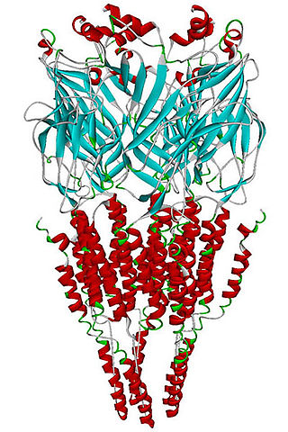
The alpha-7 nicotinic receptor, also known as the α7 receptor, is a type of nicotinic acetylcholine receptor implicated in long-term memory, consisting entirely of α7 subunits. As with other nicotinic acetylcholine receptors, functional α7 receptors are pentameric [i.e., (α7)5 stoichiometry].

<span class="mw-page-title-main">Alzheimer's disease</span> Progressive neurodegenerative disease

Alzheimer's disease (AD) is a neurodegenerative disease that usually starts slowly and progressively worsens, and is the cause of 60–70% of cases of dementia. The most common early symptom is difficulty in remembering recent events. As the disease advances, symptoms can include problems with language, disorientation, mood swings, loss of motivation, self-neglect, and behavioral issues. As a person's condition declines, they often withdraw from family and society. Gradually, bodily functions are lost, ultimately leading to death. Although the speed of progression can vary, the typical life expectancy following diagnosis is three to nine years.

Sleep onset is the transition from wakefulness into sleep. Sleep onset usually transmits into non-rapid eye movement sleep but under certain circumstances it is possible to transit from wakefulness directly into rapid eye movement sleep.

<span class="mw-page-title-main">Clinical neurochemistry</span>

Clinical neurochemistry is the field of neurological biochemistry which relates biochemical phenomena to clinical symptomatic manifestations in humans. While neurochemistry is mostly associated with the effects of neurotransmitters and similarly functioning chemicals on neurons themselves, clinical neurochemistry relates these phenomena to system-wide symptoms. Clinical neurochemistry is related to neurogenesis, neuromodulation, neuroplasticity, neuroendocrinology, and neuroimmunology in the context of associating neurological findings at both lower and higher level organismal functions.

<span class="mw-page-title-main">Nucleus basalis</span> Group of neurons in the brain

In the human brain, the nucleus basalis, also known as the nucleus basalis of Meynert or nucleus basalis magnocellularis, is a group of neurons located mainly in the substantia innominata of the basal forebrain. Most neurons of the nucleus basalis are rich in the neurotransmitter acetylcholine, and they have widespread projections to the neocortex and other brain structures.

<span class="mw-page-title-main">Neuroscience of sleep</span> Study of the neuroscientific and physiological basis of the nature of sleep

The neuroscience of sleep is the study of the neuroscientific and physiological basis of the nature of sleep and its functions. Traditionally, sleep has been studied as part of psychology and medicine. The study of sleep from a neuroscience perspective grew to prominence with advances in technology and the proliferation of neuroscience research from the second half of the twentieth century.

<span class="mw-page-title-main">Neuronal lineage marker</span> Endogenous tag expressed in different cells along neurogenesis and differentiated cells

A neuronal lineage marker is an endogenous tag that is expressed in different cells along neurogenesis and differentiated cells such as neurons. It allows detection and identification of cells by using different techniques. A neuronal lineage marker can be either DNA, mRNA or RNA expressed in a cell of interest. It can also be a protein tag, as a partial protein, a protein or an epitope that discriminates between different cell types or different states of a common cell. An ideal marker is specific to a given cell type in normal conditions and/or during injury. Cell markers are very valuable tools for examining the function of cells in normal conditions as well as during disease. The discovery of various proteins specific to certain cells led to the production of cell-type-specific antibodies that have been used to identify cells.

Robert Y. Moore is an American neurologist with interests in disorders of biological rhythms, movement disorders, and behavioral neurology. He is credited with discovering the function of the suprachiasmatic nucleus (SCN) as the circadian clock, as well as, describing its organization. He is also credited with establishing the role of the mammalian retinohypothalamic tract (RHT) as a photic entrainment pathway. Moore cin 2017 serves as a professor of neurology, with a secondary in psychiatry and neuroscience at the University of Pittsburgh, and as co-director of the National Parkinson Foundation Center of Excellence at the University of Pittsburgh.

References

  1. 1 2 3 4 5 6 7 Deurveilher S, Semba K (Feb 2011). "Basal forebrain regulation of cortical activity and sleep-wake states: Roles of cholinergic and non-cholinergic neurons". Sleep and Biological Rhythms. 9: 65–70. doi: 10.1111/j.1479-8425.2010.00465.x . S2CID   141709601.
  2. 1 2 3 4 5 6 7 8 9 10 Schliebs R, Arendt T (August 2011). "The cholinergic system in aging and neuronal degeneration". Behavioural Brain Research. 221 (2): 555–63. doi:10.1016/j.bbr.2010.11.058. PMID   21145918. S2CID   38583520.
  3. 1 2 3 4 5 6 7 8 9 10 11 Nyakas C, Granic I, Halmy LG, Banerjee P, Luiten PG (August 2011). "The basal forebrain cholinergic system in aging and dementia. Rescuing cholinergic neurons from neurotoxic amyloid-β42 with memantine" (PDF). Behavioural Brain Research. 221 (2): 594–603. doi:10.1016/j.bbr.2010.05.033. PMID   20553766. S2CID   2622145.
  4. Woolf NJ, Butcher LL (May 1986). "Cholinergic systems in the rat brain: III. Projections from the pontomesencephalic tegmentum to the thalamus, tectum, basal ganglia, and basal forebrain". Brain Research Bulletin. 16 (5): 603–37. doi:10.1016/0361-9230(86)90134-6. PMID   3742247. S2CID   39665815.
  5. Woolf NJ, Butcher LL (December 1989). "Cholinergic systems in the rat brain: IV. Descending projections of the pontomesencephalic tegmentum". Brain Research Bulletin. 23 (6): 519–40. doi:10.1016/0361-9230(89)90197-4. PMID   2611694. S2CID   4721282.
  6. 1 2 3 Zassler B, Dechant G, Humpel C (2005). "Urea enhances the nerve growth factor-induced neuroprotective effect on cholinergic neurons in organotypic rat brain slices". Neuroscience. 130 (2): 317–23. doi:10.1016/j.neuroscience.2004.09.010. PMID   15664688. S2CID   39832814.
  7. 1 2 3 4 Cuello AC, Bruno MA (June 2007). "The failure in NGF maturation and its increased degradation as the probable cause for the vulnerability of cholinergic neurons in Alzheimer's disease". Neurochemical Research. 32 (6): 1041–5. doi:10.1007/s11064-006-9270-0. PMID   17404842. S2CID   130366.
  8. 1 2 3 4 5 6 7 Hut RA, Van der Zee EA (August 2011). "The cholinergic system, circadian rhythmicity, and time memory". Behavioural Brain Research. 221 (2): 466–80. doi:10.1016/j.bbr.2010.11.039. PMID   21115064. S2CID   20338635.
  9. Smith RS, Hu R, DeSouza A, Eberly CL, Krahe K, Chan W, Araneda RC (July 2015). "Differential Muscarinic Modulation in the Olfactory Bulb". The Journal of Neuroscience. 35 (30): 10773–85. doi:10.1523/JNEUROSCI.0099-15.2015. PMC   4518052 . PMID   26224860.
  10. 1 2 Alzheimer's Association (2012). "2012 Alzheimer's disease facts and figures". Alzheimer's & Dementia. 8 (2): 131–68. doi:10.1016/j.jalz.2012.02.001. PMID   22404854. S2CID   202255258.
  11. 1 2 Auld DS, Kornecook TJ, Bastianetto S, Quirion R (October 2002). "Alzheimer's disease and the basal forebrain cholinergic system: relations to beta-amyloid peptides, cognition, and treatment strategies". Progress in Neurobiology. 68 (3): 209–45. doi:10.1016/S0301-0082(02)00079-5. PMID   12450488. S2CID   53151403.
  12. 1 2 3 Fu AL, Li Q, Dong ZH, Huang SJ, Wang YX, Sun MJ (September 2004). "Alternative therapy of Alzheimer's disease via supplementation with choline acetyltransferase". Neuroscience Letters. 368 (3): 258–62. doi:10.1016/j.neulet.2004.05.116. PMID   15364407. S2CID   23038763.
  13. Nunes-Tavares N, Santos LE, Stutz B, Brito-Moreira J, Klein WL, Ferreira ST, de Mello FG (June 2012). "Inhibition of choline acetyltransferase as a mechanism for cholinergic dysfunction induced by amyloid-β peptide oligomers". The Journal of Biological Chemistry. 287 (23): 19377–85. doi: 10.1074/jbc.m111.321448 . PMC   3365976 . PMID   22505713.
  14. Liu Y, Weick JP, Liu H, Krencik R, Zhang X, Ma L, Zhou GM, Ayala M, Zhang SC (May 2013). "Medial ganglionic eminence-like cells derived from human embryonic stem cells correct learning and memory deficits". Nature Biotechnology. 31 (5): 440–7. doi:10.1038/nbt.2565. PMC   3711863 . PMID   23604284.
  15. Szutowicz A, Bielarczyk H, Jankowska-Kulawy A, Pawełczyk T, Ronowska A (August 2013). "Acetyl-CoA the key factor for survival or death of cholinergic neurons in course of neurodegenerative diseases". Neurochemical Research. 38 (8): 1523–42. doi:10.1007/s11064-013-1060-x. PMC   3691476 . PMID   23677775.