
Arthritis is a general medical term used to describe a disorder that affects joints. Symptoms generally include joint pain and stiffness. Other symptoms may include redness, warmth, swelling, and decreased range of motion of the affected joints. In certain types of arthritis, other organs such as the skin are also affected. Onset can be gradual or sudden.

A joint or articulation is the connection made between bones, ossicles, or other hard structures in the body which link an animal's skeletal system into a functional whole. They are constructed to allow for different degrees and types of movement. Some joints, such as the knee, elbow, and shoulder, are self-lubricating, almost frictionless, and are able to withstand compression and maintain heavy loads while still executing smooth and precise movements. Other joints such as sutures between the bones of the skull permit very little movement in order to protect the brain and the sense organs. The connection between a tooth and the jawbone is also called a joint, and is described as a fibrous joint known as a gomphosis. Joints are classified both structurally and functionally.

Osteoarthritis (OA) is a type of degenerative joint disease that results from breakdown of joint cartilage and underlying bone. It is believed to be the fourth leading cause of disability in the world, affecting 1 in 7 adults in the United States alone. The most common symptoms are joint pain and stiffness. Usually the symptoms progress slowly over years. Other symptoms may include joint swelling, decreased range of motion, and, when the back is affected, weakness or numbness of the arms and legs. The most commonly involved joints are the two near the ends of the fingers and the joint at the base of the thumbs, the knee and hip joints, and the joints of the neck and lower back. The symptoms can interfere with work and normal daily activities. Unlike some other types of arthritis, only the joints, not internal organs, are affected.
Synovial fluid, also called synovia,[help 1] is a viscous, non-Newtonian fluid found in the cavities of synovial joints. With its egg white–like consistency, the principal role of synovial fluid is to reduce friction between the articular cartilage of synovial joints during movement. Synovial fluid is a small component of the transcellular fluid component of extracellular fluid.

Hyaline cartilage is the glass-like (hyaline) and translucent cartilage found on many joint surfaces. It is also most commonly found in the ribs, nose, larynx, and trachea. Hyaline cartilage is pearl-gray in color, with a firm consistency and has a considerable amount of collagen. It contains no nerves or blood vessels, and its structure is relatively simple.

A tophus is a deposit of monosodium urate crystals, in people with longstanding high levels of uric acid (urate) in the blood, a condition known as hyperuricemia. Tophi are pathognomonic for the disease gout. Most people with tophi have had previous attacks of acute arthritis, eventually leading to the formation of tophi. Chronic tophaceous gout is known as Harrison Syndrome.

Monoarthritis, or monoarticular arthritis, is inflammation (arthritis) of one joint at a time. It is usually caused by trauma, infection, or crystalline arthritis.
Connective tissue disease, also known as connective tissue disorder, or collagen vascular diseases, refers to any disorder that affects the connective tissue. The body's structures are held together by connective tissues, consisting of two distinct proteins: elastin and collagen. Tendons, ligaments, skin, cartilage, bone, and blood vessels are all made of collagen. Skin and ligaments contain elastin. The proteins and the body's surrounding tissues may suffer damage when these connective tissues become inflamed.
Calcium pyrophosphate dihydrate (CPPD) crystal deposition disease, also known as pseudogout and pyrophosphate arthropathy, is a rheumatologic disease which is thought to be secondary to abnormal accumulation of calcium pyrophosphate dihydrate crystals within joint soft tissues. The knee joint is most commonly affected. The disease is metabolic in origin and its treatment remains symptomatic.
An arthropathy is a disease of a joint.

Synovitis is the medical term for inflammation of the synovial membrane. This membrane lines joints that possess cavities, known as synovial joints. The condition is usually painful, particularly when the joint is moved. The joint usually swells due to synovial fluid collection.

Knee effusion, informally known as water on the knee, occurs when excess synovial fluid accumulates in or around the knee joint. It has many common causes, including arthritis, injury to the ligaments or meniscus, or fluid collecting in the bursa, a condition known as prepatellar bursitis.

Subacromial bursitis is a condition caused by inflammation of the bursa that separates the superior surface of the supraspinatus tendon from the overlying coraco-acromial ligament, acromion, and coracoid and from the deep surface of the deltoid muscle. The subacromial bursa helps the motion of the supraspinatus tendon of the rotator cuff in activities such as overhead work.

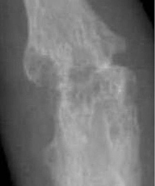
Calcific tendinitis is a common condition where deposits of calcium phosphate form in a tendon, sometimes causing pain at the affected site. Deposits can occur in several places in the body, but are by far most common in the rotator cuff of the shoulder. Around 80% of those with deposits experience symptoms, typically chronic pain during certain shoulder movements, or sharp acute pain that worsens at night. Calcific tendinitis is typically diagnosed by physical exam and X-ray imaging. The disease often resolves completely on its own, but is typically treated with non-steroidal anti-inflammatory drugs to relieve pain, rest and physical therapy to promote healing, and in some cases various procedures to breakdown and/or remove the calcium deposits.
Arthrocentesis, or joint aspiration, is the clinical procedure performed to diagnose and, in some cases, treat musculoskeletal conditions. The procedure entails using a syringe to collect synovial fluid from or inject medication into the joint capsule. Laboratory analysis of synovial fluid can further help characterize the diseased joint and distinguish between gout, arthritis, and synovial infections such as septic arthritis.

Progressive ankylosis protein homolog is a protein that in humans is encoded by the ANKH gene.
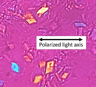
Crystal arthropathy is a class of joint disorder that is characterized by accumulation of tiny crystals in one or more joints. Polarizing microscopy and application of other crystallographic techniques have improved identification of different microcrystals including monosodium urate, calcium pyrophosphate dihydrate, calcium hydroxyapatite, and calcium oxalate.

Arthritis of the knee is typically a particularly debilitating form of arthritis. The knee may become affected by almost any form of arthritis.
Milwaukee shoulder syndrome (MSS) (apatite-associated destructive arthritis/Basic calcium phosphate (BCP) crystal arthritis/rapid destructive arthritis of the shoulder) is a rare rheumatological condition similar to pseudogout, associated with periarticular or intra-articular deposition of hydroxyapatite or basic calcium phosphate (BCP) crystals. While primarily associated with the shoulder joint, it can affect any joint in the body below the head. Along with symptomatology, the disease typically presents with positive radiologic findings, often showing marked erosion of the humeral head, cartilage, capsule, and bursae. Though rare, it is most often seen in females beginning in their 50s or 60s. Patients often have a history of joint trauma or overuse, calcium pyrophosphate dehydrate crystal deposition, neuroarthropathy, dialysis-related arthropathy or denervation.

Pain in the hip is the experience of pain in the muscles or joints in the hip/ pelvic region, a condition commonly arising from any of a number of factors. Sometimes it is closely associated with lower back pain.
