Chronaxie

Last updated
Rheobase and chronaxie are points defined on the strength-duration curve for stimulus of an excitable tissue. Rheobase chronaxie.svg
Rheobase and chronaxie are points defined on the strength-duration curve for stimulus of an excitable tissue.

Chronaxie is the minimum time required for an electric current double the strength of the rheobase to stimulate a muscle or a neuron. Rheobase is the lowest intensity with indefinite pulse duration which just stimulated muscles or nerves. [1] Chronaxie is dependent on the density of voltage-gated sodium channels in the cell, which affect that cell’s excitability. Chronaxie varies across different types of tissue: fast-twitch muscles have a lower chronaxie, slow-twitch muscles have a higher one. Chronaxie is the tissue-excitability parameter that permits choice of the optimum stimulus pulse duration for stimulation of any excitable tissue. Chronaxie (c) is the Lapicque descriptor of the stimulus pulse duration for a current of twice rheobasic (b) strength, which is the threshold current for an infinitely long-duration stimulus pulse. Lapicque showed that these two quantities (c,b) define the strength-duration curve for current: I = b(1+c/d), where d is the pulse duration. However, there are two other electrical parameters used to describe a stimulus: energy and charge. The minimum energy occurs with a pulse duration equal to chronaxie. Minimum charge (bc) occurs with an infinitely short-duration pulse. Choice of a pulse duration equal to 10c requires a current of only 10% above rheobase (b). Choice of a pulse duration of 0.1c requires a charge of 10% above the minimum charge (bc).

Contents

History

The terms chronaxie and rheobase were first coined in Louis Lapicque’s famous paper on Définition expérimentale de l’excitabilité that was published in 1909. [2]

The above I(d) curve is usually attributed to Weiss (1901) - see e.g. (Rattay 1990). It is the most simplistic of the 2 'simple' mathematical descriptors of the dependence of current strength on duration, and it leads to Weiss' linear charge progression with d:

Both Lapicque's own writings and more recent work are at odds with the linear-charge approximation. Already in 1907 Lapicque was using a linear first-order approximation of the cell membrane, modeled using a single-RC equivalent circuit. Thus:

where is the membrane time constant - in the 1st-order linear membrane model:

where

Notice that the chronaxie (c) is not explicitly present here. Notice also that - with very short duration , by the Taylor series decomposition of the exponent (around d=0):

which leads to a constant-charge approximation. The latter may fit well also more complex models of the excitable membrane, which take into account ion-channel gating mechanisms, as well as intracellular current flow, which may be the main contributors for deviations from both simple formulas.

These 'subtleties' are clearly described by Lapicque (1907, 1926 and 1931), but not too well by Geddes (2004) who emphasized the Weiss level, attributing it to Lapicque.

Measurement

An electrode is inserted into the muscle of interest, which is then stimulated using surface current. Chronaxie values increase resulting from hyperventilation can be ascribed to a change in skin impedance, the physiological factors responsible for this change being under the influence of the autonomic nervous system. This example of the preponderating influence which the condition of the skin and the underlying tissues may exert compels caution in judging the results of chronaxie measurements by percutaneous stimulation. [3] A fresh and normal sartorius placed straight in a Ringer solution and stimulated through the solution without any direct contact with the electrodes is subject to give two very distinct strength-duration curves, one of them being spread over several hundredths of a second. [4]

Values

The chronaxie values for mammalian ventricles at body temperature range from 0.5 ms (human) to 2.0 to 4.1 ms (dog); this is an 8.2/1 ratio. It has been reported that large-diameter myelinated axons have chronaxie times ranging from 50 to 100 µs and 30 to 200 µs, and neuronal cell bodies and dendrites have chronaxie times ranging from 1 to 10 ms or even up to 30 ms. The chronaxie times of grey matter were reported as being 380 +/- 191 ms and 200±700 ms. Interpretations of chronaxie times are further confounded by additional factors. The chronaxie times reported for soma and dendrites have been established using intracellular pulses that cannot be readily extrapolated to extra- cellular stimuli. Data reported in the literature use either motor response as the physiological threshold in humans or action potential generation in animals. These are largely based on stimulation through a macro-electrode, which in the case of humans is a 1.5 Â 1.2-mm DBS electrode. Data derived from micro-electrode stimulation and physiological mapping of sensory thalamus are scarce. The two stimulation methods may result in significantly different results. Few studies have attempted to correlate chronaxie times with sensory perception, although understanding the neural elements that are involved in a subjective percept, such as tingling, has important physiological implications. [5] The measurements were taken with different types of electrodes and with stimulators having unknown output impedances. The chronaxie values for human arm sensory nerves range from 0.35 to 1.17 ms, a ratio of 3.3. The values were obtained with insufficient information to establish the cause of variability. The chronaxie values for human denervated skeletal muscle ranges from 9.5 to 30 ms at body temperature, representing a ratio of 3.16. A reduction in chronaxie occurs during reinnervation. The published values for chronaxie have a wide range. If chronaxie is the best descriptor of tissue excitability in a homogeneous tissue specimen, at a known temperature, it should be determined with a constant-current stimulator providing a rectangular cathodal stimulus waveform. Chronaxie is derived from the strength-duration curve for current and it shows that if the stimulus duration is shorter than chronaxie, more current is required to stimulate, with any type or location of electrodes with a stimulator of any known or unknown output impedance. In addition, the chronaxie value, however determined, identifies the pulse duration for minimum energy. In addition, the charge delivered at chronaxie, however determined, is 2, twice the minimum charge. Therefore, if minimum charge delivery is sought to prolong the life of a battery in an implanted stimulator, a pulse duration of less than the measured chronaxie should be selected; a duration of one-tenth chronaxie provides a charge that is only 10% above the minimum charge. [6]

Stimulation

Electric and magnetic stimulation produced different sensations. For electric stimulation, sensation was typically described as localized directly below the electrodes on the surface of the skin. For magnetic stimulation, sensation was typically described as distributed throughout the palm and digits of the hand. In particular, most subjects reported sensations in either the medial or lateral digits. These observations suggest that electrical stimulation may preferentially activate cutaneous afferent nerve fibers whereas magnetic stimulation may preferentially activate deeper nerves, such as the ulnar or median nerve.

Motor vs sensory

Other studies have compared the activation of sensory and motor fibers using electric and magnetic stimulation demonstrated through stimulation of nerve and muscle tissue that magnetic activation of intramuscular nerve fibers in the arm and leg occurs at a lower threshold than for electric stimulation. Also, sensory fibers were shown to have a lower threshold for electric stimulation. Electric stimulation of the wrist by determined that when short pulses are used (less than 200 μs), motor fibers are more readily excitable, whereas for long pulse durations (greater than 1000 μs), sensory fibers are more prone to depolarization. A related observation is that electric stimulation preferentially activates sensory fibers compared to motor fibers for long pulse durations, and the inverse for short pulse durations. For magnetic stimulation, the motor fiber threshold was lower than that for sensory fibers. [7]

Significance

The main value of chronaxie is comparing excitability across different experiments and measurements using the same standard, thus making data comparisons easier. Electrical stimulation based on chronaxie could regulate myoD gene expression in denervated muscle fibers. 20 muscle contractions, induced by electrical stimulation using surface electrodes and applied on alternate days based on muscle excitability, similar to protocols used in human clinical rehabilitation, were able to reduce the accumulation of mRNA in the myoD and atrogin-1 of denervated muscles, these expressions being related to muscle growth and atrophy, respectively. The increase in myoD levels after denervation is possibly related not only to activation and proliferation of the satellite cells but also to regulation of the cell cycle. Several studies have suggested that the function of denervation-induced myoD may be to prevent the muscle atrophy induced by denervation. [8]

Medical use

Chronaxie and excitability values' medical application is electromyography, a technique for evaluating and recording the electrical activity produced by skeletal muscle. Rheobase may not necessarily be the electric current of choice. Electromyography is used to diagnose neuropathies, myopathies, and neuromuscular junction diseases. [9] [10]

Diseases

Chronaxie is increased in the tetany of hypoparathyroidism. It must be remembered, however, that it is the rheobase which corresponds to the x.c.c. of electrical reactions and that that does show a definite reduction. The rheobase depends for its value on the electrical resistance between the two electrodes as well as on the state of excitability of the stimulated motor point and therefore the decrease in the rheobase in tetany might imply no more than a decrease in the electrical resistance of the skin. It is difficult to see, however, how such an alteration of resistance could lead to the increased excitability to mechanical stimuli unless it is that these reactions are reflexes through the proprioceptive nerves. The chronaxie, on the other hand, does not depend on the interelectrode resistance but on the time relations of the excitation process, and when the chronaxie is increased, as in parathyroidectomy, it means that the intensity of twice the rheobase must act on the tissues for a longer period than is normal before the excitation process is set going. [11]

Drug interactions and toxins

Acute intoxication of rats with aldrin decreases chronaxie, whereas chronic exposure to this chlorinated hydrocarbon insecticide has the reverse effect. Chronic exposure of rats to the closely related epoxide, dieldrin, has been suggested to reduce their muscular efficiency in performing a work exercise. Dieldrin is a chlorinated hydrocarbon insecticide once widely used in crop protection and preservation. Among the diverse symptoms resulting from intoxication are muscular twitching, increasing in severity to epileptiform convulsions with loss of consciousness. Strychnine, which has a spinal locus of activity, causes tonic hind limb extension in mice, which is thought to be due to a removal of the effect of inhibitory interneurons on the nervous pathway to extensor muscles. Leptazol, on the other hand, produces a similar tonic extension by an excitatory action predominantly on cerebral structures. Diphenylhydantoin selectively elevated the threshold convulsive dose of leptazol but not that of strychnine hydrochloride, indicating an anticonvulsant activity on the nervous pathway between the predominant locus of activity of leptazol and the hind limbs. [12]

See also

Related Research Articles

<span class="mw-page-title-main">Action potential</span> Neuron communication by electric impulses

An action potential occurs when the membrane potential of a specific cell rapidly rises and falls. This depolarization then causes adjacent locations to similarly depolarize. Action potentials occur in several types of animal cells, called excitable cells, which include neurons, muscle cells, and in some plant cells. Certain endocrine cells such as pancreatic beta cells, and certain cells of the anterior pituitary gland are also excitable cells.

An evoked potential or evoked response is an electrical potential in a specific pattern recorded from a specific part of the nervous system, especially the brain, of a human or other animals following presentation of a stimulus such as a light flash or a pure tone. Different types of potentials result from stimuli of different modalities and types. Evoked potential is distinct from spontaneous potentials as detected by electroencephalography (EEG), electromyography (EMG), or other electrophysiologic recording method. Such potentials are useful for electrodiagnosis and monitoring that include detections of disease and drug-related sensory dysfunction and intraoperative monitoring of sensory pathway integrity.

<span class="mw-page-title-main">Electrical injury</span> Physiological reaction or injury caused by electric current

An electrical injury, or electrical shock is damage sustained to the skin or internal organs on direct contact with an electric current.

<span class="mw-page-title-main">Stimulus (physiology)</span> Detectable change in the internal or external surroundings

In physiology, a stimulus is a detectable change in the physical or chemical structure of an organism's internal or external environment. The ability of an organism or organ to detect external stimuli, so that an appropriate reaction can be made, is called sensitivity (excitability). Sensory receptors can receive information from outside the body, as in touch receptors found in the skin or light receptors in the eye, as well as from inside the body, as in chemoreceptors and mechanoreceptors. When a stimulus is detected by a sensory receptor, it can elicit a reflex via stimulus transduction. An internal stimulus is often the first component of a homeostatic control system. External stimuli are capable of producing systemic responses throughout the body, as in the fight-or-flight response. In order for a stimulus to be detected with high probability, its level of strength must exceed the absolute threshold; if a signal does reach threshold, the information is transmitted to the central nervous system (CNS), where it is integrated and a decision on how to react is made. Although stimuli commonly cause the body to respond, it is the CNS that finally determines whether a signal causes a reaction or not.

<span class="mw-page-title-main">Functional electrical stimulation</span> Technique that uses low-energy electrical pulses

Functional electrical stimulation (FES) is a technique that uses low-energy electrical pulses to artificially generate body movements in individuals who have been paralyzed due to injury to the central nervous system. More specifically, FES can be used to generate muscle contraction in otherwise paralyzed limbs to produce functions such as grasping, walking, bladder voiding and standing. This technology was originally used to develop neuroprostheses that were implemented to permanently substitute impaired functions in individuals with spinal cord injury (SCI), head injury, stroke and other neurological disorders. In other words, a person would use the device each time he or she wanted to generate a desired function. FES is sometimes also referred to as neuromuscular electrical stimulation (NMES).

Axonotmesis is an injury to the peripheral nerve of one of the extremities of the body. The axons and their myelin sheath are damaged in this kind of injury, but the endoneurium, perineurium and epineurium remain intact. Motor and sensory functions distal to the point of injury are completely lost over time leading to Wallerian degeneration due to ischemia, or loss of blood supply. Axonotmesis is usually the result of a more severe crush or contusion than neurapraxia.

The H-reflex is a reflectory reaction of muscles after electrical stimulation of sensory fibers in their innervating nerves. The H-reflex test is performed using an electric stimulator, which gives usually a square-wave current of short duration and small amplitude, and an EMG set, to record the muscle response. That response is usually a clear wave, called H-wave, 28-35 ms after the stimulus, not to be confused with an F-wave. An M-wave, an early response, occurs 3-6 ms after the onset of stimulation. The H and F-waves are later responses. As the stimulus increases, the amplitude of the F-wave increases only slightly, and the H-wave decreases, and at supramaximal stimulus, the H-wave will disappear. The M-wave does the opposite of the H-wave. As the stimulus increases the M-wave increases. There is a point of minimal stimulus where the M-wave is absent and the H-wave is maximal.

Pulpitis is inflammation of dental pulp tissue. The pulp contains the blood vessels, the nerves, and connective tissue inside a tooth and provides the tooth's blood and nutrients. Pulpitis is mainly caused by bacterial infection which itself is a secondary development of caries. It manifests itself in the form of a toothache.

Neural engineering is a discipline within biomedical engineering that uses engineering techniques to understand, repair, replace, or enhance neural systems. Neural engineers are uniquely qualified to solve design problems at the interface of living neural tissue and non-living constructs.

<span class="mw-page-title-main">Electrosurgery</span> Use of high-frequency, alternating polarity, electrical current in medical operations

Electrosurgery is the application of a high-frequency alternating polarity, electrical current to biological tissue as a means to cut, coagulate, desiccate, or fulgurate tissue. Its benefits include the ability to make precise cuts with limited blood loss. Electrosurgical devices are frequently used during surgical operations helping to prevent blood loss in hospital operating rooms or in outpatient procedures.

<span class="mw-page-title-main">Rheobase</span>

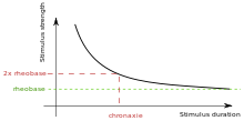
Rheobase is a measure of membrane potential excitability. In neuroscience, rheobase is the minimal current amplitude of infinite duration that results in the depolarization threshold of the cell membranes being reached, such as an action potential or the contraction of a muscle. In Greek, the root rhe translates to "current or flow", and basi means "bottom or foundation": thus the rheobase is the minimum current that will produce an action potential or muscle contraction.

In physiology, the all-or-none law is the principle that if a single nerve fibre is stimulated, it will always give a maximal response and produce an electrical impulse of a single amplitude. If the intensity or duration of the stimulus is increased, the height of the impulse will remain the same. The nerve fibre either gives a maximal response or none at all.

<span class="mw-page-title-main">Group C nerve fiber</span> One of three classes of nerve fiber in the central nervous system and peripheral nervous system

Group C nerve fibers are one of three classes of nerve fiber in the central nervous system (CNS) and peripheral nervous system (PNS). The C group fibers are unmyelinated and have a small diameter and low conduction velocity, whereas Groups A and B are myelinated. Group C fibers include postganglionic fibers in the autonomic nervous system (ANS), and nerve fibers at the dorsal roots. These fibers carry sensory information.

<span class="mw-page-title-main">Denervation</span> Loss of nerve supply

Denervation is any loss of nerve supply regardless of the cause. If the nerves lost to denervation are part of the neuronal communication to a specific function in the body then altered or a loss of physiological functioning can occur. Denervation can be caused by injury or be a symptom of a disorder like ALS, post-polio syndrome, or POTS. Additionally, it can be a useful surgical technique to alleviate major negative symptoms, such as in renal denervation. Denervation can have many harmful side effects such as increased risk of infection and tissue dysfunction.

Clinical electrophysiology is the application of electrophysiology principles to medicine. The two main branches of this discipline are electrotherapy and electrophysiologic testing Clinical electrophysiology can be utilized in the study and treatment of various physiological conditions, and most notably in clinical cardiac electrophysiology.

Repetitive nerve stimulation is a variant of the nerve conduction study where electrical stimulation is delivered to a motor nerve repeatedly several times per second. By observing the change in the muscle electrical response (CMAP) after several stimulations, a physician can assess for the presence of a neuromuscular junction disease, and differentiate between presynaptic and postsynaptic conditions. The test was first described by German neurologist Friedrich Jolly in 1895, and is also known as Jolly's test.

Electroanalgesia is a form of analgesia, or pain relief, that uses electricity to ease pain. Electrical devices can be internal or external, at the site of pain (local) or delocalized throughout the whole body. It works by interfering with the electric currents of pain signals, inhibiting them from reaching the brain and inducing a response; different from traditional analgesics, such as opiates which mimic natural endorphins and NSAIDs that help relieve inflammation and stop pain at the source. Electroanalgesia has a lower addictive potential and poses less health threats to the general public, but can cause serious health problems, even death, in people with other electrical devices such as pacemakers or internal hearing aids, or with heart problems.

A depolarizing prepulse (DPP) is an electrical stimulus that causes the potential difference measured across a neuronal membrane to become more positive or less negative, and precedes another electrical stimulus. DPPs may be of either the voltage or current stimulus variety and have been used to inhibit neural activity, selectively excite neurons, and increase the pain threshold associated with electrocutaneous stimulation.

Neurostimulation is the purposeful modulation of the nervous system's activity using invasive or non-invasive means. Neurostimulation usually refers to the electromagnetic approaches to neuromodulation.

<span class="mw-page-title-main">Shannon Criteria</span>

The Shannon criteria constitute an empirical rule in neural engineering that is used for evaluation of possibility of damage from electrical stimulation to nervous tissue.

References

  1. Irnich W (1980). "The Chronaxie Time and Its Practical Importance". Pacing and Clinical Electrophysiology. 3 (3): 292–301. doi:10.1111/j.1540-8159.1980.tb05236.x. PMID   6160523. S2CID   22590209.
  2. Irnich W (2010). "The Terms "Chronaxie" and "Rheobase" are 100 Years Old". Pacing and Clinical Electrophysiology. 33 (4): 491–496. doi: 10.1111/j.1540-8159.2009.02666.x . PMID   20132498. S2CID   27500016.
  3. Dijkstra B, Dirken MN (1939). "The effect of forced breathing on the motor chronaxie". J. Physiol. 96 (2): 109–17. doi:10.1113/jphysiol.1939.sp003761. PMC   1393855 . PMID   16995119.
  4. Lapicque L (1931). "Has the muscular substance a longer chronaxie than the nervous substance?". The Journal of Physiology. 73 (2): 189–214. doi:10.1113/jphysiol.1931.sp002806. PMC   1394320 . PMID   16994237.
  5. Anderson; et al. (2003). "Neural substrates of microstimulation-evoked tingling: a chronaxie study in human somatosensory thalamus" (PDF). European Journal of Neuroscience. 18 (3): 728–732. doi:10.1046/j.1460-9568.2003.02793.x. PMID   12911770. S2CID   27020742.
  6. Geddes L. A. (2004). "Accuracy Limitations of Chronaxie Values" (PDF). IEEE Transactions on Biomedical Engineering. 51 (1): 176–181. doi:10.1109/tbme.2003.820340. PMID   14723507. S2CID   24899734.[ permanent dead link ]
  7. Chronik, B. A., Recoskie, B. J., Scholl, T. J. (2009) The discrepancy between human peripheral nerve chronaxie times as measured using magnetic and electric field stimuli: the relevance to MRI gradient coil safety. Phys. Med. Biol. 54: 5965–5979. Retrieved from http://www.imaging.robarts.ca/scholl/sites/imaging.robarts.ca.scholl/files/2.pdf Archived 2014-02-07 at the Wayback Machine
  8. Freria; et al. (2007). "Electrical stimulation based on chronaxie reduces atrogin-1 and myod gene expression in denervated rat muscle". Muscle Nerve. 35 (1): 87–97. doi: 10.1002/mus.20668 . PMID   17034040.
  9. Adami et al. (2007) Permanent denervation of rat Tibialis Anterior after bilateral sciatectomy: Determination of chronaxie by surface electrode stimulation during progression of atrophy up to one year. Basic Appl Myol 17 (6): 237-243. Retrieved from http://www.bio.unipd.it/bam/PDF/17-6/Adami.pdf
  10. Kern H, Carraro U, Adami N, Biral D, Hofer C, Forstner C, Mödlin M, Vogelauer M, Pond A, Boncompagni S, Paolini C, Mayr W, Protasi F, Zampieri S (Oct 2010). "Home-based functional electrical stimulation rescues permanently denervated muscles in paraplegic patients with complete lower motor neuron lesion". Neurorehabil Neural Repair. 24 (8): 709–21. doi:10.1177/1545968310366129. PMID   20460493. S2CID   5963094.
  11. Buchanan D. N.; Garven H. S. D. (1926). "Chronaxie in tetany. The effect on the chronaxie of thyreoparathyreoidectomy, the administration of guanidin and of di-methyl guanidin". J Physiol. 62 (1): 115–128. doi:10.1113/jphysiol.1926.sp002343. PMC   1514884 . PMID   16993824.
  12. Natoff I. L., Reiff, B. (1967) The effect of diedrin (heod) on chronaxie and convulsion thresholds in rats and mice. Br. J. Pharmac. Chemother. 31: 197-204. Retrieved from http://onlinelibrary.wiley.com/doi/10.1111/j.1476-5381.1967.tb01990.x/pdf