Myelinogenesis

Last updated
Myelination of a peripheral nerve by a Schwann cell Periferal nerve myelination.jpg
Myelination of a peripheral nerve by a Schwann cell

Myelinogenesis is the formation and development of myelin sheaths in the nervous system, typically initiated in late prenatal neurodevelopment and continuing throughout postnatal development. [1] Myelinogenesis continues throughout the lifespan to support learning and memory via neural circuit plasticity as well as remyelination following injury. [2] Successful myelination of axons increases action potential speed by enabling saltatory conduction, which is essential for timely signal conduction between spatially separate brain regions, as well as provides metabolic support to neurons. [3]

Contents

Stages

Myelin is formed by oligodendrocytes in the central nervous system and Schwann cells in the peripheral nervous system. Therefore, the first stage of myelinogenesis is often defined as the differentiation of oligodendrocyte progenitor cells (OPCs) or Schwann cell progenitors into their mature counterparts, [4] followed by myelin formation around axons. [5]

The oligodendrocyte lineage can be further classified into four stages based on their relation to the onset of myelination: [6]

  1. Differentiation: OPCs exit their proliferative, self-renewing state and begin to express genes and proteins associated with oligodendrocyte fate commitment.
  2. Preoligodendrocyte: These cells express the O4 antigen and develop multiple processes which extend radially with no particular organization. [7]
  3. Immature oligodendrocyte: Sometimes referred to as premyelinating oligodendrocytes, these cells extend "pioneer processes" which contact axons and anchor premyelinating oligodendrocytes to neurons such that they are poised to commence myelinogenesis in response to axonal signals. These pioneer processes grow longitudinally along their target axons. [7]
  4. Mature oligodendrocyte: After myelinogenesis, mature oligodendrocytes surround axons in organized, multilamellar myelin sheaths that contain myelin basic protein (MBP) and myelin proteolipid protein (PLP).

Myelinogenesis thus encompasses the process of transition between phases 3 and 4. [6] Upon initiation of myelinogenesis, each pioneer process forms lamellar extensions which extend and elaborate circumferentially around the target axon. This forms the first turn of the myelin sheath. [7] The sheath continues to expand along the length of the target axon while new membrane is synthesized at the leading edge of the inner tongue of the developing myelin sheath, which begins to take on a spiral cross-sectional structure.

CNS myelin sheath formation.svg
Myelination in the central nervous system by an oligodendrocyte

To drive proper assembly of membrane layers, PLP is inserted into the membrane to stabilize interactions between external leaflets of the myelin membranes; MBP is locally translated and inserted into the cytoplasmic membrane leaflets to strengthen myelin membranes internally. [8] In concert with the formation of axonal nodes of Ranvier, the myelin sheath's edges form paranodal loops. [9]

Mechanism

Transmission electron micrograph of a myelinated axon Myelinated neuron.jpg
Transmission electron micrograph of a myelinated axon
Neuron with oligodendrocyte and myelin sheath showing cytoskeletal structures at a node of Ranvier Neuron with oligodendrocyte and myelin sheath.svg
Neuron with oligodendrocyte and myelin sheath showing cytoskeletal structures at a node of Ranvier

The basic helix–loop–helix transcription factor OLIG1 plays an integral role in the process of oligodendrocyte myelinogenesis by regulating expression of myelin-related genes. OLIG1 is necessary in order to initiate myelination by oligodendrocytes in the brain, but is somewhat dispensable in the spinal cord. [10]

Axon-derived signals regulate the onset of myelinogenesis. Researchers studied regenerating PNS axons for 28 weeks in order to investigate whether or not peripheral axons stimulate oligodendrocytes to begin myelination. Experimental induction of myelination by regenerating peripheral axons demonstrated that Schwann cells and oligodendrocytes have a shared mechanism to stimulate myelination. [11] A similar study working to provide evidence for neuronal regulation of myelinogenesis suggested that myelin formation was due to Schwann cells that were controlled by an undefined property of an associated axon. [11]

Recent research in rats has suggested that apotransferrin and thyroid hormone act both separately and synergistically to promote myelinogenesis, as apotransferrin promotes expression of thyroid hormone receptor alpha. [12]

Peripheral myelinogenesis

1. Axon 2. Nucleus of Schwann cell 3. Schwann cell 4. Myelin sheath 5. Neurilemma Myelin sheath (1).svg
1. Axon 2. Nucleus of Schwann cell 3. Schwann cell 4. Myelin sheath 5. Neurilemma

Peripheral myelinogenesis is controlled by the synthesis of proteins P1, P2, and P0. [13] By using SDS-PAGE, researchers revealed distinct bands with band sizes of 27,000 daltons (P1), 19,000 daltons (P2), and 14,000 daltons (P0). Studies have also shown that P1 and P2 are active before P0 since this protein comes from the peripheral nervous system. [13] In the process of regeneration, Schwann cells re-synthesize proteins associated with myelin-specific proteins when axonal presence is re-established. Synthesis of myelin-specific proteins only occurs in Schwann cells associated with axons. [13] Furthermore, membrane-membrane interactions between axons may be required to promote the synthesis of P1, P2, and P0.

Myelinogenesis in the optic nerve

The process and mechanistic function of myelinogenesis has traditionally been studied using ultrastructure and biochemical techniques in rat optic nerves. The implementation of this method of study has long allowed for experimental observation of myelinogenesis in a model organism nerve that consists entirely of unmyelinated axons. Furthermore, the use of the rat optic nerve helped provide insight for early myelinogenesis researchers into improper and atypical courses of myelinogenesis. [14]

One early study showed that in the developing rat optic nerves, formation of oligodendrocytes and subsequent myelination occurs postnatal. In the optic nerve, the oligodendrocyte cells divided for the final time at five days, with the onset of myelin formation occurring on or around day 6 or 7. However, the exact process by which the oligodendrocytes were stimulated to produce myelin was not yet fully understood, but early myelination in the optic nerve has been linked to a rise in the production of various lipids – cholesterol, cerebroside, and sulfatide. [14]

As researchers began to do postnatal research, they found that myelinogenesis in the rat optic nerve initially commences with axons the largest diameters before proceeding to the remaining smaller axons. In the second week postnatal, oligodendrocyte formation slowed – at this point, 15% of axons have been myelinated – however, myelinogenesis continued to rapidly increase. During the fourth week postnatal, nearly 85% of the axons in the rat optic had been myelinated. [14] During the fifth week and onward toward week sixteen, the myelination decelerated and the remaining unmyelinated axons were ensheathed in myelin. [15] Through the rat optic nerve, early research made significant contributions to knowledge in the field of myelinogenesis.

Role of sulfatides

Studies on the developing optic nerve revealed that galactocerebroside (which forms sulfatide) appeared on the 9th post-natal day and reached a peak on the 15th post-natal day. [14] This expression was similar to a period where the optic nerve showed a maximal myelination period of the axon. As the activity of axon myelination decreased, and one could conclude that the activity of the enzyme is paralleled with the incorporation of sulfate ([35S]) into sulfatide in vivo.

The studies on a rat optic nerve revealed that 15 days post-natal is when an increase in myelination is observed. Before this time period, most of the axons, roughly about 70%, are not myelinated. At this time, [35S] Sulfate was incorporated into sulfatide and the activity of cerebroside, sulfotransferase reached a peak in enzyme activity. This time frame also showed a period of maximal myelination based on the biochemical data. [14]

In the CNS, sulfatide, sulfated glycoproteins, and sulfated mucopolysaccharides appear to be associated with neurons rather than myelin. When graphing the amount of sulfatide made from [35S] and the activity of sulfotransferase, we get to distinguished peaks. [14] The peaks occur on the 15th post-natal day. These peaks corresponded with the maximal myelination period of the optic nerve that has been seen throughout the experiment. [14]

In conclusion, the early phase of myelination was correlated with the increases synthesis of lipids, cholesterol, cerebroside, and sulfatide. [14] It is likely that these compounds are synthesized and packaged in the Golgi Apparatus of oligodendroglia. [14] Even though the transport of these lipids is unknown, it appears that myelination is delayed without their synthesis.

Clinical significance

Because myelin forms an electrically insulating layer that surrounds the axon of some nerve cells, any demyelinating disease can affect the functioning of the nervous system. One such disease is multiple sclerosis (MS), where demyelination occurs in the central nervous system (CNS). [16] Although research is being conducted on protecting oligodendrocytes and promoting remyelination in MS, [17] current therapies mainly address the role of the immune system in demyelination. [18]

Research History

Primary somatosensory cortex (CP: posterior centrale) and primary motor cortex (CA: anterior centrale) of a 7-month-old human fetus. Nissl-stained parasagittal section (Flechsig 1921) FlechsigSaggital4.jpg
Primary somatosensory cortex (CP: posterior centrale) and primary motor cortex (CA: anterior centrale) of a 7-month-old human fetus. Nissl-stained parasagittal section (Flechsig 1921)

Another researcher, Paul Flechsig spent most of his career studying and publishing the details of the process in the cerebral cortex of humans. This takes place mostly between two months before and after birth. He identified 45 separate cortical areas and, in fact, mapped the cerebral cortex by the myelination pattern. The first cortical region to myelinate is in the motor cortex (part of Brodmann's area 4), the second is the olfactory cortex and the third is part of the somatosensory cortex (BA 3,1,2).

The last areas to myelinate are the anterior cingulate cortex (F#43), the inferior temporal cortex (F#44) and the dorsolateral prefrontal cortex (F#45).

In the cerebral convolutions, as in all other parts of the central nervous system, the nerve-fibres do not develop everywhere simultaneously, but step by step in a definite succession, this order of events being particularly maintained in regard to the appearance of the medullary substance. In the convolutions of the cerebrum the investment with medullary substance (myelinisation) has already begun in some places three months before the maturity of the foetus, whilst in other places numerous fibres are devoid of medullary substance even three months after birth. The order of succession in the convolutions is governed by a law identical with the law which I have shown holds good for the spinal cord, the medulla oblongata, and the mesocephalon, and which may be stated somewhat in this way- that, speaking approximately, equally important nerve-fibres are developed simultaneously, but those of dissimilar importance are developed one after another in a succession defined by an imperative law (Fundamental Law of Myelogenesis). The formation of medullary substance is almost completed in certain convolutions at a time when in some it is not even begun and in others has made only slight progress. [19]

Related Research Articles

<span class="mw-page-title-main">Axon</span> Long projection on a neuron that conducts signals to other neurons

An axon, or nerve fiber, is a long, slender projection of a nerve cell, or neuron, in vertebrates, that typically conducts electrical impulses known as action potentials away from the nerve cell body. The function of the axon is to transmit information to different neurons, muscles, and glands. In certain sensory neurons, such as those for touch and warmth, the axons are called afferent nerve fibers and the electrical impulse travels along these from the periphery to the cell body and from the cell body to the spinal cord along another branch of the same axon. Axon dysfunction can be the cause of many inherited and acquired neurological disorders that affect both the peripheral and central neurons. Nerve fibers are classed into three types – group A nerve fibers, group B nerve fibers, and group C nerve fibers. Groups A and B are myelinated, and group C are unmyelinated. These groups include both sensory fibers and motor fibers. Another classification groups only the sensory fibers as Type I, Type II, Type III, and Type IV.

<span class="mw-page-title-main">Central nervous system</span> Brain and spinal cord

The central nervous system (CNS) is the part of the nervous system consisting primarily of the brain and spinal cord. The CNS is so named because the brain integrates the received information and coordinates and influences the activity of all parts of the bodies of bilaterally symmetric and triploblastic animals—that is, all multicellular animals except sponges and diploblasts. It is a structure composed of nervous tissue positioned along the rostral to caudal axis of the body and may have an enlarged section at the rostral end which is a brain. Only arthropods, cephalopods and vertebrates have a true brain, though precursor structures exist in onychophorans, gastropods and lancelets.

<span class="mw-page-title-main">Myelin</span> Fatty substance that surrounds nerve cell axons to insulate them and increase transmission speed

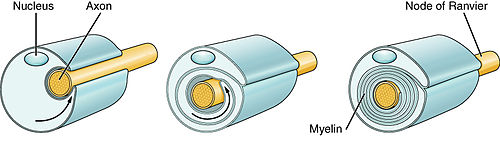
Myelin is a lipid-rich extramembranous structure found on the axons and dendrites of neuron in many bilaterian animals, mainly vertebrates, as well as some arthropods and annelids. A circumferential wrapping of myelin, known as a myelin sheath, increases the conduction speed of electrical impulses passing along the axon by generating saltatory conductions, which are much faster than conduction along an unmyelinated axon, and also reduce signal loss due to extrinsic disturbances.

<span class="mw-page-title-main">Schwann cell</span> Glial cell type

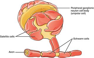
Schwann cells or neurolemmocytes are the principal glia of the peripheral nervous system (PNS). Glial cells function to support neurons and in the PNS, also include satellite cells, olfactory ensheathing cells, enteric glia and glia that reside at sensory nerve endings, such as the Pacinian corpuscle. The two types of Schwann cells are myelinating and nonmyelinating. Myelinating Schwann cells wrap around axons of motor and sensory neurons to form the myelin sheath. The Schwann cell promoter is present in the downstream region of the human dystrophin gene that gives shortened transcript that are again synthesized in a tissue-specific manner.

<span class="mw-page-title-main">Neurilemma</span> Layer present on Schwann cells of PNS neurons

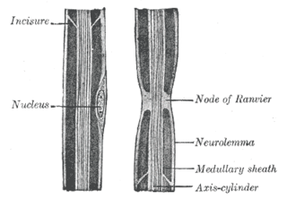
Neurilemma is the outermost nucleated cytoplasmic layer of Schwann cells that surrounds the axon of the neuron. It forms the outermost layer of the nerve fiber in the peripheral nervous system.

<span class="mw-page-title-main">Nervous tissue</span> Main component of the nervous system

Nervous tissue, also called neural tissue, is the main tissue component of the nervous system. The nervous system regulates and controls body functions and activity. It consists of two parts: the central nervous system (CNS) comprising the brain and spinal cord, and the peripheral nervous system (PNS) comprising the branching peripheral nerves. It is composed of neurons, also known as nerve cells, which receive and transmit impulses, and neuroglia, also known as glial cells or glia, which assist the propagation of the nerve impulse as well as provide nutrients to the neurons.

<span class="mw-page-title-main">Oligodendrocyte</span> Neural cell type

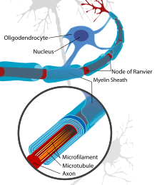
Oligodendrocytes, also known as oligodendroglia, are a type of neuroglia whose main functions are to provide support and insulation to axons within the central nervous system (CNS) of jawed vertebrates. Their function is similar to that of Schwann cells, which perform the same task in the peripheral nervous system (PNS). Oligodendrocytes accomplish this by forming the myelin sheath around axons. Unlike Schwann cells, a single oligodendrocyte can extend its processes to cover around 50 axons, with each axon being wrapped in approximately 1 μm of myelin sheath. Furthermore, an oligodendrocyte can provide myelin segments for multiple adjacent axons.

<span class="mw-page-title-main">Node of Ranvier</span> Gaps between myelin sheaths on the axon of a neuron

In neuroscience and anatomy, nodes of Ranvier, also known as myelin-sheath gaps, occur along a myelinated axon where the axolemma is exposed to the extracellular space. Nodes of Ranvier are uninsulated and highly enriched in ion channels, allowing them to participate in the exchange of ions required to regenerate the action potential. Nerve conduction in myelinated axons is referred to as saltatory conduction due to the manner in which the action potential seems to "jump" from one node to the next along the axon. This results in faster conduction of the action potential.

<span class="mw-page-title-main">Wallerian degeneration</span> Biological process of axonal degeneration

Wallerian degeneration is an active process of degeneration that results when a nerve fiber is cut or crushed and the part of the axon distal to the injury degenerates. A related process of dying back or retrograde degeneration known as 'Wallerian-like degeneration' occurs in many neurodegenerative diseases, especially those where axonal transport is impaired such as ALS and Alzheimer's disease. Primary culture studies suggest that a failure to deliver sufficient quantities of the essential axonal protein NMNAT2 is a key initiating event.

Oligodendrocyte progenitor cells (OPCs), also known as oligodendrocyte precursor cells, NG2-glia, O2A cells, or polydendrocytes, are a subtype of glia in the central nervous system named for their essential role as precursors to oligodendrocytes. They are typically identified in the human by co-expression of PDGFRA and CSPG4.

Sulfatide, also known as 3-O-sulfogalactosylceramide, SM4, or sulfated galactocerebroside, is a class of sulfolipids, specifically a class of sulfoglycolipids, which are glycolipids that contain a sulfate group. Sulfatide is synthesized primarily starting in the endoplasmic reticulum and ending in the Golgi apparatus where ceramide is converted to galactocerebroside and later sulfated to make sulfatide. Of all of the galactolipids that are found in the myelin sheath, one fifth of them are sulfatide. Sulfatide is primarily found on the extracellular leaflet of the myelin plasma membrane produced by the oligodendrocytes in the central nervous system and in the Schwann cells in the peripheral nervous system. However, sulfatide is also present on the extracellular leaflet of the plasma membrane of many cells in eukaryotic organisms.

<span class="mw-page-title-main">Myelin incisure</span>

Myelin incisures are small pockets of cytoplasm left behind during the Schwann cell myelination process.

Remyelination is the process of propagating oligodendrocyte precursor cells to form oligodendrocytes to create new myelin sheaths on demyelinated axons in the CNS. This is a process naturally regulated in the body and tends to be very efficient in a healthy CNS. The process creates a thinner myelin sheath than normal, but it helps to protect the axon from further damage, from overall degeneration, and proves to increase conductance once again. The processes underlying remyelination are under investigation in the hope of finding treatments for demyelinating diseases, such as multiple sclerosis.

<span class="mw-page-title-main">Myelin-associated glycoprotein</span> Protein-coding gene in the species Homo sapiens

Myelin-associated glycoprotein is a type 1 transmembrane protein glycoprotein localized in periaxonal Schwann cell and oligodendrocyte membranes, where it plays a role in glial-axonal interactions. MAG is a member of the SIGLEC family of proteins and is a functional ligand of the NOGO-66 receptor, NgR. MAG is believed to be involved in myelination during nerve regeneration in the PNS and is vital for the long-term survival of the myelinated axons following myelinogenesis. In the CNS MAG is one of three main myelin-associated inhibitors of axonal regeneration after injury, making it an important protein for future research on neurogenesis in the CNS.

Neuroregeneration involves the regrowth or repair of nervous tissues, cells or cell products. Neuroregenerative mechanisms may include generation of new neurons, glia, axons, myelin, or synapses. Neuroregeneration differs between the peripheral nervous system (PNS) and the central nervous system (CNS) by the functional mechanisms involved, especially in the extent and speed of repair. When an axon is damaged, the distal segment undergoes Wallerian degeneration, losing its myelin sheath. The proximal segment can either die by apoptosis or undergo the chromatolytic reaction, which is an attempt at repair. In the CNS, synaptic stripping occurs as glial foot processes invade the dead synapse.

<span class="mw-page-title-main">Myelin protein zero</span> Protein-coding gene in the species Homo sapiens

Myelin protein zero is a single membrane glycoprotein which in humans is encoded by the MPZ gene. P0 is a major structural component of the myelin sheath in the peripheral nervous system (PNS). Myelin protein zero is expressed by Schwann cells and accounts for over 50% of all proteins in the peripheral nervous system, making it the most common protein expressed in the PNS. Mutations in myelin protein zero can cause myelin deficiency and are associated with neuropathies like Charcot–Marie–Tooth disease and Dejerine–Sottas disease.

<span class="mw-page-title-main">GJB1</span> Protein-coding gene in humans

Gap junction beta-1 protein (GJB1), also known as connexin 32 (Cx32), is a transmembrane protein that in humans is encoded by the GJB1 gene. Gap junction beta-1 protein is a member of the gap junction connexin family of proteins that regulates and controls the transfer of communication signals across cell membranes, primarily in the liver and peripheral nervous system. However, the protein is expressed in multiple organs, including in oligodendrocytes in the central nervous system.

In neurobiology, a mesaxon is a pair of parallel plasma membranes of a Schwann cell. It marks the point of edge-to-edge contact by the Schwann cell encircling the axon. A single Schwann cell of the peripheral nervous system will wrap around and support only one individual axon, while the oligodendrocytes found in the central nervous system can wrap around and support 5-8 axons. Thin unmyelinated axons are often bundled, with several unmyelinated axons to a single mesaxon.

<span class="mw-page-title-main">LINGO1</span> Protein-coding gene in the species Homo sapiens

Leucine rich repeat and Immunoglobin-like domain-containing protein 1 also known as LINGO-1 is a protein which is encoded by the LINGO1 gene in humans. It belongs to the family of leucine-rich repeat proteins which are known for playing key roles in the biology of the central nervous system. LINGO-1 is a functional component of the Nogo receptor also known as the reticulon 4 receptor.

Anti-MAG peripheral neuropathy is a specific type of peripheral neuropathy in which the person's own immune system attacks cells that are specific in maintaining a healthy nervous system. As these cells are destroyed by antibodies, the nerve cells in the surrounding region begin to lose function and create many problems in both sensory and motor function. Specifically, antibodies against myelin-associated glycoprotein (MAG) damage Schwann cells. While the disorder occurs in only 10% of those afflicted with peripheral neuropathy, people afflicted have symptoms such as muscle weakness, sensory problems, and other motor deficits usually starting in the form of a tremor of the hands or trouble walking. There are, however, multiple treatments that range from simple exercises in order to build strength to targeted drug treatments that have been shown to improve function in people with this type of peripheral neuropathy.

References

  1. Eilam, R.; Bar-Lev, D.D.; Levin-Zaidman, S.; Tsoory, M.; LoPresti, P.; Sela, M.; Arnon, R.; Aharoni, R. (2014). "Oligodendrogenesis and myelinogenesis during postnatal development effect of glatiramer acetate". Glia. 62 (4): 649–665. doi:10.1002/glia.22632. PMID   24481644. S2CID   25559134.
  2. Williamson JM, Lyons DA (2018). "Myelin Dynamics Throughout Life: An Ever-Changing Landscape?". Front Cell Neurosci. 12: 424. doi: 10.3389/fncel.2018.00424 . PMC   6252314 . PMID   30510502.
  3. Philips T, Rothstein JD (2017). "Oligodendroglia: metabolic supporters of neurons". J Clin Invest. 127 (9): 3271–3280. doi:10.1172/JCI90610. PMC   5669561 . PMID   28862639.
  4. Serrano-Regal MP, Luengas-Escuza I, Bayón-Cordero L, Ibarra-Aizpurua N, Alberdi E, Pérez-Samartín A; et al. (2020). "Oligodendrocyte Differentiation and Myelination Is Potentiated via GABAB Receptor Activation". Neuroscience. 439: 163–180. doi:10.1016/j.neuroscience.2019.07.014. PMID   31349008. S2CID   198934117.{{cite journal}}: CS1 maint: multiple names: authors list (link)
  5. Watkins, T., Mulinyawe, S., Emery, B., Barres, B. (2008). Distinct Stages of Myelination Regulated by Y-Secretase and Astrocytes in a Rapidly Myelinating CNS Coculture System. 555-569
  6. 1 2 Kinney, H. C., & Volpe, J. J. (2018). Myelination Events. Volpe’s Neurology of the Newborn, 176–188. doi:10.1016/b978-0-323-42876-7.00008-9
  7. 1 2 3 Friedrich, VL., Hardy, RJ., (1996). Progressive Remodeling of the Oligodendrocyte Process Arbor during Myelinogenesis. 243-54.
  8. Baron W, Hoekstra D (2010). "On the biogenesis of myelin membranes: sorting, trafficking and cell polarity". FEBS Lett. 584 (9): 1760–70. doi: 10.1016/j.febslet.2009.10.085 . PMID   19896485.
  9. Snaidero N, Möbius W, Czopka T, Hekking LH, Mathisen C, Verkleij D; et al. (2014). "Myelin membrane wrapping of CNS axons by PI(3,4,5)P3-dependent polarized growth at the inner tongue". Cell. 156 (1–2): 277–90. doi:10.1016/j.cell.2013.11.044. PMC   4862569 . PMID   24439382.{{cite journal}}: CS1 maint: multiple names: authors list (link)
  10. Xin, M. (2005). Myelinogenesis and Axonal Recognition by Oligodendrocytes in Brain Are Uncoupled in Olig1-Null Mice. Journal of Neuroscience, 25(6), 1354-1365. doi:10.1523/jneurosci.3034-04.2005
  11. 1 2 Weinberg, E., & Spencer, P. (1979). Studies on the control of myelinogenesis. 3. Signaling of oligodendrocyte myelination by regenerating peripheral axons. Brain Research, 162(2), 273-279. doi:10.1016/0006-8993(79)90289-0
  12. Marziali, L.N., Garcia, C.I., Pasquini, J.M. (2015). Transferrin and thyroid hormone converge in the control of myelinogenesis. Experimental Neurology. Vol 265. 129–141.
  13. 1 2 3 Politis, MJ, N. Sternberger, Kathy Ederle, and Peter S. Spencer. "Studies on the Control of Myelinogenesis." The Journal of Neuroscience 2.9 (1982): 1252-266.
  14. 1 2 3 4 5 6 7 8 9 Tennekoon, GI., Cohen, SR., Price, DL., McKhann, GM. (1977). Myelinogenesis in optic nerve. A morphological, autoradiographic, and biochemical analysis. Journal of Cell Biology, 72(3), 604-616.
  15. Dangata, Y., Kaufman, M. (1997). Myelinogenesis in the Optic Nerve of (C57BL x CBA) F1 Hybrid Mice: A Morphometric Analysis.European Journal of Morphology, 35(1), 3-18.
  16. "NINDS Multiple Sclerosis Information Page". National Institute of Neurological Disorders and Stroke. 19 November 2015. Archived from the original on 13 February 2016. Retrieved 6 March 2016.
  17. Rodgers, Jane M.; Robinson, Andrew P.; Miller, Stephen D. (2013). "Strategies for protecting oligodendrocytes and enhancing remyelination in multiple sclerosis". Discovery Medicine. 86 (86): 53–63. PMC   3970909 . PMID   23911232.
  18. Cohen JA (July 2009). "Emerging therapies for relapsing multiple sclerosis". Arch. Neurol. 66 (7): 821–8. doi:10.1001/archneurol.2009.104. PMID   19597083.
  19. Flechsig, Paul (1901-10-19). "Developmental (myelogenetic) localisation of the cerebral cortex in the human subject". The Lancet . 158 (4077): 1028. doi:10.1016/s0140-6736(01)01429-5.