Related Research Articles

An eyelid is a thin fold of skin that covers and protects an eye. The levator palpebrae superioris muscle retracts the eyelid, exposing the cornea to the outside, giving vision. This can be either voluntarily or involuntarily. "Palpebral" means relating to the eyelids. Its key function is to regularly spread the tears and other secretions on the eye surface to keep it moist, since the cornea must be continuously moist. They keep the eyes from drying out when asleep. Moreover, the blink reflex protects the eye from foreign bodies. A set of specialized hairs known as lashes grow from the upper and lower eyelid margins to further protect the eye from dust and debris.

The lacrimal glands are paired exocrine glands, one for each eye, found in most terrestrial vertebrates and some marine mammals, that secrete the aqueous layer of the tear film. In humans, they are situated in the upper lateral region of each orbit, in the lacrimal fossa of the orbit formed by the frontal bone. Inflammation of the lacrimal glands is called dacryoadenitis. The lacrimal gland produces tears which are secreted by the lacrimal ducts, and flow over the ocular surface, and then into canals that connect to the lacrimal sac. From that sac, the tears drain through the lacrimal duct into the nose.

Xanthelasma is a sharply demarcated yellowish deposit of cholesterol underneath the skin. It usually occurs on or around the eyelids. While they are neither harmful to the skin nor painful, these minor growths may be disfiguring and can be removed. There is a growing body of evidence for the association between xanthelasma deposits and blood low-density lipoprotein levels and increased risk of atherosclerosis.

The canthus is either corner of the eye where the upper and lower eyelids meet. More specifically, the inner and outer canthi are, respectively, the medial and lateral ends/angles of the palpebral fissure.

The orbicularis oculi is a muscle in the face that closes the eyelids. It arises from the nasal part of the frontal bone, from the frontal process of the maxilla in front of the lacrimal groove, and from the anterior surface and borders of a short fibrous band, the medial palpebral ligament.
Von Graefe's sign is the lagging of the upper eyelid on downward rotation of the eye, indicating exophthalmic goiter. It is a dynamic sign, whereas lid lag is a static sign which may also be present in cicatricial eyelid retraction or congenital ptosis.

The tarsi or tarsal plates are two comparatively thick, elongated plates of dense connective tissue, about 10 mm (0.39 in) in length for the upper eyelid and 5 mm for the lower eyelid; one is found in each eyelid, and contributes to its form and support. They are located directly above the lid margins. The tarsus has a lower and upper part making up the palpebrae.

Vernal keratoconjunctivitis is a recurrent, bilateral, and self-limiting type of conjunctivitis having a periodic seasonal incidence.
The medial palpebral ligament is a ligament of the face. It attaches to the frontal process of the maxilla, the lacrimal groove, and the tarsus of each eyelid. It has a superficial (anterior) and a deep (posterior) layer, with many surrounding attachments. It connects the medial canthus of each eyelid to the medial part of the orbit. It is a useful point of fixation during eyelid reconstructive surgery.

The medial palpebral arteries are arteries of the head that contribute arterial blood supply to the eyelids. They are derived from the ophthalmic artery; a single medial palpebral artery issues from the ophthalmic artery before splitting into a superior and an inferior medial palpebral artery, each supplying one eyelid.

In anatomy, the orbital septum is a membranous sheet that acts as the anterior (frontal) boundary of the orbit. It extends from the orbital rims to the eyelids. It forms the fibrous portion of the eyelids.

Graves' ophthalmopathy, also known as thyroid eye disease (TED), is an autoimmune inflammatory disorder of the orbit and periorbital tissues, characterized by upper eyelid retraction, lid lag, swelling, redness (erythema), conjunctivitis, and bulging eyes (exophthalmos). It occurs most commonly in individuals with Graves' disease, and less commonly in individuals with Hashimoto's thyroiditis, or in those who are euthyroid.
The palpebral fissure is the elliptic space between the medial and lateral canthi of the two open eyelids. In simple terms, it is the opening between the eyelids. In adult humans, this measures about 10 mm vertically and 30 mm horizontally.
Oculoplastics, or oculoplastic surgery, includes a wide variety of surgical procedures that deal with the orbit, eyelids, tear ducts, and the face. It also deals with the reconstruction of the eye and associated structures.

Blepharophimosis is a congenital anomaly in which the eyelids are underdeveloped such that they cannot open as far as usual and permanently cover part of the eyes. Both the vertical and horizontal palpebral fissures are shortened; the eyes also appear spaced more widely apart as a result, known as telecanthus.
Cryptophthalmos is a rare congenital anomaly in which the skin is continuous over the eyeball with absence of palpebral fissures and presence of eyelashes. It is classified into three types: complete, incomplete and abortive. Failure of eyelid separation can be associated with maldevelopment of the underlying cornea and microphthalmia. Cryptophthalmos usually occurs on both sides and occurs in association with multiple other malformations collectively referred to as Fraser syndrome. Along with microphthalmia, it may be associated with a tuft of hair.
Stellwag's sign is a sign of infrequent or incomplete blinking associated with exophthalmos or Graves orbitopathy. It is accompanied by Dalrymple's sign, which is a retraction of the upper eyelids resulting in an apparent widening of the palpebral opening.
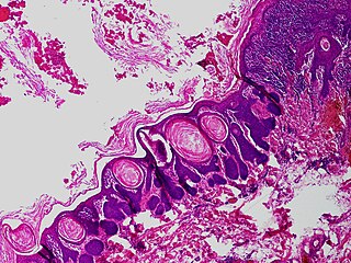
Trichofolliculoma is a cutaneous condition characterized by a benign, highly structured tumor of the pilosebaceous unit. Trichofolliculoma is a rare tumor of the eyelid. It can be suspected by the “cotton bag sign”
Concretion in the palpebral conjunctiva, is called conjunctival concretion, that is a small, hard, yellowish-white calcified matter, superficially buried beneath the palpebral conjunctiva. Most of concretions in the eye form in the palpebral conjunctiva, which is a clear membrane to surround the inside of the eyelid; fewer can be located in the cornea and retina.
Ankyloblepharon is a medical condition, defined as the adhesion of the edges of the upper eyelid with the lower eyelid. Ankyloblepharon must be differentiated from blepharophimosis, in which palpebral aperture is reduced and there is telecanthus, but the eyelid margins are normal. Another condition similar to ankyloblepharon is symblepharon, in which the palpebral conjunctiva is attached to the bulbar conjunctiva. Recognition of ankyloblepharon necessitates systemic examination to detect associated abnormalities such as genitourinary and cardiac abnormalities and syndactyly.
References
- ↑ Cline D; Hofstetter HW; Griffin JR. Dictionary of Visual Science. 4th ed. Butterworth-Heinemann, Boston 1997. ISBN 0-7506-9895-0