Brushfield spots

Last updated
The Brushfield spots are the spots between the inner and outer red circles. Brushfield eyes magnified.jpg
The Brushfield spots are the spots between the inner and outer red circles.

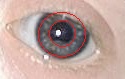
Brushfield spots are small, white or greyish/brown spots on the periphery of the iris in the human eye due to aggregation of connective tissue, a normal constituent of the iris stroma. The spots are named after the physician Thomas Brushfield, who first described them in his 1924 M.D. thesis. [1]

Brushfield spots are a characteristic feature of the chromosomal disorder Down syndrome or trisomy 21. They occur in 35–78% of newborn infants with Down syndrome. [2] Brushfield spots tend to be obscured by pigmentation of the anterior border layer of the iris in patients with darker irides. Hence, they are much more likely to be observed in children with lightly pigmented eyes. Brushfield spots are more commonly found in Down syndrome patients of European descent than similarly affected children of Asian heritage. [3]

Brushfield spots comprise focal areas of iris stromal hyperplasia, surrounded by relative hypoplasia.

Similar spots described by Krückmann [4] and Wolfflin [5] are found in individuals without Down syndrome. Termed Krückmann-Wolfflin bodies, these spots typically are less well defined, fewer in number and more peripherally located than the Brushfield spots of trisomy 21. [6]

Related Research Articles

<span class="mw-page-title-main">Down syndrome</span> Genetic disorder

Down syndrome or Down's syndrome, also known as trisomy 21, is a genetic disorder caused by the presence of all or part of a third copy of chromosome 21. It is usually associated with developmental delays, mild to moderate intellectual disability, and characteristic physical features. There are three types of Down syndrome, all with the same features: Trisomy 21, the most common type; Mosaic Down syndrome, and Translocation Down syndrome.

<span class="mw-page-title-main">Williams syndrome</span> Medical condition

Williams syndrome (WS), also Williams–Beuren syndrome (WBS), is a genetic disorder that affects many parts of the body. Facial features frequently include a broad forehead, underdeveloped chin, short nose, and full cheeks. Mild to moderate intellectual disability is observed in people with WS, with particular challenges with visual spatial tasks such as drawing. Verbal skills are relatively unaffected. Many people with WS have an outgoing personality, an openness to engaging with other people, and a happy disposition. Medical issues with teeth, heart problems, and periods of high blood calcium are common.

<span class="mw-page-title-main">Patau syndrome</span> Chromosomal disorder in which there are three copies of chromosome 13

Patau syndrome is a syndrome caused by a chromosomal abnormality, in which some or all of the cells of the body contain extra genetic material from chromosome 13. The extra genetic material disrupts normal development, causing multiple and complex organ defects.

<span class="mw-page-title-main">Trisomy 18</span> Chromosomal disorder in which there are three copies of chromosome 18

Trisomy 18, also known as Edwards syndrome, is a genetic disorder caused by the presence of a third copy of all or part of chromosome 18. Many parts of the body are affected. Babies are often born small and have heart defects. Other features include a small head, small jaw, clenched fists with overlapping fingers, and severe intellectual disability.

<span class="mw-page-title-main">Heterochromia iridum</span> Difference in coloration, usually of the iris but also of hair or skin

Heterochromia is a variation in coloration most often used to describe color differences of the iris, but can also be applied to color variation of hair or skin. Heterochromia is determined by the production, delivery, and concentration of melanin. It may be inherited, or caused by genetic mosaicism, chimerism, disease, or injury. It occurs in humans and certain breeds of domesticated animals.

Hypotonia is a state of low muscle tone, often involving reduced muscle strength. Hypotonia is not a specific medical disorder, but a potential manifestation of many different diseases and disorders that affect motor nerve control by the brain or muscle strength. Hypotonia is a lack of resistance to passive movement, whereas muscle weakness results in impaired active movement. Central hypotonia originates from the central nervous system, while peripheral hypotonia is related to problems within the spinal cord, peripheral nerves and/or skeletal muscles. Severe hypotonia in infancy is commonly known as floppy baby syndrome. Recognizing hypotonia, even in early infancy, is usually relatively straightforward, but diagnosing the underlying cause can be difficult and often unsuccessful. The long-term effects of hypotonia on a child's development and later life depend primarily on the severity of the muscle weakness and the nature of the cause. Some disorders have a specific treatment but the principal treatment for most hypotonia of idiopathic or neurologic cause is physical therapy and/or occupational therapy for remediation.

Crouzon syndrome is an autosomal dominant genetic disorder known as a branchial arch syndrome. Specifically, this syndrome affects the first branchial arch, which is the precursor of the maxilla and mandible. Since the branchial arches are important developmental features in a growing embryo, disturbances in their development create lasting and widespread effects.

<span class="mw-page-title-main">Coloboma</span> Hole in one of the structures of the eye

A coloboma is a hole in one of the structures of the eye, such as the iris, retina, choroid, or optic disc. The hole is present from birth and can be caused when a gap called the choroid fissure, which is present during early stages of prenatal development, fails to close up completely before a child is born. Ocular coloboma is relatively uncommon, affecting less than one in every 10,000 births.

<span class="mw-page-title-main">Imperforate anus</span> Birth defect of malformed rectum

An imperforate anus or anorectal malformations (ARMs) are birth defects in which the rectum is malformed. ARMs are a spectrum of different congenital anomalies which vary from fairly minor lesions to complex anomalies. The cause of ARMs is unknown; the genetic basis of these anomalies is very complex because of their anatomical variability. In 8% of patients, genetic factors are clearly associated with ARMs. Anorectal malformation in Currarino syndrome represents the only association for which the gene HLXB9 has been identified.

<span class="mw-page-title-main">Atrioventricular septal defect</span> Medical condition

Atrioventricular septal defect (AVSD) or atrioventricular canal defect (AVCD), also known as "common atrioventricular canal" or "endocardial cushion defect" (ECD), is characterized by a deficiency of the atrioventricular septum of the heart that creates connections between all four of its chambers. It is a very specific combination of 3 defects:

<span class="mw-page-title-main">Rumination syndrome</span> Medical condition

Rumination syndrome, or merycism, is a chronic motility disorder characterized by effortless regurgitation of most meals following consumption, due to the involuntary contraction of the muscles around the abdomen. There is no retching, nausea, heartburn, odour, or abdominal pain associated with the regurgitation as there is with typical vomiting, and the regurgitated food is undigested. The disorder has been historically documented as affecting only infants, young children, and people with cognitive disabilities . It is increasingly being diagnosed in a greater number of otherwise healthy adolescents and adults, though there is a lack of awareness of the condition by doctors, patients, and the general public.

The obsolete medical terms Mongolian idiocy and Mongolism referred to a specific type of mental deficiency, associated with the genetic disorder now known as Down syndrome. The obsolete term for a person with this syndrome was Mongolian idiot.

<span class="mw-page-title-main">Dermatoglyphics</span>

Dermatoglyphics is the scientific study of fingerprints, lines, mounts and shapes of hands, as distinct from the superficially similar pseudoscience of palmistry.

<span class="mw-page-title-main">Micrognathism</span> Condition in which the jaw is small

Micrognathism is a condition where the jaw is undersized. It is also sometimes called mandibular hypoplasia. It is common in infants, but is usually self-corrected during growth, due to the jaws' increasing in size. It may be a cause of abnormal tooth alignment and in severe cases can hamper feeding. It can also, both in adults and children, make intubation difficult, either during anesthesia or in emergency situations.

The red reflex refers to the reddish-orange reflection of light from the back of the eye, or fundus, observed when using an ophthalmoscope or retinoscope. The reflex relies on the transparency of optical media and reflects off the fundus back through media into the aperture of the ophthalmoscope. The red reflex is considered abnormal if there is any asymmetry between the eyes, dark spots, or white reflex (Leukocoria).

<span class="mw-page-title-main">Cat eye syndrome</span> Genetic partial duplication of chromosome 22

Cat-eye syndrome (CES) or Schmid–Fraccaro syndrome is a rare condition caused by an abnormal extra chromosome, i.e. a small supernumerary marker chromosome. This chromosome consists of the entire short arm and a small section of the long arm of chromosome 22. In consequence, individuals with the cat-eye syndrome have three (trisomic) or four (tetrasomic) copies of the genetic material contained in the abnormal chromosome instead of the normal two copies. The prognosis for patients with CES varies depending on the severity of the condition and their associated signs and symptoms, especially when heart or kidney abnormalities are seen.

<span class="mw-page-title-main">Trisomy 22</span> Medical condition

Trisomy 22 is a chromosomal disorder in which three copies of chromosome 22 are present rather than two. It is a frequent cause of spontaneous abortion during the first trimester of pregnancy. Progression to the second trimester and live birth are rare. This disorder is found in individuals with an extra copy or a variation of chromosome 22 in some or all cells of their bodies.

PHACE syndrome is a medical condition involving the uncommon association between large infantile hemangiomas, usually of the face, and birth defects of the brain, heart, eyes, skin and/or arteries. PHACE is an acronym that stands for the medical names of the parts of the body it often impacts:

<span class="mw-page-title-main">Tetrasomy X</span> Chromosomal disorder with 4 X chromosomes

Tetrasomy X, also known as 48,XXXX, is a chromosomal disorder in which a female has four, rather than two, copies of the X chromosome. It is associated with intellectual disability of varying severity, characteristic "coarse" facial features, heart defects, and skeletal anomalies such as increased height, clinodactyly, and radioulnar synostosis. Tetrasomy X is a rare condition, with few medically recognized cases; it is estimated to occur in approximately 1 in 50,000 females.

<span class="mw-page-title-main">Trisomy X</span> Chromosome disorder in women

Trisomy X, also known as triple X syndrome and characterized by the karyotype 47,XXX, is a chromosome disorder in which a female has an extra copy of the X chromosome. It is relatively common and occurs in 1 in 1,000 females, but is rarely diagnosed; fewer than 10% of those with the condition know they have it.

References

  1. Wallis, Hugh R.E. "The Significance of Brushfield's Spots in the Diagnosis of Mongolism in Infancy", Archives of Disease in Childhood, 1951 December; 26 (130): 495-500. Retrieved on 21 March 2009.
  2. Sanez, R.B. (January 1999), "Primary care of infants and young children with Down syndrome", American Family Physician, vol. 59, no. 2, p. 381, archived from the original on 2011-06-06, retrieved 2007-10-18
  3. Kim, J.H.; et al. (November 2002), "Characteristic ocular findings in Asian children with Down syndrome", Nature, vol. 16, no. 6, pp. 710–714, doi: 10.1038/sj.eye.6700208 , PMID   12439664
  4. Krückmann, E. (1907), "Die Erkrankungen des Uvealtractus und des Glaskopers Regenbogenhaut", in Graefe, A. von; Saemisch, T. (eds.), Handbuch der gesamten Augenheilkunde, vol. 5, pp. 1–11
  5. Wolfflin, E. (1902), "Ein klinischer Beitrag zur Kenntniss der Structur der Iris", Arch. F. Augenh, 45: 1–4
  6. Donaldson, DD (1961), "The Significance of Spotting of the Iris in Mongoloids: Brushfield Spots", Arch. Ophthalmol., 65 (1): 26–31, doi:10.1001/archopht.1961.01840020028007