
The Papanicolaou test is a method of cervical screening used to detect potentially precancerous and cancerous processes in the cervix or, more rarely, anus. Abnormal findings are often followed up by more sensitive diagnostic procedures and, if warranted, interventions that aim to prevent progression to cervical cancer. The test was independently invented in the 1920s by the Greek physician Georgios Papanikolaou and named after him. A simplified version of the test was introduced by the Canadian obstetrician Anna Marion Hilliard in 1957.

Anatomical pathology (Commonwealth) or anatomic pathology (U.S.) is a medical specialty that is concerned with the diagnosis of disease based on the macroscopic, microscopic, biochemical, immunologic and molecular examination of organs and tissues. Over the 20th century, surgical pathology has evolved tremendously: from historical examination of whole bodies (autopsy) to a more modernized practice, centered on the diagnosis and prognosis of cancer to guide treatment decision-making in oncology. Its modern founder was the Italian scientist Giovanni Battista Morgagni from Forlì.

Georgios Nikolaou Papanikolaou was a Greek physician, zoologist and microscopist who was a pioneer in cytopathology and early cancer detection, and inventor of the pap smear for detection of cervical cancer.

Cytopathology is a branch of pathology that studies and diagnoses diseases on the cellular level. The discipline was founded by George Nicolas Papanicolaou in 1928. Cytopathology is generally used on samples of free cells or tissue fragments, in contrast to histopathology, which studies whole tissues. Cytopathology is frequently, less precisely, called "cytology", which means "the study of cells".

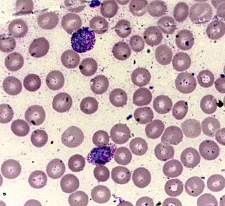
Romanowsky staining is a prototypical staining technique that was the forerunner of several distinct but similar stains widely used in hematology and cytopathology. Romanowsky-type stains are used to differentiate cells for microscopic examination in pathological specimens, especially blood and bone marrow films, and to detect parasites such as malaria within the blood.

Staining is a technique used to enhance contrast in samples, generally at the microscopic level. Stains and dyes are frequently used in histology, in cytology, and in the medical fields of histopathology, hematology, and cytopathology that focus on the study and diagnoses of diseases at the microscopic level. Stains may be used to define biological tissues, cell populations, or organelles within individual cells.

A blood smear, peripheral blood smear or blood film is a thin layer of blood smeared on a glass microscope slide and then stained in such a way as to allow the various blood cells to be examined microscopically. Blood smears are examined in the investigation of hematological (blood) disorders and are routinely employed to look for blood parasites, such as those of malaria and filariasis.

Wright's stain is a hematologic stain that facilitates the differentiation of blood cell types. It is classically a mixture of eosin (red) and methylene blue dyes. It is used primarily to stain peripheral blood smears, urine samples, and bone marrow aspirates, which are examined under a light microscope. In cytogenetics, it is used to stain chromosomes to facilitate diagnosis of syndromes and diseases.

Giemsa stain, named after German chemist and bacteriologist Gustav Giemsa, is a nucleic acid stain used in cytogenetics and for the histopathological diagnosis of malaria and other parasites.

Fibroadenomas are benign breast tumours characterized by an admixture of stromal and epithelial tissue. Breasts are made of lobules and ducts. These are surrounded by glandular, fibrous and fatty tissues. Fibroadenomas develop from the lobules. The glandular tissue and ducts grow over the lobule to form a solid lump.

Fine-needle aspiration (FNA) is a diagnostic procedure used to investigate lumps or masses. In this technique, a thin, hollow needle is inserted into the mass for sampling of cells that, after being stained, are examined under a microscope (biopsy). The sampling and biopsy considered together are called fine-needle aspiration biopsy (FNAB) or fine-needle aspiration cytology (FNAC). Fine-needle aspiration biopsies are very safe minor surgical procedures. Often, a major surgical biopsy can be avoided by performing a needle aspiration biopsy instead, eliminating the need for hospitalization. In 1981, the first fine-needle aspiration biopsy in the United States was done at Maimonides Medical Center. Today, this procedure is widely used in the diagnosis of cancer and inflammatory conditions. Fine needle aspiration is generally considered a safe procedure. Complications are infrequent.

Eosin Y, also called C.I. 45380 or C.I. Acid Red 87, is a member of the triarylmethane dyes. It is produced from fluorescein by bromination.

Papanicolaou stain is a multichromatic (multicolored) cytological staining technique developed by George Papanicolaou in 1942. The Papanicolaou stain is one of the most widely used stains in cytology, where it is used to aid pathologists in making a diagnosis. Although most notable for its use in the detection of cervical cancer in the Pap test or Pap smear, it is also used to stain non-gynecological specimen preparations from a variety of bodily secretions and from small needle biopsies of organs and tissues. Papanicolaou published three formulations of this stain in 1942, 1954, and 1960.
Leishman stain, also known as Leishman's stain, is used in microscopy for staining blood smears. It is generally used to differentiate between and identify white blood cells, malaria parasites, and trypanosomas. It is based on a methanolic mixture of "polychromed" methylene blue and eosin. The methanolic stock solution is stable and also serves the purpose of directly fixing the smear eliminating a prefixing step. If a working solution is made by dilution with an aqueous buffer, the resulting mixture is very unstable and cannot be used for long. Leishman stain is named after its inventor, the Scottish pathologist William Boog Leishman. It is a version of the Romanowsky stain, and is thus similar to and partially replaceable by Giemsa stain, Jenner's stain, and Wright's stain.

A koilocyte is a squamous epithelial cell that has undergone a number of structural changes, which occur as a result of infection of the cell by human papillomavirus (HPV). Identification of these cells by pathologists can be useful in diagnosing various HPV-associated lesions.
In diagnostic pathology, a hematoxylin body, or LE body, is a dense, homogeneous, basophilic particle, easily stainable with hematoxylin. It consists of degraded nuclear material from an injured cell, along with autoantibodies and a limited amount of cytoplasm.
Vaginal cytology is a microscopic examination of cells from the vaginal epithelium. In veterinary medicine, it helps differentiate the stages of the mammalian estrous cycle because the vaginal epithelium changes in response to sex hormone levels; practically, it is used to distinguish when a female canine is at a particular point in the estrous cycle. In a normal vaginal smear, lactational cells, navicular cells, endocervical cells, endometrial cells, trophoblastic cells, and leucocytes may be present.

Dmitri Leonidovich Romanowsky was a Russian physician who is best known for his invention of an eponymous histological stain called Romanowsky stain. It paved the way for the discovery and diagnosis of microscopic pathogens, such as malarial parasites, and later developments of new histological stains that became fundamental to microbiology and physiology.
Liu's stain (劉氏染色法) is a staining technique used to stain animal cells. It is an improved staining based on Romanowsky stain, and was introduced by professor Chen-Hui Liu(劉禎輝), faculty of National Taiwan University, in 1953. The method sees a wide variety of usage in Taiwan. Comparing to other staining methods, Liu's stain is relatively fast, taking no more than 3 minutes to complete the process. In pathology, Liu's stain is primarily used to distinguish blood cells, but it can also apply on vaginal discharge, sputum, and pus as a simple stain.

A cytocentrifuge, sometimes referred to as a cytospin, is a specialized centrifuge used to concentrate cells in fluid specimens onto a microscope slide so that they can be stained and examined. Cytocentrifuges are used in various areas of the clinical laboratory, such as cytopathology, hematology and microbiology, as well as in biological research. The method can be used on many different types of specimens, including fine needle aspirates, cerebrospinal fluid, serous and synovial fluid, and urine.
