Encephalomalacia

Last updated
Cerebral softening
Other namesEncephalomalacia
FIGURA 03.jpg
Stroke brain (similar to cerebral softening)
Specialty Neurology

Cerebral softening, also known as encephalomalacia, is a localized softening of the substance of the brain, due to bleeding or inflammation. Three varieties, distinguished by their color and representing different stages of the disease progress, are known respectively as red, yellow, and white softening. [1] [2]

Contents

Causes

Stroke

Ischemia: A decrease or restriction of circulating blood flow to a region of the brain which deprives neurons of the necessary substrates (primarily glucose); represents 80% of all strokes. A thrombus or embolus plugs an artery so there is a reduction or cessation of blood flow. This hypoxia or anoxia leads to neuronal injury, which is known as a stroke. The death of neurons leads to a so-called softening of the cerebrum in the affected area.[ citation needed ] [3]

Hemorrhage: Intracerebral hemorrhage occurs in deep penetrating vessels and disrupts the connecting pathways, causing a localized pressure injury and in turn injury to brain tissue in the affected area. Hemorrhaging can occur in instances of embolic ischemia, in which the previously obstructed region spontaneously restores blood flow. This is known as a hemorrhagic infarction and a resulting red infarct occurs, which points to a type of cerebral softening known as red softening. [1] [4]

Circle of Willis

In a study on the circle of Willis and its relation to cerebral vascular disorders, a comparison on various anomalies between normal brains (those without the condition of cerebral softening) and brains with cerebral softening were looked at to observe trends in the differences of the anatomical structure of the circle of Willis. Statistically significant results were found in the percentage of normal brains that had a normal circle of Willis and those that had cerebral softening and had a normal circle of Willis. The results yielded 52% of normal brains having a normal circle of Willis, while only 33% of brains with cerebral softening had a normal circle of Willis. There were also a higher number of string-like vessels in brains with cerebral softening (42%), than there were in normal brains (27%). These results point to an assumption of a higher incidence rate of anomalies in brains with cerebral softening versus those that do not have cerebral softening. [5]

Types of softening

Red softening

Red softening is one of the three types of cerebral softening. As its name suggests, certain regions of cerebral softening result in a red color. This is due to a hemorrhagic infarct, in which blood flow is restored to an area of the brain that was previously restricted by an embolism. This is termed a "red infarct" or also known as red softening. [1]

Upon autopsy of several subjects, Dr. Cornelio Fazio found that the most common areas of this type of softening occurred where there was a hemorrhage of the middle cerebral artery or the superior or deep branches to it. The subjects' softened area was not always near the arteries but where the capillaries perfused the brain tissue. The symptoms were similar to that of a stroke. [6]

White softening

White softening is another form of cerebral softening. This type of softening occurs in areas that continue to be poorly perfused, with little to no blood flow. These are known as "pale" or "anemic infarcts" and are areas that contain dead neuronal tissue, which result in a softening of the cerebrum. [1]

Yellow softening

Yellow softening is the third type of cerebral softening. As its name implies, the affected softened areas of the brain have a yellow appearance. This yellow appearance is due to atherosclerotic plaque build-up in interior brain arteries coupled with yellow lymph around the choroid plexus, which occurs in specific instances of brain trauma. [2]

Stages

Early life

Newborn cerebral softening has traditionally been attributed to trauma at birth and its effect on brain tissue into adulthood. [7] However, more recent research shows that cerebral softening in newborns and the degeneration of white matter is caused by asphyxia and/or later infection. There is no causal evidence to support the hypothesis that problems in labor contribute to the development of softening in infant white matter. [8] Also, further evidence shows a possible connection between low sugar and high protein levels in cerebral spinal fluid that can contribute to disease or virus susceptibility leading to cerebral softening. [9]

Later life

Cases of cerebral softening in infancy versus in adulthood are much more severe due to an infant's inability to sufficiently recover brain tissue loss or compensate the loss with other parts of the brain. Adults can more easily compensate and correct for the loss of tissue use and therefore the mortality likelihood in an adult with cerebral softening is less than in an infant. [10]

Documented cases

In this late 19th-century case study, a 10-year-old boy was found to have cerebral softening in specific parts of the brain, limiting specific sensory function. The identifiable softening enabled researchers to detect and partition cerebral areas related to different sensory perceptions. [11]

Another case in the late 19th century showed that cerebral softening, when caused by hemorrhaging, can affect various neural pathways leading to convulsions, spasms, coma and death. [12]

A third case in 1898 followed the ten-day decline of a 66-year-old woman with cerebral softening. She had yellow softening which led to symptoms that started slowly with transient facial paralysis on the right side of the face. The limbs later became cold and the right side of the body transiently progressed between semi-paralysis and tetanus. Her heart rate and respiration rate became slow by days three and four. Later she developed a yellow jaundiced appearance in the sclera and skin that the doctor proposed as paroxysmal hemoglobinuria upon autopsy. On the last days, the paralysis became more frequent, respiration rose and she developed tachycardia. She died on the evening of the tenth day. The autopsy revealed that the top of the brain down to the lateral ventricle were healthy, but below that there was a 2.5 × 2 × 1 inch area on the left side of the brain that was softened and yellow. The choroid plexus was also matted and surrounded by vessels filled with yellow lymph. The floor of the left lateral ventricle, thalamus, and corpus striatum were softened to the point of unrecognition. These physical abnormalities match the symptoms mentioned and are a prime example of yellow cerebral softening. [13]

In 1858 doctor Thomas Inman described four of thirty discovered cases with cerebral softening. Each case was similar to the previous article. There was some atheroma in the internal brain arteries that led to the cerebral softening of the left side of the brain around the left lateral ventricle, thalamus and corpus striatum. There were similar right sided numbness in some patients, coldness of the extremities, and impairments in vision. In some cases, the lungs and the pleura were stuck together as well as the intestines to the peritoneal wall. This again matches yellow cerebral softening. [14]

Related Research Articles

<span class="mw-page-title-main">Cerebral arteriovenous malformation</span> Medical condition

A cerebral arteriovenous malformation is an abnormal connection between the arteries and veins in the brain—specifically, an arteriovenous malformation in the cerebrum.

Porencephaly is an extremely rare cephalic disorder involving encephalomalacia. It is a neurological disorder of the central nervous system characterized by cysts or cavities within the cerebral hemisphere. Porencephaly was termed by Heschl in 1859 to describe a cavity in the human brain. Derived from Greek roots, the word porencephaly means 'holes in the brain'. The cysts and cavities are more likely to be the result of destructive (encephaloclastic) cause, but can also be from abnormal development (malformative), direct damage, inflammation, or hemorrhage. The cysts and cavities cause a wide range of physiological, physical, and neurological symptoms. Depending on the patient, this disorder may cause only minor neurological problems, without any disruption of intelligence, while others may be severely disabled or die before the second decade of their lives. However, this disorder is far more common within infants, and porencephaly can occur both before or after birth.

<span class="mw-page-title-main">Cerebrovascular disease</span> Condition that affects the arteries that supply the brain

Cerebrovascular disease includes a variety of medical conditions that affect the blood vessels of the brain and the cerebral circulation. Arteries supplying oxygen and nutrients to the brain are often damaged or deformed in these disorders. The most common presentation of cerebrovascular disease is an ischemic stroke or mini-stroke and sometimes a hemorrhagic stroke. Hypertension is the most important contributing risk factor for stroke and cerebrovascular diseases as it can change the structure of blood vessels and result in atherosclerosis. Atherosclerosis narrows blood vessels in the brain, resulting in decreased cerebral perfusion. Other risk factors that contribute to stroke include smoking and diabetes. Narrowed cerebral arteries can lead to ischemic stroke, but continually elevated blood pressure can also cause tearing of vessels, leading to a hemorrhagic stroke.

<span class="mw-page-title-main">Cerebral edema</span> Excess accumulation of fluid (edema) in the intracellular or extracellular spaces of the brain

Cerebral edema is excess accumulation of fluid (edema) in the intracellular or extracellular spaces of the brain. This typically causes impaired nerve function, increased pressure within the skull, and can eventually lead to direct compression of brain tissue and blood vessels. Symptoms vary based on the location and extent of edema and generally include headaches, nausea, vomiting, seizures, drowsiness, visual disturbances, dizziness, and in severe cases, death.

<span class="mw-page-title-main">Infarction</span> Tissue death due to inadequate blood supply

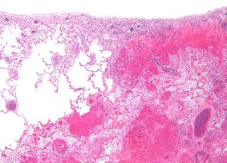
Infarction is tissue death (necrosis) due to inadequate blood supply to the affected area. It may be caused by artery blockages, rupture, mechanical compression, or vasoconstriction. The resulting lesion is referred to as an infarct (from the Latin infarctus, "stuffed into").

<span class="mw-page-title-main">Stroke</span> Death of a region of brain cells due to poor blood flow

Stroke is a medical condition in which poor blood flow to the brain causes cell death. There are two main types of stroke: ischemic, due to lack of blood flow, and hemorrhagic, due to bleeding. Both cause parts of the brain to stop functioning properly.

<span class="mw-page-title-main">Subarachnoid hemorrhage</span> Bleeding into the subarachnoid space

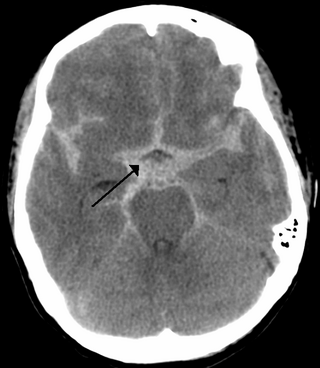
Subarachnoid hemorrhage (SAH) is bleeding into the subarachnoid space—the area between the arachnoid membrane and the pia mater surrounding the brain. Symptoms may include a severe headache of rapid onset, vomiting, decreased level of consciousness, fever, weakness, numbness, and sometimes seizures. Neck stiffness or neck pain are also relatively common. In about a quarter of people a small bleed with resolving symptoms occurs within a month of a larger bleed.

<span class="mw-page-title-main">Intracranial hemorrhage</span> Hemorrhage, or bleeding, within the skull

Intracranial hemorrhage (ICH), also known as intracranial bleed, is bleeding within the skull. Subtypes are intracerebral bleeds, subarachnoid bleeds, epidural bleeds, and subdural bleeds.

A thunderclap headache is a headache that is severe and has a sudden onset. It is defined as a severe headache that takes seconds to minutes to reach maximum intensity. Although approximately 75% are attributed to "primary" headaches—headache disorder, non-specific headache, idiopathic thunderclap headache, or uncertain headache disorder—the remainder are secondary to other causes, which can include some extremely dangerous acute conditions, as well as infections and other conditions. Usually, further investigations are performed to identify the underlying cause.

<span class="mw-page-title-main">Cerebral hypoxia</span> Oxygen shortage of the brain

Cerebral hypoxia is a form of hypoxia, specifically involving the brain; when the brain is completely deprived of oxygen, it is called cerebral anoxia. There are four categories of cerebral hypoxia; they are, in order of increasing severity: diffuse cerebral hypoxia (DCH), focal cerebral ischemia, cerebral infarction, and global cerebral ischemia. Prolonged hypoxia induces neuronal cell death via apoptosis, resulting in a hypoxic brain injury.

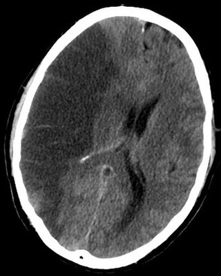
Intracerebral hemorrhage (ICH), also known as hemorrhagic stroke, is a sudden bleeding into the tissues of the brain, into its ventricles, or into both. An ICH is a type of bleeding within the skull and one kind of stroke. Symptoms can vary dramatically depending on the severity, acuity, and location (anatomically) but can include headache, one-sided weakness, numbness, tingling, or paralysis, speech problems, vision or hearing problems, memory loss, attention problems, coordination problems, balance problems, dizziness or lightheadedness or vertigo, nausea/vomiting, seizures, decreased level of consciousness or total loss of consciousness, neck stiffness, and fever.

<span class="mw-page-title-main">Intraparenchymal hemorrhage</span> Medical condition

Intraparenchymal hemorrhage (IPH) is one form of intracerebral bleeding in which there is bleeding within brain parenchyma. The other form is intraventricular hemorrhage (IVH).

<span class="mw-page-title-main">Cerebral infarction</span> Stroke resulting from lack of blood flow

Cerebral infarction, also known as an ischemic stroke, is the pathologic process that results in an area of necrotic tissue in the brain. In mid to high income countries, a stroke is the main reason for disability among people and the 2nd cause of death. It is caused by disrupted blood supply (ischemia) and restricted oxygen supply (hypoxia). This is most commonly due to a thrombotic occlusion, or an embolic occlusion of major vessels which leads to a cerebral infarct. In response to ischemia, the brain degenerates by the process of liquefactive necrosis.

<span class="mw-page-title-main">Periventricular leukomalacia</span> Degeneration of white matter near the lateral ventricles of the brain

Periventricular leukomalacia (PVL) is a form of white-matter brain injury, characterized by the necrosis of white matter near the lateral ventricles. It can affect newborns and fetuses; premature infants are at the greatest risk of neonatal encephalopathy which may lead to this condition. Affected individuals generally exhibit motor control problems or other developmental delays, and they often develop cerebral palsy or epilepsy later in life. The white matter in preterm born children is particularly vulnerable during the third trimester of pregnancy when white matter developing takes place and the myelination process starts around 30 weeks of gestational age.

<span class="mw-page-title-main">Watershed stroke</span> Medical condition

A watershed stroke is defined as a brain ischemia that is localized to the vulnerable border zones between the tissues supplied by the anterior, posterior and middle cerebral arteries. The actual blood stream blockage/restriction site can be located far away from the infarcts. Watershed locations are those border-zone regions in the brain supplied by the major cerebral arteries where blood supply is decreased. Watershed strokes are a concern because they comprise approximately 10% of all ischemic stroke cases. The watershed zones themselves are particularly susceptible to infarction from global ischemia as the distal nature of the vasculature predisposes these areas to be most sensitive to profound hypoperfusion.

<span class="mw-page-title-main">Brain ischemia</span> Medical condition

Brain ischemia is a condition in which there is insufficient bloodflow to the brain to meet metabolic demand. This leads to poor oxygen supply or cerebral hypoxia and thus leads to the death of brain tissue or cerebral infarction/ischemic stroke. It is a sub-type of stroke along with subarachnoid hemorrhage and intracerebral hemorrhage.

<span class="mw-page-title-main">Lacunar stroke</span> Medical condition

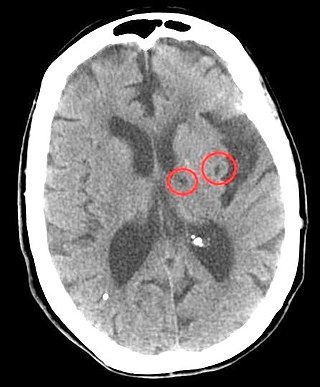
Lacunar stroke or lacunar cerebral infarct (LACI) is the most common type of ischemic stroke, resulting from the occlusion of small penetrating arteries that provide blood to the brain's deep structures. Patients who present with symptoms of a lacunar stroke, but who have not yet had diagnostic imaging performed, may be described as having lacunar stroke syndrome (LACS).

<span class="mw-page-title-main">Intraventricular hemorrhage</span> Medical condition

Intraventricular hemorrhage (IVH), also known as intraventricular bleeding, is a bleeding into the brain's ventricular system, where the cerebrospinal fluid is produced and circulates through towards the subarachnoid space. It can result from physical trauma or from hemorrhagic stroke.

<span class="mw-page-title-main">Anterior cerebral artery syndrome</span> Medical condition

Anterior cerebral artery syndrome is a condition whereby the blood supply from the anterior cerebral artery (ACA) is restricted, leading to a reduction of the function of the portions of the brain supplied by that vessel: the medial aspects of the frontal and parietal lobes, basal ganglia, anterior fornix and anterior corpus callosum.

A silent stroke is a stroke that does not have any outward symptoms associated with stroke, and the patient is typically unaware they have suffered a stroke. Despite not causing identifiable symptoms, a silent stroke still causes damage to the brain and places the patient at increased risk for both transient ischemic attack and major stroke in the future. In a broad study in 1998, more than 11 million people were estimated to have experienced a stroke in the United States. Approximately 770,000 of these strokes were symptomatic and 11 million were first-ever silent MRI infarcts or hemorrhages. Silent strokes typically cause lesions which are detected via the use of neuroimaging such as MRI. The risk of silent stroke increases with age but may also affect younger adults. Women appear to be at increased risk for silent stroke, with hypertension and current cigarette smoking being amongst the predisposing factors.

References

  1. 1 2 3 4 Shah, Sid. "Stroke Pathophysiology." Foundation for Education and Research in Neurological Emergencies. 1-7. http://www.uic%5B%5D. edu/com/ferne/pdf/pathophys0501.pdf.
  2. 1 2 Br Med J 1898;1:140
  3. "Vertebrobasilar circulatory disorders: Symptoms and causes". www.medicalnewstoday.com. 27 June 2018.
  4. Martín A, Macé E, Boisgard R, Montaldo G, Thézé B, Tanter M, Tavitian B (2012). "Imaging of perfusion, angiogenesis, and tissue elasticity after stroke". Journal of Cerebral Blood Flow and Metabolism. 32 (8): 1496–507. doi:10.1038/jcbfm.2012.49. PMC   3421095 . PMID   22491156.
  5. Alpers, BJ; Berry, RG (1963). "Circle of Willis in cerebral vascular disorders: the anatomical structure". Archives of Neurology. 8: 398–402. doi:10.1001/archneur.1963.00460040068006. ISSN   0003-9942. PMID   14012272.
  6. Fazio, Cornelio."Red Softening of the Brain1".Journal of Neuropathology & Experimental Neurology:January 1949 - Volume 8 - Issue 1 - ppg 43-60.Genoa, Italy. Web. 13 Feb. 2014.
  7. Diamond IB (1934). "Encephalomalacia in Infants". Archives of Neurology & Psychiatry. 31 (6): 1153–1164. doi:10.1001/archneurpsyc.1934.02250060039003.
  8. Young Ethel F., Courvile Cyril B. (1962). "Central Softening Of The Cerebral Hemispheres In The Newborn". Clinical Pediatrics. 1 (2): 95–102.
  9. Chutorian AM, Michener RC, Defendini R, Hilal SK, Gamboa ET (1979). "Neonatal polycystic encephalomalacia: four new cases and review of the literature". J Neurol Neurosurg Psychiatry. 42 (2): 154–160. doi:10.1136/jnnp.42.2.154. PMC   490181 . PMID   422963.
  10. Orejón de Luna G, Mateos Beato F, Simón de las Heras R, Miralles Molina M. Unidad de Neurología Pediátrica, Hospital Doce de Octubre, Madrid. Anales Espanoles de Pediatria [1997, 46(1):33-39]. Journal Article, English Abstract (lang: spa)
  11. Finger, Stanley, Randy L. Buckner, and Hugh Buckingham. "Does The Right Hemisphere Take Over After Damage To Broca’S Area? The Barlow Case Of 1877 And Its History." Brain & Language 85.3 (2003): 385. Academic Search Complete. Web. 13 Feb. 2014.
  12. Parsons John H (1891). "Note On aCase Of Cerebral Softening". The British Medical Journal. 2 (1617): 1352–1353. JSTOR   20244675.
  13. Ord-Mackenzie S. A. (1993). "Transient And Recurring Paresis in Acute Cerebral Softening". Br Med J. 1 (1933): 140–141. doi:10.1136/bmj.1.1933.140. PMC   2410420 . PMID   20757554.
  14. Inman Thomas (1858). "Cases Of Cerebral Softening, Etc.: With Remarks". The British Medical Journal. 1 (67): 289–290. doi: 10.1136/bmj.s4-1.67.289-a . JSTOR   25192210.