
Bluetongue disease is a noncontagious, insect-borne, viral disease of ruminants, mainly sheep and less frequently cattle, yaks, goats, buffalo, deer, dromedaries, and antelope. It is caused by Bluetongue virus (BTV). The virus is transmitted by the midges Culicoides imicola, Culicoides variipennis, and other culicoids.

The rhinovirus is a positive-sense, single-stranded RNA virus belonging to the genus Enterovirus in the family Picornaviridae. Rhinovirus is the most common viral infectious agent in humans and is the predominant cause of the common cold.
Coxsackie B4 virus are enteroviruses that belong to the Picornaviridae family. These viruses can be found worldwide. They are positive-sense, single-stranded, non-enveloped RNA viruses with icosahedral geometry. Coxsackieviruses have two groups, A and B, each associated with different diseases. Coxsackievirus group A is known for causing hand-foot-and-mouth diseases while Group B, which contains six serotypes, can cause a varying range of symptoms like gastrointestinal distress myocarditis. Coxsackievirus B4 has a cell tropism for natural killer cells and pancreatic islet cells. Infection can lead to beta cell apoptosis which increases the risk of insulitis.
Poliovirus, the causative agent of polio, is a serotype of the species Enterovirus C, in the family of Picornaviridae. There are three poliovirus serotypes, numbered 1, 2, and 3.

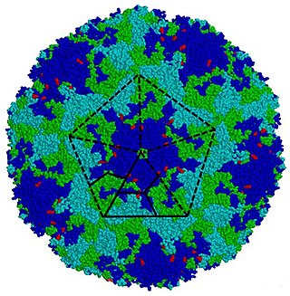
Picornaviruses are a group of related nonenveloped RNA viruses which infect vertebrates including fish, mammals, and birds. They are viruses that represent a large family of small, positive-sense, single-stranded RNA viruses with a 30 nm icosahedral capsid. The viruses in this family can cause a range of diseases including the common cold, poliomyelitis, meningitis, hepatitis, and paralysis.
Enterovirus is a genus of positive-sense single-stranded RNA viruses associated with several human and mammalian diseases. Enteroviruses are named by their transmission-route through the intestine.

Orbivirus is a genus of double-stranded RNA viruses in the family Reoviridae and subfamily Sedoreovirinae. Unlike other reoviruses, orbiviruses are arboviruses. They can infect and replicate within a wide range of arthropod and vertebrate hosts. Orbiviruses are named after their characteristic doughnut-shaped capsomers.

Aphthovirus is a viral genus of the family Picornaviridae. Aphthoviruses infect split-hooved animals, and include the causative agent of foot-and-mouth disease, Foot-and-mouth disease virus (FMDV). There are seven FMDV serotypes: A, O, C, SAT 1, SAT 2, SAT 3 and Asia 1, and four non-FMDV serotypes belonging to three additional species Bovine rhinitis A virus (BRAV), Bovine rhinitis B virus (BRBV) and Equine rhinitis A virus (ERAV).

Dependoparvovirus is a genus in the subfamily Parvovirinae of the virus family Parvoviridae; they are Group II viruses according to the Baltimore classification. Some dependoparvoviruses are also known as adeno-associated viruses because they cannot replicate productively in their host cell without the cell being coinfected by a helper virus such as an adenovirus, a herpesvirus, or a vaccinia virus.

Pleconaril (Picovir) is an antiviral drug that was being developed by Schering-Plough for prevention of asthma exacerbations and common cold symptoms in patients exposed to picornavirus respiratory infections. Pleconaril, administered either orally or intranasally, is active against viruses in the Picornaviridae family, including Enterovirus and Rhinovirus. It has shown useful activity against the dangerous enterovirus D68.
Gyrovirus is a genus of viruses, in the family Anelloviridae. Until 2011, chicken anemia virus was the only Gyrovirus identified, but since then gyroviruses have also been identified in humans. Diseases associated with this genus include: chicken infectious anemia, which is associated with depletion of cortical thymocytes and erythroblastoid cells.
Bocaparvovirus is a genus of viruses in the subfamily Parvovirinae of the virus family Parvoviridae. Humans, cattle, and dogs serve as natural hosts. There are 28 species in this genus. Diseases associated with this genus include, in humans, acute respiratory illness, and in cattle, diarrhea and mild respiratory symptoms.

Murine polyomavirus is an unenveloped double-stranded DNA virus of the polyomavirus family. The first member of the family discovered, it was originally identified by accident in the 1950s. A component of mouse leukemia extract capable of causing tumors, particularly in the parotid gland, in newborn mice was reported by Ludwik Gross in 1953 and identified as a virus by Sarah Stewart and Bernice Eddy at the National Cancer Institute, after whom it was once called "SE polyoma". Stewart and Eddy would go on to study related polyomaviruses such as SV40 that infect primates, including humans. These discoveries were widely reported at the time and formed the early stages of understanding of oncoviruses.

Picornain 3C is a protease found in picornaviruses, which cleaves peptide bonds of non-terminal sequences. Picornain 3C’s endopeptidase activity is primarily responsible for the catalytic process of selectively cleaving Gln-Gly bonds in the polyprotein of poliovirus and with substitution of Glu for Gln, and Ser or Thr for Gly in other picornaviruses. Picornain 3C are cysteine proteases related by amino acid sequence to trypsin-like serine proteases. Picornain 3C is encoded by enteroviruses, rhinoviruses, aphtoviruses and cardioviruses. These genera of picoviruses cause a wide range of infections in humans and mammals.
Major capsid protein VP1 is a viral protein that is the main component of the polyomavirus capsid. VP1 monomers are generally around 350 amino acids long and are capable of self-assembly into an icosahedral structure consisting of 360 VP1 molecules organized into 72 pentamers. VP1 molecules possess a surface binding site that interacts with sialic acids attached to glycans, including some gangliosides, on the surfaces of cells to initiate the process of viral infection. The VP1 protein, along with capsid components VP2 and VP3, is expressed from the "late region" of the circular viral genome.
Aichivirus A formerly Aichi virus (AiV) belongs to the genus Kobuvirus in the family Picornaviridae. Six species are part of the genus Kobuvirus, Aichivirus A-F. Within Aichivirus A, there are six different types including human Aichi virus, canine kobuvirus, murine kobuvirus, Kathmandu sewage kobuvirus, roller kobuvirus, and feline kobuvirus. Three different genotypes are found in human Aichi virus, represented as genotype A, B, and C.
Epizootic hemorrhagic disease virus, often abbreviated to EHDV, is a species of the genus Orbivirus, a member of the family Reoviridae. It is the causative agent of epizootic hemorrhagic disease, an acute, infectious, and often fatal disease of wild ruminants. In North America, the most severely affected ruminant is the white-tailed deer, although it may also infect mule deer, black-tailed deer, elk, bighorn sheep, and pronghorn antelope. It is often mistakenly referred to as “bluetongue virus” (BTV), another Orbivirus that like EHDV causes the host to develop a characteristic blue tongue due to systemic hemorrhaging and lack of oxygen in the blood. Despite showing clinical similarities, these two viruses are genetically distinct.
Hamster polyomavirus is an unenveloped double-stranded DNA virus of the polyomavirus family whose natural host is the hamster. It was originally described in 1967 by Arnold Graffi as a cause of epithelioma in Syrian hamsters.
Minor capsid protein VP2 and minor capsid protein VP3 are viral proteins that are components of the polyomavirus capsid. Polyomavirus capsids are composed of three proteins; the major component is major capsid protein VP1, which self-assembles into pentamers that in turn self-assemble into enclosed icosahedral structures. The minor components are VP2 and VP3, which bind in the interior of the capsid.
Triatoma virus (TrV) is a virus belonging to the insect virus family Dicistroviridae. Within this family, there are currently 3 genera and 15 species of virus. Triatoma virus belongs to the genus Cripavirus. It is non-enveloped and its genetic material is positive-sense, single-stranded RNA. The natural hosts of triatoma virus are invertebrates. TrV is a known pathogen to Triatoma infestans, the major vector of Chagas disease in Argentina which makes triatoma virus a major candidate for biological vector control as opposed to chemical insecticides. Triatoma virus was first discovered in 1984 when a survey of pathogens of triatomes was conducted in the hopes of finding potential biological control methods for T. infestans.
