Picornavirus

Last updated • 12 min readFrom Wikipedia, The Free Encyclopedia

Picornaviridae
Polioviruses.jpg
Electronmicrograph of poliovirus
Rhinovirus.PNG
Isosurface of a human rhinovirus showing protein spikes
Virus classification OOjs UI icon edit-ltr.svg
(unranked): Virus
Realm: Riboviria
Kingdom: Orthornavirae
Phylum: Pisuviricota
Class: Pisoniviricetes
Order: Picornavirales
Family:Picornaviridae
Genera [1]

See text

Picornaviruses are a group of related nonenveloped RNA viruses which infect vertebrates including fish, [2] mammals, and birds. They are viruses that represent a large family of small, positive-sense, single-stranded RNA viruses with a 30 nm icosahedral capsid. The viruses in this family can cause a range of diseases including the common cold, poliomyelitis, meningitis, hepatitis, and paralysis. [3] [4] [5] [6]

Contents

Picornaviruses constitute the family Picornaviridae, order Picornavirales , and realm Riboviria . There are 158 species in this family, assigned to 68 genera. Notable examples are genera Enterovirus (including Rhinovirus and Poliovirus ), Aphthovirus , Cardiovirus , and Hepatovirus . [1] [7]

Etymology

The name "picornavirus" has a dual etymology. Firstly, the name derives from picorna- which is an acronym for "poliovirus, insensitivity to ether, coxsackievirus, orphan virus, rhinovirus, and ribonucleic acid". Secondly, the name derives from pico-, which designates a very small unit of measurement (equivalent to 10−12), combined with rna to describe this group of very small RNA viruses. [8]

History

The first animal virus discovered (1897) was the foot-and-mouth disease virus (FMDV). It is the prototypic member of the genus Aphthovirus in the Picornaviridae family. [5] The plaque assay was developed using poliovirus; the discovery of viral replication in culture was also with poliovirus in 1949. This was the first time that infectious virus had been produced in cultured cells. [9] Polyprotein synthesis, internal ribosome entry sites, and uncapped mRNA were all discovered by studying poliovirus infected cells, and a poliovirus clone was the first infectious DNA clone made of an RNA virus in animals. Along with rhinovirus, poliovirus was the first animal virus to have its structure determined by x-ray crystallography. RNA dependent RNA polymerase was discovered in Mengovirus , a genus of picornaviruses. [10]

Virology

Structure

FMDV structural proteins VP1, VP2, VP3, and VP4 form the biological protomer and icosahedral capsid. FMDV icosahedral capsid.jpg
FMDV structural proteins VP1, VP2, VP3, and VP4 form the biological protomer and icosahedral capsid.

Picornaviruses are nonenveloped, with an icosahedral capsid. [4] The capsid is an arrangement of 60 protomers in a tightly packed icosahedral structure. Each protomer consists of four polypeptides known as VP (viral protein) 1, 2, 3 and 4. VP2 and VP4 polypeptides originate from one protomer known as VP0 that is cleaved to give the different capsid components. The icosahedral capsid is said to have a triangulation number of 3, this means that in the icosahedral structure each of the 60 triangles that make up the capsid are split into three little triangles with a subunit on the corner.[ citation needed ]

Many picornaviruses have a deep cleft formed by around each of the 12 vertices of icosahedrons. The outer surface of the capsid is composed of regions of VP1, VP2, and VP3. Around each of the vertices is a canyon lined with the C termini of VP1 and VP3. The interior surface of the capsid is composed of VP4 and the N termini of VP1. J. Esposito and Frederick A. Murphy demonstrates cleft structure referred to as canyons, using X-ray crystallography and cryoelectron microscopy. [9]

Depending on the type and degree of dehydration, the viral particle is around 30–32 nm in diameter. [7] The viral genome is around 2500 nm in length, so it is tightly packaged within the capsid along with substances such as sodium ions to balance the negative charges on the RNA caused by the phosphate groups.[ citation needed ]

Genome

Genome organization and proteins of enteroviruses and aphthoviruses Genome of enteroviruses and aphthoviruses.png
Genome organization and proteins of enteroviruses and aphthoviruses

Picornaviruses are classed under Baltimore's viral classification system as group IV viruses, as they contain a single-stranded, positive-sense RNA genome. Their genome ranges between 6.7 and 10.1 (kilobases) in length. [7] Like most positive-sense RNA genomes, the genetic material alone is infectious; although substantially less virulent than if contained within the viral particle, the RNA can have increased infectivity when transfected into cells. The genome RNA is unusual because it has a protein on the 5' end that is used as a primer for transcription by RNA polymerase. This primer is called VPg genome, and it ranges between 2 and 3 kb. VPg contain tyrosine residue at the 3' end. Tyrosine as a –OH source for covalently linked to 5' end of RNA. [9] [11]

The genome is not segmented and positive-sense (the same sense as mammalian mRNA, being read 5' to 3'). Unlike mammalian mRNA, picornaviruses do not have a 5' cap, but a virally encoded protein known as VPg. However, like mammalian mRNA, the genome does have a poly(A) tail at the 3' end. An untranslated region (UTR) is found at both ends of the picornavirus genome. The 5' UTR is usually longer, being around 500–1200 nucleotides (nt) in length, compared to that of the 3' UTR, which is around 30–650 nt. The 5' UTR is thought to be important in translation, and the 3' in negative-strand synthesis; however, the 5' end may also have a role to play in virulence of the virus. The rest of the genome encodes structural proteins at the 5' end and nonstructural proteins at the 3' end in a single polyprotein.[ citation needed ]

The polyprotein is organised as: L-1ABCD-2ABC-3ABCD with each letter representing a protein, but variations to this layout exist.[ citation needed ]

The 1A, 1B, 1C, and 1D proteins are the capsid proteins VP4, VP2, VP3, and VP1, respectively. Virus-coded proteases perform the cleavages, some of which are intramolecular. The polyprotein is first cut to yield P1, P2, and P3. P1 becomes myristylated at the N terminus before being cleaved to VP0, VP3, and VP1, the proteins that will form procapsids; VP0 will later be cleaved to produce VP2 and VP4. Other cleavage products include 3B (VPg), 2C (an ATPase), and 3D (the RNA polymerase). [9] [12]

Replication

RNA elements

Foot-and-mouth disease virus genome and RNA structural elements 12985 2016 561 Fig1A HTML.jpg
Foot-and-mouth disease virus genome and RNA structural elements

Genomic RNAs of picornaviruses possess multiple RNA elements, and they are required for both negative- and positive-strand RNA synthesis. The cis-acting replication element (CRE) is required for replication. The stem-loop-structure that contains the CRE is independent of position, but changes with location between virus types when it has been identified. Also, the 3' end elements of viral RNA are significant and efficient for RNA replication of picornaviruses. The 3' end of picornavirus contains a poly(A) tract, which is required for infectivity. RNA synthesis, though, is hypothesized to occur in this region. The 3' end NCR of poliovirus is not necessary for negative-strand synthesis, but is important element for positive–strand synthesis. Additionally, the 5' end NCR that contains secondary structural elements is required for RNA replication and poliovirus translation initiation. Internal ribosome entry sites are RNA structures that allow cap-independent initiation of translation, and are able to initiate translation in the middle of a messenger RNA. [13]

Lifecycle

Picornavirus life cycle Fcimb-09-00283-g002.jpg
Picornavirus life cycle

The viral particle binds to cell surface receptors. Cell surface receptors are characterized for each serotype of picornaviruses. For example, poliovirus receptor is glycoprotein CD155, which is special receptor for human and some other primate species. For this reason, poliovirus could not be made in many laboratories until transgenic mice having a CD155 receptor on their cell surfaces were developed in the 1990s. These animals can be infected and used for studies of replication and pathogenesis. [9] Binding causes a conformational change in the viral capsid proteins, and myristic acid is released. The acid forms a pore in the cell membrane through which RNA is injected. [14]

Once inside the cell, the RNA uncoats and the (+) strand RNA genome is replicated through a double-stranded RNA intermediate that is formed using viral RNA-dependent RNA polymerase. Translation by host-cell ribosomes is not initiated by a 5' G cap as usual, but rather is initiated by an internal ribosome entry site. The viral life cycle is very rapid, with the whole process of replication being completed on average within 8 hours. As little as 30 minutes after initial infection, though, cell protein synthesis declines to almost zero output – essentially the macromolecular synthesis of cell proteins is shut off. Over the next 1–2 hours, a loss of margination of chromatin and homogeneity occurs in the nucleus, before the viral proteins start to be synthesized and a vacuole appears in the cytoplasm close to the nucleus that gradually starts to spread as the time after infection reaches around 3 hours. After this time, the cell plasma membrane becomes permeable; at 4–6 hours, the virus particles assemble, and can sometimes be seen in the cytoplasm. Around 8 hours, the cell is effectively dead, and lyses to release the viral particles.[ citation needed ]

Experimental data from single-step growth curve-like experiments have allowed observation of the replication of the picornaviruses in great detail. The whole of replication occurs within the host-cell cytoplasm and infection can even happen in cells that do not contain a nucleus (enucleated) and those treated with actinomycin D (this antibiotic would inhibit viral replication if this occurred in the nucleus.)[ citation needed ]

Translation takes place by -1 ribosomal frameshifting, viral initiation, and ribosomal skipping. The virus exits in host cell by lysis, and viroporins. Vertebrates serve as the natural hosts. Transmission routes are fecal-oral, contact, ingestion, and air-borne particles. [4]

Viral protein (VPg)

Picornaviruses have a viral protein (VPg) covalently linked to 5' end of their genomes instead of 7-methylguanosine cap like cellular mRNAs. Virus RNA polymerases use VPg as primer. VPg as primer uses both positive- and negative-strand RNA synthesis. Picornavirus replication is initiated by the uridylylation of VPg. It is uridylylated at the hydroxyl group of a tyrosine residue. [3] A VPg primer mechanism is used by the picornavirus (entero- aphtho-, and others), additional virus groups (poty-, como-, calici-, and others) and picornavirus-like (coronavirus, notavirus, etc.) supergroup of RNA viruses. The mechanism has been best studied for the enteroviruses (which include many human pathogens, such as poliovirus and coxsackie viruses), as well as for the aphthovirus, an animal pathogen causing foot-and-mouth disease.[ citation needed ]

In this group, primer-dependent RNA synthesis uses a small 22– to 25-amino acid-long viral protein linked to the VPg [15] to initiate polymerase activity, where the primer is covalently bound to the 5' end of the RNA template. [16] The uridylylation occurs at a tyrosine residue at the third position of the VPg. A CRE, which is a RNA stem loop structure, serves as a template for the uridylylation of VPg, resulting in the synthesis of VPgpUpUOH. Mutations within the CRE-RNA structure prevent VPg uridylylation, and mutations within the VPg sequence can severely diminish RdRp catalytic activity. [17] While the tyrosine hydroxyl of VPg can prime negative-strand RNA synthesis in a CRE- and VPgpUpUOH-independent manner, CRE-dependent VPgpUpUOH synthesis is absolutely required for positive-strand RNA synthesis. CRE-dependent VPg uridylylation lowers the K of UTP required for viral RNA replication and CRE-dependent VPgpUpUOH synthesis, and is required for efficient negative-strand RNA synthesis, especially when UTP concentrations are limiting. [18] The VPgpUpUOH primer is transferred to the 3’ end of the RNA template for elongation, which can continue by addition of nucleotide bases by RdRp. Partial crystal structures for VPgs of foot and mouth disease virus [19] and coxsackie virus B3 [20] suggest that there may be two sites on the viral polymerase for the small VPgs of the picornaviruses. NMR solution structures of poliovirus VPg [21] and VPgpU [22] show that uridylylation stabilizes the structure of the VPg, which is otherwise quite flexible in solution. The second site may be used for uridylylation,v after which the VPgpU can initiate RNA synthesis. The VPg primers of caliciviruses, whose structures are only beginning to be revealed, [23] are much larger than those of the picornaviruses. Mechanisms for uridylylation and priming may be quite different in all of these groups.[ citation needed ]

VPg uridylylation may include the use of precursor proteins, allowing for the determination of a possible mechanism for the location of the diuridylylated, VPg-containing precursor at the 3' end of positive- or negative-strand RNA for production of full-length RNA. Determinants of VPg uridylylation efficiency suggest formation and/or collapse or release of the uridylylated product as the rate-limiting step in vitro depending upon the VPg donor employed. [24] Precursor proteins also have an effect on VPg-CRE specificity and stability. [25] The upper RNA stem loop, to which VPg binds, has a significant impact on both retention, and recruitment, of VPg and Pol. The stem loop of CRE will partially unwind, allowing the precursor components to bind and recruit VPg and Pol4. The CRE loop has a defined consensus sequence to which the initiation components bind, but no consensus sequence exists for the supporting stem, which suggests that only the structural stability of the CRE is important. [26]

Assembly and organization of the picornavirus VPg ribonucleoprotein complex

  1. Two 3CD (VPg complex) molecules bind to CRE with the 3C domains (VPg domain) contacting the upper stem and the 3D domains (VPg domain) contacting the lower stem.
  2. The 3C dimer opens the RNA stem by forming a more stable interaction with single strands forming the stem.
  3. 3Dpol is recruited to and retained in this complex by a physical interaction between the back-of-the-thumb subdomain of 3Dpol and a surface of one or both 3C subdomains of 3CD.

VPg may also play an important role in specific recognition of viral genome by movement proteins (MP), which are nonstructural proteins encoded by many, if not all, plant viruses to enable their movement from one infected cell to neighboring cells. [27] MP and VPg interact to provide specificity for the transport of viral RNA from cell to cell. To fulfill energy requirements, MP also interacts with P10, which is a cellular ATPase.[ citation needed ]

Diseases

Picornaviruses cause a range of diseases. Enteroviruses of the picornavirus family infect the enteric tract, which is reflected in their name. Rhinoviruses infect primarily the nose and the throat. Enteroviruses replicate at 37 °C, whereas rhinoviruses grow better at 33 °C, as this is the lower temperature of the nose. Enteroviruses are stable under acidic conditions, thus they are able to survive exposure to gastric acid. In contrast, rhinoviruses are acid-labile (inactivated or destroyed by low pH conditions), so rhinovirus infections are restricted to the nose and throat.[ citation needed ]

Taxonomy

These genera are recognized: [1]

See also

Related Research Articles

<span class="mw-page-title-main">RNA virus</span> Subclass of viruses

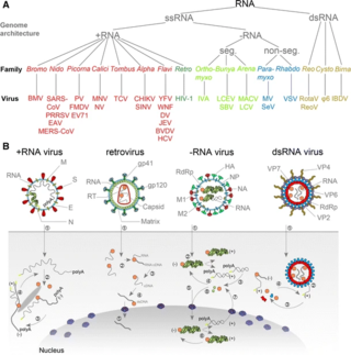
An RNA virus is a virus—other than a retrovirus—that has ribonucleic acid (RNA) as its genetic material. The nucleic acid is usually single-stranded RNA (ssRNA) but it may be double-stranded (dsRNA). Notable human diseases caused by RNA viruses include the common cold, influenza, SARS, MERS, COVID-19, Dengue virus, hepatitis C, hepatitis E, West Nile fever, Ebola virus disease, rabies, polio, mumps, and measles.

<span class="mw-page-title-main">Rhinovirus</span> Genus of viruses (Enterovirus)

The rhinovirus is a positive-sense, single-stranded RNA virus belonging to the genus Enterovirus in the family Picornaviridae. Rhinovirus is the most common viral infectious agent in humans and is the predominant cause of the common cold.

Coxsackie B4 virus are enteroviruses that belong to the Picornaviridae family. These viruses can be found worldwide. They are positive-sense, single-stranded, non-enveloped RNA viruses with icosahedral geometry. Coxsackieviruses have two groups, A and B, each associated with different diseases. Coxsackievirus group A is known for causing hand-foot-and-mouth diseases while Group B, which contains six serotypes, can cause a varying range of symptoms like gastrointestinal distress myocarditis. Coxsackievirus B4 has a cell tropism for natural killer cells and pancreatic islet cells. Infection can lead to beta cell apoptosis which increases the risk of insulitis.

<span class="mw-page-title-main">Poliovirus</span> Enterovirus

Poliovirus, the causative agent of polio, is a serotype of the species Enterovirus C, in the family of Picornaviridae. There are three poliovirus serotypes, numbered 1, 2, and 3.

<i>Hepadnaviridae</i> Family of viruses

Hepadnaviridae is a family of viruses. Humans, apes, and birds serve as natural hosts. There are currently 18 species in this family, divided among 5 genera. Its best-known member is hepatitis B virus. Diseases associated with this family include: liver infections, such as hepatitis, hepatocellular carcinomas, and cirrhosis. It is the sole accepted family in the order Blubervirales.

<i>Enterovirus</i> Genus of viruses

Enterovirus is a genus of positive-sense single-stranded RNA viruses associated with several human and mammalian diseases. Enteroviruses are named by their transmission-route through the intestine.

<span class="mw-page-title-main">Viral replication</span> Formation of biological viruses during the infection process

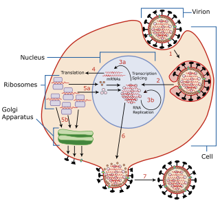
Viral replication is the formation of biological viruses during the infection process in the target host cells. Viruses must first get into the cell before viral replication can occur. Through the generation of abundant copies of its genome and packaging these copies, the virus continues infecting new hosts. Replication between viruses is greatly varied and depends on the type of genes involved in them. Most DNA viruses assemble in the nucleus while most RNA viruses develop solely in cytoplasm.

VPg is a protein that is covalently attached to the 5′ end of positive strand viral RNA and acts as a primer during RNA synthesis in a variety of virus families including Picornaviridae, Potyviridae, Astroviridae and Caliciviridae. There are some studies showing that a possible VPg protein is also present in astroviridae, however, experimental evidence for this has not yet been provided and requires further study. The primer activity of VPg occurs when the protein becomes uridylated, providing a free hydroxyl that can be extended by the virally encoded RNA-dependent RNA polymerase. For some virus families, VPg also has a role in translation initiation by acting like a 5' mRNA cap.

<i>Aphthovirus</i> Genus of viruses

Aphthovirus is a viral genus of the family Picornaviridae. Aphthoviruses infect split-hooved animals, and include the causative agent of foot-and-mouth disease, Foot-and-mouth disease virus (FMDV). There are seven FMDV serotypes: A, O, C, SAT 1, SAT 2, SAT 3 and Asia 1, and four non-FMDV serotypes belonging to three additional species Bovine rhinitis A virus (BRAV), Bovine rhinitis B virus (BRBV) and Equine rhinitis A virus (ERAV).

<i>Cardiovirus</i> Genus of viruses

Cardiovirus are a group of viruses within order Picornavirales, family Picornaviridae. Vertebrates serve as natural hosts for these viruses.

Parechovirus B, formerly called the Ljungan virus, was first discovered in the mid-1990s after being isolated from a bank vole near the Ljungan river in Medelpad county, Sweden. It has since been established that Parechovirus B, which is also found in several places in Europe and America, causes serious illness in wild as well as laboratory animals. Several scientific articles have recently reported findings indicating that Parechovirus B is associated with malformations, intrauterine fetal death, and sudden infant death syndrome in humans. In addition, studies are being conducted worldwide to investigate the possible connection of the virus to diabetes, neurological and other illnesses in humans.

<span class="mw-page-title-main">RNA-dependent RNA polymerase</span> Enzyme that synthesizes RNA from an RNA template

RNA-dependent RNA polymerase (RdRp) or RNA replicase is an enzyme that catalyzes the replication of RNA from an RNA template. Specifically, it catalyzes synthesis of the RNA strand complementary to a given RNA template. This is in contrast to typical DNA-dependent RNA polymerases, which all organisms use to catalyze the transcription of RNA from a DNA template.

Erbovirus is a genus of viruses in the order Picornavirales, in the family Picornaviridae. Horses serve as natural hosts. There is only one species in this genus: Erbovirus A. Diseases associated with this genus include: upper respiratory tract disease with viremia and fecal shedding. Viruses belonging to the genus Erbovirus have been isolated in horses with acute upper febrile respiratory disease. The structure of the Erbovirus virion is icosahedral, having a diameter of 27–30 nm.

<span class="mw-page-title-main">Hepatitis A virus internal ribosome entry site (IRES)</span>

This family represents the internal ribosome entry site (IRES) of the hepatitis A virus. HAV IRES is a 450 nucleotide long sequence located in the 735 nt long 5’ UTR of Hepatitis A viral RNA genome. IRES elements allow cap and end-independent translation of mRNA in the host cell. The IRES achieves this by mediating the internal initiation of translation by recruiting a ribosomal 40S pre-initiation complex directly to the initiation codon and eliminates the requirement for eukaryotic initiation factor, eIF4F.

<i>Picornavirales</i> Order of viruses

Picornavirales is an order of viruses with vertebrate, invertebrate, protist and plant hosts. The name has a dual etymology. First, picorna- is an acronym for poliovirus, insensitivity to ether, coxsackievirus, orphan virus, rhinovirus, and ribonucleic acid. Secondly, pico-, meaning extremely small, combines with RNA to describe these very small RNA viruses. The order comprises viruses that historically are referred to as picorna-like viruses.

Eckard Wimmer is a German American virologist, organic chemist and distinguished professor of molecular genetics and microbiology at Stony Brook University. He is best known for his seminal work on the molecular biology of poliovirus and the first chemical synthesis of a viral genome capable of infection and subsequent production of live viruses.

<span class="mw-page-title-main">Picornain 3C</span>

Picornain 3C is a protease found in picornaviruses, which cleaves peptide bonds of non-terminal sequences. Picornain 3C’s endopeptidase activity is primarily responsible for the catalytic process of selectively cleaving Gln-Gly bonds in the polyprotein of poliovirus and with substitution of Glu for Gln, and Ser or Thr for Gly in other picornaviruses. Picornain 3C are cysteine proteases related by amino acid sequence to trypsin-like serine proteases. Picornain 3C is encoded by enteroviruses, rhinoviruses, aphtoviruses and cardioviruses. These genera of picoviruses cause a wide range of infections in humans and mammals.

Aichivirus A formerly Aichi virus (AiV) belongs to the genus Kobuvirus in the family Picornaviridae. Six species are part of the genus Kobuvirus, Aichivirus A-F. Within Aichivirus A, there are six different types including human Aichi virus, canine kobuvirus, murine kobuvirus, Kathmandu sewage kobuvirus, roller kobuvirus, and feline kobuvirus. Three different genotypes are found in human Aichi virus, represented as genotype A, B, and C.

<span class="mw-page-title-main">Positive-strand RNA virus</span> Class of viruses in the Baltimore classification

Positive-strand RNA viruses are a group of related viruses that have positive-sense, single-stranded genomes made of ribonucleic acid. The positive-sense genome can act as messenger RNA (mRNA) and can be directly translated into viral proteins by the host cell's ribosomes. Positive-strand RNA viruses encode an RNA-dependent RNA polymerase (RdRp) which is used during replication of the genome to synthesize a negative-sense antigenome that is then used as a template to create a new positive-sense viral genome.

Triatoma virus (TrV) is a virus belonging to the insect virus family Dicistroviridae. Within this family, there are currently 3 genera and 15 species of virus. Triatoma virus belongs to the genus Cripavirus. It is non-enveloped and its genetic material is positive-sense, single-stranded RNA. The natural hosts of triatoma virus are invertebrates. TrV is a known pathogen to Triatoma infestans, the major vector of Chagas disease in Argentina which makes triatoma virus a major candidate for biological vector control as opposed to chemical insecticides. Triatoma virus was first discovered in 1984 when a survey of pathogens of triatomes was conducted in the hopes of finding potential biological control methods for T. infestans.

References

  1. 1 2 3 "Virus Taxonomy: 2020 Release". International Committee on Taxonomy of Viruses (ICTV). March 2021. Retrieved 20 May 2021.
  2. Altan E, Kubiski SV, Boros Á, Reuter G, Sadeghi M, Deng X, Creighton EK, Crim MJ, Delwart E (2019). "A highly divergent picornavirus infecting the Gut Epithelia of Zebrafish (Danio rerio) in research institutions worldwide". Zebrafish. 16 (3): 291–299. doi: 10.1089/zeb.2018.1710 . PMID   30939077. S2CID   92999901.
  3. 1 2 Ryu WS (2016). "Chapter 11 – Picornavirus". Molecular virology of human pathogenic viruses. Korea: Academic Press. pp. 153–64. doi:10.1016/b978-0-12-800838-6.00011-4. ISBN   978-0-12-800838-6.
  4. 1 2 3 "Viral Zone". ExPASy. Retrieved 15 June 2015.
  5. 1 2 Martinez-Salas E, Saiz M, Sobrino F (2008). "Foot-and-Mouth Disease Virus". In Mettenleiter TC, Sobrino F (eds.). Animal Viruses: Molecular Biology. Norfolk, UK: Caister Academic Press. ISBN   978-1-904455-22-6.
  6. Lau SK, Woo PC, Lai KK, Huang Y, Yip CC, Shek CT, Lee P, Lam CS, Chan KH, Yuen KY (September 2011). "Complete genome analysis of three novel picornaviruses from diverse bat species". Journal of Virology. 85 (17): 8819–28. doi:10.1128/JVI.02364-10. PMC   3165794 . PMID   21697464.
  7. 1 2 3 "Picornaviridae - Picornaviridae - Picornavirales". International Committee on Taxonomy of Viruses (ICTV). Archived from the original on 21 September 2017. Retrieved 12 June 2020.
  8. "Picornaviridae". International Committee on Taxonomy of Viruses (ICTV). October 2017. Archived from the original on 21 September 2017. Retrieved 5 February 2019.
  9. 1 2 3 4 5 Carter JB, Saunders VA (2007). "Picornaviruses (and other plus-strand RNA viruses)". Virology: Principles and applications. Chichester, England: John Wiley & Sons. pp. 160–65. ISBN   978-0-470-02386-0.
  10. Knipe DM, Howley P (2013). Fields Virology. Lippincott Williams & Wilkins. ISBN   978-1-4698-3066-7.
  11. Zabel P, Moerman M, Lomonossoff G, Shanks M, Beyreuther K (July 1984). "Cowpea mosaic virus VPg: sequencing of radiochemically modified protein allows mapping of the gene on B RNA". The EMBO Journal. 3 (7): 1629–34. doi:10.1002/j.1460-2075.1984.tb02021.x. PMC   557569 . PMID   16453534.
  12. Acheson NH (2011). Fundamentals of Molecular Virology (2nd ed.). John Wiley & Sons, Inc. ISBN   978-0470900598.
  13. Daijogo S, Semler BL (2011). "Mechanistic intersections between picornavirus translation and RNA replication". Advances in Virus Research. 80: 1–24. doi:10.1016/B978-0-12-385987-7.00001-4. ISBN   9780123859877. PMID   21762819.
  14. "Pathology, Microbiology and Immunology - School of Medicine Columbia | University of South Carolina".
  15. Flanegan JB, Baltimore D (September 1977). "Poliovirus-specific primer-dependent RNA polymerase able to copy poly(A)". Proceedings of the National Academy of Sciences of the United States of America. 74 (9): 3677–80. Bibcode:1977PNAS...74.3677F. doi: 10.1073/pnas.74.9.3677 . PMC   431685 . PMID   198796.
  16. Ambros V, Baltimore D (August 1978). "Protein is linked to the 5' end of poliovirus RNA by a phosphodiester linkage to tyrosine". The Journal of Biological Chemistry. 253 (15): 5263–66. doi: 10.1016/S0021-9258(17)30361-7 . PMID   209034.
  17. Gu C, Zeng T, Li Y, Xu Z, Mo Z, Zheng C (October 2009). "Structure-function analysis of mutant RNA-dependent RNA polymerase complexes with VPg". Biochemistry. Biokhimiia. 74 (10): 1132–41. doi:10.1134/S0006297909100095. PMID   19916926. S2CID   24968119.
  18. Steil BP, Barton DJ (October 2008). "Poliovirus cis-acting replication element-dependent VPg Uridylylation lowers the Km of the initiating nucleoside triphosphate for viral RNA replication". Journal of Virology. 82 (19): 9400–08. doi:10.1128/JVI.00427-08. PMC   2546976 . PMID   18653453.
  19. Ferrer-Orta C, Arias A, Agudo R, Pérez-Luque R, Escarmís C, Domingo E, Verdaguer N (February 2006). "The structure of a protein primer-polymerase complex in the initiation of genome replication". The EMBO Journal. 25 (4): 880–88. doi:10.1038/sj.emboj.7600971. PMC   1383552 . PMID   16456546.
  20. Gruez A, Selisko B, Roberts M, Bricogne G, Bussetta C, Jabafi I, et al. (October 2008). "The crystal structure of coxsackievirus B3 RNA-dependent RNA polymerase in complex with its protein primer VPg confirms the existence of a second VPg binding site on Picornaviridae polymerases". Journal of Virology. 82 (19): 9577–90. doi:10.1128/JVI.00631-08. PMC   2546979 . PMID   18632861.
  21. Schein CH, Oezguen N, Volk DE, Garimella R, Paul A, Braun W (July 2006). "NMR structure of the viral peptide linked to the genome (VPg) of poliovirus". Peptides. 27 (7): 1676–84. doi:10.1016/j.peptides.2006.01.018. PMC   1629084 . PMID   16540201.
  22. Schein CH, Oezguen N, van der Heden van Noort GJ, Filippov DV, Paul A, Kumar E, Braun W (August 2010). "NMR solution structure of poliovirus uridylyated peptide linked to the genome (VPgpU)". Peptides. 31 (8): 1441–48. doi:10.1016/j.peptides.2010.04.021. PMC   2905501 . PMID   20441784.
  23. Leen EN, Kwok KY, Birtley JR, Simpson PJ, Subba-Reddy CV, Chaudhry Y, et al. (May 2013). "Structures of the compact helical core domains of feline calicivirus and murine norovirus VPg proteins" (PDF). Journal of Virology. 87 (10): 5318–30. doi:10.1128/JVI.03151-12. PMC   3648151 . PMID   23487472.
  24. Pathak HB, Oh HS, Goodfellow IG, Arnold JJ, Cameron CE (November 2008). "Picornavirus genome replication: roles of precursor proteins and rate-limiting steps in oriI-dependent VPg uridylylation". The Journal of Biological Chemistry. 283 (45): 30677–88. doi: 10.1074/jbc.M806101200 . PMC   2576561 . PMID   18779320.
  25. Shen M, Wang Q, Yang Y, Pathak HB, Arnold JJ, Castro C, Lemon SM, Cameron CE (November 2007). "Human Rhinovirus Type 14 Gain-of-Function Mutants for oriI Utilization Define Residues of 3C(D) and 3Dpol That Contribute to Assembly and Stability of the Picornavirus VPg Uridylylation Complex". J. Virol. 81 (22): 12485–95. doi:10.1128/JVI.00972-07. PMC   2169002 . PMID   17855535.
  26. Yang Y, Rijnbrand R, McKnight KL, Wimmer E, Paul A, Martin A, Lemon SM (August 2002). "Sequence requirements for viral RNA replication and VPg uridylylation directed by the internal cis-acting replication element (CRE) of human rhinovirus type 14". Journal of Virology. 76 (15): 7485–94. doi:10.1128/JVI.76.15.7485-7494.2002. PMC   136355 . PMID   12097561.
  27. Roy Chowdhury S, Savithri HS (January 2011). Pfeffer S (ed.). "Interaction of Sesbania mosaic virus movement protein with VPg and P10: implication to specificity of genome recognition". PLOS ONE. 6 (1): e15609. Bibcode:2011PLoSO...615609R. doi: 10.1371/journal.pone.0015609 . PMC   3016346 . PMID   21246040.

Further reading