Poliovirus, the causative agent of polio, is a serotype of the species Enterovirus C, in the family of Picornaviridae. There are three poliovirus serotypes: types 1, 2, and 3.

Hepadnaviridae is a family of viruses. Humans, apes, and birds serve as natural hosts. There are currently 18 species in this family, divided among 5 genera. Its best-known member is hepatitis B virus. Diseases associated with this family include: liver infections, such as hepatitis, hepatocellular carcinomas, and cirrhosis. It is the sole accepted family in the order Blubervirales.

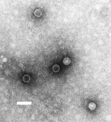
Picornaviruses are a group of related nonenveloped RNA viruses which infect vertebrates including fish, mammals, and birds. They are viruses that represent a large family of small, positive-sense, single-stranded RNA viruses with a 30 nm icosahedral capsid. The viruses in this family can cause a range of diseases including the common cold, poliomyelitis, meningitis, hepatitis, and paralysis.

Rubella virus (RuV) is the pathogenic agent of the disease rubella, transmitted only between humans via the respiratory route, and is the main cause of congenital rubella syndrome when infection occurs during the first weeks of pregnancy.

Dicistroviridae is a family of viruses in the order Picornavirales. Invertebrates, including aphids, leafhoppers, flies, bees, ants, and silkworms, serve as natural hosts. There are 15 species in this family, assigned to three genera. Diseases associated with this family include: DCV: increased reproductive potential. extremely pathogenic when injected with high associated mortality. CrPV: paralysis and death.

Aphthovirus is a viral genus of the family Picornaviridae. Aphthoviruses infect split-hooved animals, and include the causative agent of foot-and-mouth disease, Foot-and-mouth disease virus (FMDV). There are seven FMDV serotypes: A, O, C, SAT 1, SAT 2, SAT 3 and Asia 1, and four non-FMDV serotypes belonging to three additional species Bovine rhinitis A virus (BRAV), Bovine rhinitis B virus (BRBV) and Equine rhinitis A virus (ERAV).

Astroviruses (Astroviridae) are a type of virus that was first discovered in 1975 using electron microscopes following an outbreak of diarrhea in humans. In addition to humans, astroviruses have now been isolated from numerous mammalian animal species and from avian species such as ducks, chickens, and turkey poults. Astroviruses are 28–35 nm diameter, icosahedral viruses that have a characteristic five- or six-pointed star-like surface structure when viewed by electron microscopy. Along with the Picornaviridae and the Caliciviridae, the Astroviridae comprise a third family of nonenveloped viruses whose genome is composed of plus-sense, single-stranded RNA. Astrovirus has a non-segmented, single stranded, positive sense RNA genome within a non-enveloped icosahedral capsid. Human astroviruses have been shown in numerous studies to be an important cause of gastroenteritis in young children worldwide. In animals, Astroviruses also cause infection of the gastrointestinal tract but may also result in encephalitis, hepatitis (avian) and nephritis (avian).
Parechovirus B, formerly called the Ljungan virus, was first discovered in the mid-1990s after being isolated from a bank vole near the Ljungan river in Medelpad county, Sweden. It has since been established that Parechovirus B, which is also found in several places in Europe and America, causes serious illness in wild as well as laboratory animals. Several scientific articles have recently reported findings indicating that Parechovirus B is associated with malformations, intrauterine fetal death, and sudden infant death syndrome in humans. In addition, studies are being conducted worldwide to investigate the possible connection of the virus to diabetes, neurological and other illnesses in humans.
Kobuvirus is a genus of viruses in the order Picornavirales, in the family Picornaviridae. Humans and cattle serve as natural hosts. There are six species in this genus. Diseases associated with this genus include: gastroenteritis. The genus was named because of the virus particles' lumpy appearance by electron microscopy; "kobu" means "knob" in Japanese.

Mengovirus, also known as Columbia SK virus, mouse Elberfield virus, and Encephalomyocarditisvirus (EMCV), belongs to the genus Cardiovirus which is a member of the Picornaviridae. Its genome is a single stranded positive-sense RNA molecule, making the Mengoviruses a class IV virus under the Baltimore classification system. The genome is approximately 8400nt in length, and has 5’ VG protein and a 3’ polyadenine tail. Mengovirus was isolated by George W. A. Dick in 1948, in the Mengo district of Entebbe in Uganda, from a captive rhesus monkey that had developed hind limb paralysis.
Teschovirus is a genus of viruses in the order Picornavirales, in the family Picornaviridae. Pigs serve as natural hosts. There are two species in this genus, including Teschovirus A, which is responsible for the porcine enteroviral encephalomyelitis disease caused in pigs. The genus name comes from this species and the disease it causes: Teschen disease, which itself was named for the town Teschen in Poland/Czechoslovakia where the disease was first recognised in 1929.

The Coronavirus packaging signal is a conserved cis-regulatory element found in Betacoronavirus. It has an important role in regulating the packaging of the viral genome into the capsid. As part of the viral life cycle, within the infected cell, the viral genome becomes associated with viral proteins and assembles into new infective progeny viruses. This process is called packaging and is vital for viral replication.

This family represents the internal ribosome entry site (IRES) of the hepatitis A virus. HAV IRES is a 450 nucleotide long sequence located in the 735 nt long 5’ UTR of Hepatitis A viral RNA genome. IRES elements allow cap and end-independent translation of mRNA in the host cell. The IRES achieves this by mediating the internal initiation of translation by recruiting a ribosomal 40S pre-initiation complex directly to the initiation codon and eliminates the requirement for eukaryotic initiation factor, eIF4F.

Iflaviridae is a family of positive sense RNA viruses insect-infecting viruses. Some of the insects commonly infected by iflaviruses include aphids, leafhoppers, flies, bees, ants, silkworms and wasps. The name "Ifla" is derived from the name "Infectious flacherie virus", a member species. There is one genus (Iflavirus) and 16 species in this family.
Theiler's murine encephalomyelitis virus (TMEV) is a single-stranded RNA murine cardiovirus from the family Picornaviridae. It has been used as a mouse model for studying virally induced paralysis, as well as encephalomyelitis comparable to multiple sclerosis. Depending on the mouse and viral strain, viral pathogenesis can range from negligible, to chronic or acute encephalomyelitis.
Saffold virus (SAFV) is a single-stranded RNA human virus belonging to the family Picornaviridae. Discovered in 2007, it is the first human virus in the genus Cardiovirus and may provide a link to the development of multiple sclerosis or other serious diseases in humans.
Aichivirus A formerly Aichi virus (AiV) belongs to the genus Kobuvirus in the family Picornaviridae. Six species are apart of the genus Kobuvirus, Aichivirus A-F. Within Aichivirus A, there are six different types including human Aichi virus, canine kobuvirus, murine kobuvirus, Kathmandu sewage kobuvirus, roller kobuvirus, and feline kobuvirus.Three different genotypes are found in human Aichi virus, represented as genotype A, B, and C.

Pneumoviridae is a family of negative-strand RNA viruses in the order Mononegavirales. Humans, cattle, and rodents serve as natural hosts. Respiratory tract infections are associated with member viruses such as human respiratory syncytial virus. There are five species in the family which are divided between the genera Metapneumovirus and Orthopneumovirus. The family used to be considered as a sub-family of Paramyxoviridae, but has been reclassified as of 2016.

The black queen cell virus (BQCV) is a virus that infects honey bees, specifically Apis mellifera, Apis florea, and Apis dorsata. Infection of the latter two species is more recent and can be attributed to genetic similarity and geographical closeness. It is important to learn about this virus because it is one of the most common bee viruses and bees are the most important pollinators. The agricultural industry depends on the bee's pollination to increase its economic value.

Flavivirus 5' UTR are untranslated regions in the genome of viruses in the genus Flavivirus.
