Exercise-induced pulmonary hemorrhage

Last updated

Exercise-induced pulmonary hemorrhage (EIPH), also known as "bleeding" or a "bleeding attack", refers to the presence of blood in the airways of the lung in association with exercise. EIPH is common in horses undertaking intense exercise, but it has also been reported in human athletes, racing camels and racing greyhounds. Horses that experience EIPH may also be referred to as "bleeders" or as having "broken a blood vessel". In the majority of cases, EIPH is not apparent unless an endoscopic examination of the airways is performed following exercise. This is distinguished from other forms of bleeding from the nostrils, called epistaxis.

Contents

Epidemiology

EIPH often occurs in horses that race at high speeds, with the number of affected race horses increasing in proportion to the speed and intensity of the exercise. It may occur in racing Thoroughbreds (flat racing, steeplechasing or jump racing), American Quarter Horses (incidence of 50–75%), Standardbreds (incidence of 40–60%), Arabians, and Appaloosas. EIPH also occurs in eventers, jumpers, polo ponies, endurance horses, draft horses that pull competitively, [1] and horses taking part in Western speed events such as barrel racing, [2] reining and cutting. It is rare in endurance horses or draft breeds. [3]

The lowest intensities of exercise which may cause EIPH are intense trotting (40–60% maximal oxygen uptake) [4] and cantering at speeds of 16–19 miles per hour (26–31 km/h). [5]

EIPH occurs less frequently in stallions than mares or geldings. [6]

Prevalence

Approximately 43 to 75% of horses have blood in the trachea and bronchi following a single post-race endoscopic examination. [7] In one study, all horses endoscoped on at least three separate occasions following racing had EIPH at least once. [8]

Epistaxis (blood coming from one or both nostrils) is much less common, occurring in 0.25–13% of cases. [1] [9] In a survey of over 220,000 horse starts in UK Flat and National Hunt (jump) racing, 185 cases of epistaxis were identified (0.83 per 1000 starts). Similar frequencies have been reported in Japan (1.5 per 1000 starts) and South Africa (1.65 per 1000 starts), whereas a higher frequency has been reported in Korean (8.4 per 1000 starts). [10]

Clinical signs

Unless a horse has severe EIPH, with blood present at the nostrils (known as epistaxis), the main sign is usually poor athletic performance; other signs are generally subtle and not easy to detect. [11] Frequent swallowing and coughing in the immediate post-exercise recovery period, and poor appetite post-performance may be suggestive of EIPH. A definitive diagnosis can only be made by endoscopic examination of the trachea. In the case where no blood is visible in the trachea, EIPH in the small airways may still be present and can be confirmed by a bronchoalveolar lavage. Impaired arterial blood gas (oxygen) tensions during intense exercise, increased blood lactate, and rarely death have been noted (likely due to ruptured chordae tendinae or a different mechanism of lung hemorrhage). Epistaxis is diagnosed when blood is visible at either or both nostrils during or following exercise. To confirm whether the blood is coming from the upper or lower airway requires further examination by endoscopy, although in some cases it is not possible to determine the location. In the majority of epistaxis cases, the blood originates from the lung. Epistaxis during or following exercise can less commonly occur as a result of upper airway hemorrhage, for example following head trauma, subepiglottic cysts, atrial fibrillation, or guttural pouch mycoses.

Effects on performance

EIPH reduces a horse's racing performance. Severe EIPH (epistaxis) shortens a horse's racing career. Moderate to severe EIPH is associated with a worsened finishing position in a race, and finishing farther behind the winner. Horses with mild EIPH earn more prize money than those with more severe EIPH. [12] While single bouts of EIPH may not even be apparent to the rider, owner or trainer of a horse unless an endoscopic examination is undertaken, the effect on performance within a single race appears to be significant but relatively subtle. [13] In a 2005 study, horses finishing races with grade 4 EIPH were on average 6 metres behind those finishing with grade 0. [13] However, the effect of repeated bouts of EIPH that occur with daily training may lead to more significant changes and a greater degree of tissue damage over time [14] with consequent loss of lung function.

Diagnosis

As only one in ten cases of EIPH have epistaxis (bleeding from the nostrils), [15] and as epistaxis can have causes other than EIPH, [16] various diagnostic tools are used to diagnose EIPH.

Endoscopy

EIPH is most commonly diagnosed by endoscopic examination of the trachea and larger bronchi, with the optimal timing for endoscopy being 60–90 minutes after hard exercise. [15] This post-exercise delay allows time for blood within the lungs to travel to the trachea. [17] Blood can usually be detected in the trachea or bronchi for 1–3 days after an episode of EIPH, but may be present for up to a week. [18] The amount of blood visible in the trachea at the time of examination is most commonly graded on a scale of 0 (no blood) to 4 (airways awash with blood). [19] In some mildly-affected horses, endoscopy must be performed on multiple occasions to confirm a diagnosis of EIPH. [20]

The level of restraint required to allow endoscopy to be performed depends on the horse; a nose twitch or sedation may be required. [18]

Cytopathology

Cytopathology (examination under a microscope) of either a tracheal wash or a bronchoalveolar lavage sample can determine whether EIPH has occurred.

  1. For acute hemorrhage the number of red blood cells present can be quantified using a hemocytometer. A normal lung wash sample contains fewer than 10 red blood cells/μl of fluid. In the case of EIPH, the numbers will be several magnitudes or more higher.
  2. For chronic hemorrhage the presence of high numbers of hemosiderophages, with a lot of iron inside their vesicles, indicate that hemorrhage has occurred in the lung at some time in the past. Hemosiderophages are alveolar macrophages that have ingested and digested red blood cells from previous episodes of EIPH. The end product of the digestion of the red blood cells is an iron-storage complex known as hemosiderin. [21]
    Schematic of the continuous hemosiderin content in alveolar macrophages stained with Perl's Prussian blue. According to the scoring system by Doucet and Viel, macrophages have to be classified into 5 discrete grades. (a) Grade 0. (b) Borderline between Grades 0 and 1. (c) Grade 1. (d) Borderline between Grades 1 and 2. (e) Grade 2. (f) Borderline between Grades 2 and 3. (g) Grade 3. (h) Borderline between Grades 3 and 4. (i) Grade 4. EIPH hemosiderin content in alveolar macrophages.jpg
    Schematic of the continuous hemosiderin content in alveolar macrophages stained with Perl’s Prussian blue. According to the scoring system by Doucet and Viel, macrophages have to be classified into 5 discrete grades. (a) Grade 0. (b) Borderline between Grades 0 and 1. (c) Grade 1. (d) Borderline between Grades 1 and 2. (e) Grade 2. (f) Borderline between Grades 2 and 3. (g) Grade 3. (h) Borderline between Grades 3 and 4. (i) Grade 4.

Bronchoalveolar lavage

Bronchoalveolar lavage (BAL) is a procedure whereby a small volume of fluid is put into the airways in order sample the cells and fluids of the alveoli and epithelium of the bronchi. [23] BAL may be performed using a BAL tube, which allows fluid to be added to and removed from the bronchi, or may be performed during endoscopy, if the endoscope has an irrigation channel. [24] To perform BAL, the horse is usually sedated, and local anaesthetic is usually instilled into the airways to reduce coughing. [24]

BAL is less useful when severe hemorrhage has occurred. BAL results only reflect the situation in the localized region of the lung which has been lavaged, and may not be representative of the whole lung. [24] The fluid obtained by BAL is submitted for cytological examination.

Imaging

Radiography

Imaging the lungs by taking a radiograph (x-ray) of the chest cannot be used to diagnose EIPH, as the lungs of affected and unaffected horses share the same characteristics. [20]

Pulmonary scintigraphy

Scintigraphy of the lungs may detect moderate to severe alterations in the perfusion and possibly ventilation of the dorso-caudal lung. [25] The use of radio-labelled red blood cells and scintigraphy to localise and or quantify hemorrhage is not useful due to general sequestration of labeled RBC by the lung, even in the absence of hemorrhage. [26]

Post-mortem examination

EIPH seldom causes the death of a horse, but if an affected horse dies and undergoes a post-mortem examination, repeated episodes of EIPH will have caused a characteristic blue-gray-brown staining in the lungs. [27] The staining is due to the presence of hemosiderin. The staining is usually most intense in the dorso-caudal region of the left and right diaphragmatic lobes which often progresses cranioventral with repetitive damage. There are often distinct borders between healthy lung tissue and those parts of the lungs that have been affected by EIPH. Other histopathologic findings include fibrosis, bronchial artery neovascularization, venous remodeling, bronchiolitis, hemosiderin accumulation, increased tissue cellularity (i.e. hemosiderophages), multifocal areas of inflammation, and increased thickness of vascular and airway walls.

Causes

EIPH occurs when blood enters the air passages of a horse's lung, due to fractured capillaries. A variety of causes have been proposed, but EIPH is most likely a multi-factorial condition, involving airway, vascular, inflammatory, blood, cardiac, locomotory, and remodelling components.

The primary mechanism is likely to be high pulmonary vascular pressures with concurrent negative airway pressures, causing extreme stress across the pulmonary capillary membrane (the fragile membrane separating blood in the pulmonary capillaries from the air-filled alveoli) and consequent hemorrhage into the air spaces of the lung. Other contributing factors may include upper airway obstruction, increased blood viscosity, abnormalities of cardiac origin (small cross-sectional area of atrioventricular valves, stiff valves, slow left ventricular relaxation time, right tricuspid valve regurgitation), preferential distribution of blood flow to the dorsocaudal lung regions, mechanical trauma, lower airway obstruction, inflammation, abnormalities of blood coagulation, inhomogeneity of ventilation and locomotory trauma. EIPH begins in the dorso-caudal region of the lung and progresses in a cranioventral direction over time.

High pulmonary blood pressures

The most widely accepted theory is that high transmural pressures lead to pulmonary capillary stress failure. There may be contributions from the bronchial circulation. Pulmonary capillary transmural pressure is determined by pulmonary capillary pressure and airway pressure. The horse has very high pulmonary vascular pressures during intense exercise, exceeding 100 mmHg in the pulmonary artery during intense exercise. During expiration the high positive pressures in the pulmonary blood vessels pushing out are opposed by high positive airway pressures pushing back and this does not place undue stress on the thin blood vessel walls. During inspiration, the high positive pressures in the pulmonary blood vessels pushing out are met by negative pressures distending the blood vessel and placing increased stress on the walls. Studies in vitro show that significant disruption of the pulmonary capillaries occurs at pressures of approximately 80 mmHg. In vivo, significant EIPH occurs above a mean pulmonary artery pressure of around 80–95 mmHg. [28] [29] On the basis of this theory, any factor or disease that would increase pulmonary vascular pressures (e.g. hypervolaemia) or increase the magnitude of the negative pressures in the lung during inspiration (e.g. dynamic upper airway obstruction) would increase the severity of EIPH; however neither experimentally induced laryngeal hemiplegia nor dorsal displacement of the soft palate increase pulmonary capillary transmural pressure. [30] [31] Furthermore, the magnitude of exercise-induced pulmonary arterial, capillary and venous hypertension is reportedly similar in horses either with or without EIPH.

Locomotory associated trauma

This theory is based on the fact that, during galloping, the absence of any bone attachment of the forelegs to the spine in the horse causes the shoulder to compress the cranial rib cage. [32] The compression of the chest initiates a pressure wave of compression and expansion which spreads outwards. However, due to the shape of the lung and reflections off the chest wall, the wave of expansion and compression becomes focussed and amplified in the dorso-caudal lung. [33] The alternate expansion and compression at the microscopic level in adjacent areas of lung tissue creates shear stress and capillary disruption. The theory predicts that hemorrhage would be more severe on hard track surfaces, but it does not explain why EIPH can occur in horses during swimming exercise.

Veno-occlusive remodelling

This theory proposes how high pulmonary venous pressures may lead to the capillary rupture and the tissue changes observed in EIPH. [14] Regional veno-occlusive remodeling, especially within the caudodorsal lung fields, contributes to the pathogenesis of EIPH, with the venous remodeling leading to regional vascular congestion and hemorrhage, hemosiderin accumulation, fibrosis, and bronchial angiogenesis.

Risk factors

While all horses undertaking intense exercise experience some degree of EIPH, some horses consistently experience greater haemorrhage and other horses experience isolated episodes of increased EIPH. In the case of horses that consistently demonstrate greater severity of EIPH this is most likely due to congenital factors, such as very high pulmonary vascular pressures. In horses that experience isolated episodes of increased severity of EIPH, possible contributing factors may include, amongst others, pulmonary infection or atrial fibrillation, inflammation, longer distances, longer duration of exercise, hard surfaces, steeplechasing/hurdling, increased length of career, breed (i.e. Thoroughbred greater than Standardbred), time in training/racing, genetics, and cold temperatures.

Management and treatment

Furosemide (trade name: Lasix) administered prior to racing or strenuous exercise in Thoroughbred and Standardbred racehorses reduces the severity EIPH in 68% of horses. [34] Up to 85% of Thoroughbred racehorses in the United States have been administered furosemide at least once during their racing career. [7] Furosemide decreases pulmonary arterial pressure via its diuretic effects, bronchodilates, and redistributes blood flow during exercise. It reduces EIPH ranging from 90% at sub-maximal exercise and about 50% at maximal exercise intensities. However, over time, it causes electrolyte imbalances and has reduced effectiveness. Furosemide is prohibited in competing horses in some countries, and by the International Olympic Committee. The United States and Canada are the only countries which permit furosemide use during racing.

Other vascular agents such as nitric oxide (NO), n-nitro-l-arginine methyl ester (L-Name), nitroglycerin, NO + phosphodiesterase inhibitors (e.g., sildenafil), and endothelin receptor antagonists have no effect, and in some cases worsen of the EIPH.

A non-pharmacological nasal strip, applied prophylactically, variably reduces EIPH, depending on the severity of EIPH in an individual horse, and the duration and intensity of the exercise bout. The strip minimizes the increase in resistance to breathing, and the work of breathing as exercise intensity increases. There is synergy with the nasal strip in combination with furosemide. The strip has a spring-like action that mechanically supports and maintains the size of the nasal passage at its narrowest part, the nasal valve. Resistance to breathing doubles during intense or long-duration endurance exercise, with >50% of the total resistance originating at the nasal passages. Decreased resistance reduces the airway forces across the pulmonary capillary membrane, reducing the EIPH. Use of the strip is permitted by the FEI, and in U.S. horse racing.

Bronchodilatation is already maximized in the exercising horse, therefore bronchodilatory agents such as ipratropium, [35] albuterol, [35] or clenbuterol are not effective in reducing EIPH.

Coagulation dysfunction is not a cause of EIPH, and anticoagulatory agents, including carbazochrome salicylate, aspirin, Premarin, Amicar, and vitamin K, are not effective in reducing EIPH, and in some cases may worsen it.

Omega-3 fatty acids (DHA and EPA) reduce EIPH, presumably via increasing the functionality of the white blood cells (WBCs) in removing the blood from the lungs. Concentrated equine serum reduced EIPH by 53% through a combined mechanism of a 30% reduction in inflammation and an increased functionality of the WBCs. Other anti-inflammatory agents, such as hesperidin-citrus bioflavinoids, vitamin C, NSAIDs such as phenylbutazone, corticosteroids, heated water vapor therapy, cromoglicic acid or nedocromil, have no beneficial effects in reducing EIPH severity. Phenylbutazone can partially reverse the beneficial effects of furosemide.

Other ineffective treatments include leukocyte elastase protease inhibitors, the EIPH Patch, hyperbaric oxygen therapy, pentoxyfylline, guanabenz, clonidine, snake venom, and enalapril.

Horses that undergo surgical correction for upper airway dysfunction are rested, and are under environmentally controlled environments with reduced dust may see some benefit.

Related Research Articles

Diffusing capacity of the lung (DL) measures the transfer of gas from air in the lung, to the red blood cells in lung blood vessels. It is part of a comprehensive series of pulmonary function tests to determine the overall ability of the lung to transport gas into and out of the blood. DL, especially DLCO, is reduced in certain diseases of the lung and heart. DLCO measurement has been standardized according to a position paper by a task force of the European Respiratory and American Thoracic Societies.

<span class="mw-page-title-main">Pulmonary edema</span> Fluid accumulation in the tissue and air spaces of the lungs

Pulmonary edema, also known as pulmonary congestion, is excessive liquid accumulation in the tissue and air spaces of the lungs. It leads to impaired gas exchange and may cause hypoxemia and respiratory failure. It is due to either failure of the left ventricle of the heart to remove oxygenated blood adequately from the pulmonary circulation, or an injury to the lung tissue directly or blood vessels of the lung.

<span class="mw-page-title-main">Furosemide</span> Loop diuretic medication

Furosemide is a loop diuretic medication used to treat edema due to heart failure, liver scarring, or kidney disease. It had many trade names including Discoid, Frusemide, Lasix and Uremide. Furosemide may also be used for the treatment of high blood pressure. It can be taken intravenously or orally. When given intravenously, furosemide typically takes effect within five minutes; when taken orally, it typically metabolizes within an hour.
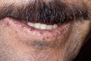
<span class="mw-page-title-main">Pulmonary hemorrhage</span> Medical condition

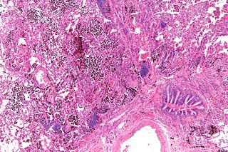
Pulmonary hemorrhage is an acute bleeding from the lung, from the upper respiratory tract and the trachea, and the pulmonary alveoli. When evident clinically, the condition is usually massive. The onset of pulmonary hemorrhage is characterized by a cough productive of blood (hemoptysis) and worsening of oxygenation leading to cyanosis. Treatment should be immediate and should include tracheal suction, oxygen, positive pressure ventilation, and correction of underlying abnormalities such as disorders of coagulation. A blood transfusion may be necessary.

<span class="mw-page-title-main">Flunixin</span> NSAID analgesic veterinary drug

Flunixin is a nonsteroidal anti-inflammatory drug (NSAID), analgesic, and antipyretic used in horses, cattle and pigs. It is often formulated as the meglumine salt. In the United States, it is regulated by the U.S. Food and Drug Administration (FDA), and may only be lawfully distributed by order of a licensed veterinarian. There are many trade names for the product.

<span class="mw-page-title-main">Hypoxemia</span> Abnormally low level of oxygen in the blood

Hypoxemia is an abnormally low level of oxygen in the blood. More specifically, it is oxygen deficiency in arterial blood. Hypoxemia has many causes, and often causes hypoxia as the blood is not supplying enough oxygen to the tissues of the body.

Cardiac asthma is the medical condition of intermittent wheezing, coughing, and shortness of breath that is associated with underlying congestive heart failure (CHF). Symptoms of cardiac asthma are related to the heart's inability to effectively and efficiently pump blood in a CHF patient. This can lead to accumulation of fluid in and around the lungs, disrupting the lung's ability to oxygenate blood.

<span class="mw-page-title-main">Cribbing (horse)</span> Stable vice in horses

Cribbing is a form of stereotypy (equine oral stereotypic behaviour), otherwise known as wind sucking or crib-biting. Cribbing is considered to be an abnormal, compulsive behavior seen in some horses, and is often labelled a stable vice. The major factors that cause cribbing include stress, stable management, genetic and gastrointestinal irritability.

Robert Cook is an equine veterinarian. He has published many papers, mainly on diseases of the horse's mouth, ear, nose and throat both in scientific and horseman's journals, covering various topics:

Positive end-expiratory pressure (PEEP) is the pressure in the lungs above atmospheric pressure that exists at the end of expiration. The two types of PEEP are extrinsic PEEP and intrinsic PEEP. Pressure that is applied or increased during an inspiration is termed pressure support.PEEP is a therapeutic parameter set in the ventilator, or a complication of mechanical ventilation with air trapping (auto-PEEP).

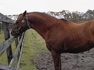
Equine polysaccharide storage myopathy is a hereditary glycogen storage disease of horses that causes exertional rhabdomyolysis. It is currently known to affect the following breeds American Quarter Horses, American Paint Horses, Warmbloods, Cobs, Dales Ponies, Thoroughbreds, Arabians, New Forest ponies, and a large number of Heavy horse breeds. While incurable, PSSM can be managed with appropriate diet and exercise. There are currently 2 subtypes, known as Type 1 PSSM and Type 2 PSSM.

<span class="mw-page-title-main">Tongue-tie (tack)</span>

A tongue-tie is a piece of equipment used by equestrians to prevent a horse from getting its tongue over the bit, which would make the animal very difficult to control. It is usually a strip of cloth or rubber, passed through the mouth and tied below the chin.

<span class="mw-page-title-main">Respiratory system of the horse</span> Biological system by which a horse circulates air for the purpose of gaseous exchange

The respiratory system of the horse is the biological system by which a horse circulates air for the purpose of gaseous exchange.

Lameness is an abnormal gait or stance of an animal that is the result of dysfunction of the locomotor system. In the horse, it is most commonly caused by pain, but can be due to neurologic or mechanical dysfunction. Lameness is a common veterinary problem in racehorses, sport horses, and pleasure horses. It is one of the most costly health problems for the equine industry, both monetarily for the cost of diagnosis and treatment, and for the cost of time off resulting in loss-of-use.

Colitis X, equine colitis X or peracute toxemic colitis is a catchall term for various fatal forms of acute or peracute colitis found in horses, but particularly a fulminant colitis where clinical signs include sudden onset of severe diarrhea, abdominal pain, shock, and dehydration. Death is common, with 90–100% mortality, usually in less than 24 hours. The causative factor may be Clostridium difficile, but it also may be caused by other intestinal pathogens. Horses under stress appear to be more susceptible to developing colitis X, and like the condition pseudomembranous colitis in humans, an association with prior antibiotic use also exists. Immediate and aggressive treatment can sometimes save the horse, but even in such cases, 75% mortality is considered a best-case scenario.

<span class="mw-page-title-main">Guttural pouch</span>

Guttural pouches are large, auditory-tube diverticula that contain between 300 and 600 ml of air. They are present in odd-toed mammals, some bats, hyraxes, and the American forest mouse. They are paired bilaterally just below the ears, behind the skull and connect to the nasopharynx.

Racehorse injuries and fatalities are a side effect of training and competition. The problem with equine injuries is that they so often result in death. A 2005 study by the United States Department of Agriculture found that injuries are the second leading cause of death in horses, second only to old age. Nureyev's recovery from a broken leg while retired at stud in 1987 typifies the struggle horses have after being injured.

The treatment of equine lameness is a complex subject. Lameness in horses has a variety of causes, and treatment must be tailored to the type and degree of injury, as well as the financial capabilities of the owner. Treatment may be applied locally, systemically, or intralesionally, and the strategy for treatment may change as healing progresses. The end goal is to reduce the pain and inflammation associated with injury, to encourage the injured tissue to heal with normal structure and function, and to ultimately return the horse to the highest level of performance possible following recovery.

Susan Marie Stover is a professor of veterinary anatomy at the University of California, Davis School of Veterinary Medicine and director of the J.D. Wheat Veterinary Orthopedic Research Laboratory. One of the focuses of her wide-ranging research is musculoskeletal injuries in racehorses, particularly catastrophic breakdowns. Her identification of risk factors has resulted in improved early detection and changes to horse training and surgical repair methods. On July 30, 2016, Stover received the Lifetime Excellence in Research Award from the American Veterinary Medical Association. In August 2016, she was selected for induction into the University of Kentucky Equine Research Hall of Fame.

<span class="mw-page-title-main">Horse tongue</span> Anatomy of the equine tongue

The horse tongue, like that of most mammals, is pink in color and plays an important role in taste perception. With its long, narrow shape typical of herbivorous animals, it enables the horse to grasp its vegetable food, with the help of its lips and teeth. This tongue is sensitive to pressure and temperature, and is involved in licking and chewing behaviors. Although the mare licks its foal for a long time immediately after birth, there is little research into the gustatory sensitivity of horses and the social use these animals make of their tongues.

References

  1. 1 2 Riegal, Ronald; Susan Hakola (2004). The Illustrated Atlas of Clinical Equine Anatomy and Common Disorders of the Horse. Vol. 2. Equistar Publications. p. [ page needed ]. ISBN   978-0-9654461-1-2.
  2. Léguillette, R; Steinmann, M; Bond, SL; Stanton, B (July 2016). "Tracheobronchoscopic assessment of exercise-induced pulmonary hemorrhage and airway inflammation in barrel racing horses". Journal of Veterinary Internal Medicine. 30 (4): 1327–32. doi:10.1111/jvim.13959. PMC   5089608 . PMID   27278854.
  3. Hinchcliff, Kenneth W. "Exercise-Induced Pulmonary Hemorrhage". Pulmonary Hemorrhage (PDF). Versailles, Kentucky: Kentucky Equine Research, Inc. Archived from the original (PDF) on 7 January 2010.
  4. Epp, TS; McDonough, P; Padilla, DJ; Gentile, JM; Edwards, KL; Erickson, HH; Poole, DC (August 2006). "Exercise-induced pulmonary haemorrhage during submaximal exercise". Equine Veterinary Journal. Supplement. 38 (S36): 502–7. doi:10.1111/j.2042-3306.2006.tb05595.x. PMID   17402474. S2CID   24739288.
  5. Oikawa, M (November 1999). "Exercise-induced haemorrhagic lesions in the dorsocaudal extremities of the caudal lobes of the lungs of young thoroughbred horses". Journal of Comparative Pathology. 121 (4): 339–47. doi:10.1053/jcpa.1999.0331. PMID   10542123.
  6. Hillidge, CJ; Whitlock, TW (May 1986). "Sex variation in the prevalence of exercise-induced pulmonary haemorrhage in racing quarter horses". Research in Veterinary Science. 40 (3): 406–7. doi:10.1016/S0034-5288(18)30559-9. PMID   3738238.
  7. 1 2 Hinchcliff, Kenneth (2009). "Exercise-Induced Pulmonary Hemorrhage" (PDF). Advances in Equine Nutrition. 4: 367–374 via Google Scholar.
  8. Birks, EK; Shuler, KM; Soma, LR; Martin, BB; Marconato, L; Del Piero, F; Teleis, DC; Schar, D; Hessinger, AE; Uboh, CE (September 2002). "EIPH: postrace endoscopic evaluation of Standardbreds and Thoroughbreds". Equine Veterinary Journal. Supplement. 34 (34): 375–8. doi: 10.1111/j.2042-3306.2002.tb05451.x . PMID   12405719.
  9. Merck (2008). "Exercise-induced Pulmonary Hemorrhage (Epistaxis, "Bleeder")". The Merck Veterinary Manual . Retrieved 24 August 2010.
  10. Kim, BS; Hwang, YK; Kwon, CJ; Lim, YJ (1998). "Survey on Incidence of Exercise Induced Pulmonary Hemorrhage (EIPH) of Thoroughbred Race horses in Seoul Race course". Korean Journal of Veterinary Clinical Medicine. 15: 417–426.
  11. Wilkins, Pamela A. (2014). "Chapter 31: Diseases of the respiratory system: Exercise induced pulmonary hemorrhage". In Smith, Bradford P. (ed.). Large animal internal medicine (5 ed.). Elsevier Health Sciences. pp. 543–549. ISBN   9780323088404.
  12. Hinchcliff, K.W.; Couetil, L.L.; Knight, P.K.; Morley, P.S.; Robinson, N.E.; Sweeney, C.R.; van Erck, E. (May 2015). "Exercise induced pulmonary hemorrhage in horses: American College of Veterinary Internal Medicine consensus statement". Journal of Veterinary Internal Medicine. 29 (3): 743–758. doi:10.1111/jvim.12593. PMC   4895427 . PMID   25996660.
  13. 1 2 Hinchcliff, KW; Jackson, MA; Morley, PS; Brown, JA; Dredge, AE; O'Callaghan, PA; McCaffrey, JP; Slocombe, RE; Clarke, AE (1 September 2005). "Association between exercise-induced pulmonary hemorrhage and performance in Thoroughbred racehorses". Journal of the American Veterinary Medical Association. 227 (5): 768–74. doi: 10.2460/javma.2005.227.768 . PMID   16178399.
  14. 1 2 Derksen, FJ; Williams, KJ; Pannirselvam, RR; de Feijter-Rupp, H; Steel, CM; Robinson, NE (July 2009). "Regional distribution of collagen and haemosiderin in the lungs of horses with exercise-induced pulmonary haemorrhage". Equine Veterinary Journal. 41 (6): 586–91. doi: 10.2746/042516409X429419 . PMID   19803055.
  15. 1 2 Barakzai, Safia (2006). "Chapter 7: Trachea and bronchi". Handbook of equine respiratory endoscopy . Edinburgh: Elsevier Saunders. pp.  89–104. ISBN   9780702028182.
  16. Ramzan, Peter H.L. (2014). "Respiratory conditions: lower airway disease". The racehorse: a veterinary manual. CRC Press. pp. 222–234. ISBN   9781482221916.
  17. Weese, Scott; Munroe, Graham, eds. (2011). "Chapter 3: Respiratory system". Equine clinical medicine, surgery, and reproduction. London: Manson Publishing/The Veterinary Press. pp. 381–480. ISBN   9781840766080.
  18. 1 2 Hinchcliff, KW (2013). "Chapter 29: Exercise-induced pulmonary hemorrhage (EIPH)". In Hinchcliff, KW; Kaneps, AJ; Geor, RJ (eds.). Equine Sports Medicine and Surgery E-Book (2nd, revised ed.). Elsevier Health Sciences. pp. 633–648. ISBN   9780702054228.
  19. Léguillette, R.; Steinmann, M.; Bond, S.L.; Stanton, B. (July 2016). "Tracheobronchoscopic assessment of exercise-induced pulmonary hemorrhage and airway inflammation in barrel racing horses". Journal of Veterinary Internal Medicine. 30 (4): 1327–1332. doi:10.1111/jvim.13959. PMC   5089608 . PMID   27278854.
  20. 1 2 Reed, SM; Bayly, WM; Sellon, DC, eds. (2009). "Disorders of the respiratory system: Exercise-induced pulmonary hemorrhage". Equine internal medicine (3rd ed.). St. Louis, MO: Saunders/Elsevier. pp. 346–349. ISBN   9781437708851.
  21. 1 2 Bertram, Christof A.; Marzahl, Christian; Bartel, Alexander; Stayt, Jason; Bonsembiante, Federico; Beeler-Marfisi, Janet; Barton, Ann K.; Brocca, Ginevra; Gelain, Maria E.; Gläsel, Agnes; Preez, Kelly du; Weiler, Kristina; Weissenbacher-Lang, Christiane; Breininger, Katharina; Aubreville, Marc; Maier, Andreas; Klopfleisch, Robert; Hill, Jenny (17 November 2022). "Cytologic scoring of equine exercise-induced pulmonary hemorrhage: Performance of human experts and a deep learning-based algorithm". Veterinary Pathology. 60 (1): 75–85. doi:10.1177/03009858221137582. ISSN   0300-9858. PMC   9827485 . PMID   36384369.
  22. Doucet, Michele Y.; Viel, Laurent (May 2002). "Alveolar Macrophage Graded Hemosiderin Score from Bronchoalveolar Lavage in Horses with Exercise-Induced Pulmonary Hemorrhage and Controls". Journal of Veterinary Internal Medicine. 16 (3): 281–286. doi:10.1111/j.1939-1676.2002.tb02370.x. PMID   12041658.
  23. McKane, SA; Rose, RJ (10 June 2010). "Effects of exercise intensity and training on bronchoalveolar lavage cytology". Equine Veterinary Journal. 27 (S18): 58–62. doi:10.1111/j.2042-3306.1995.tb04891.x. S2CID   73156946.
  24. 1 2 3 Stack, A (2014). "Chapter 58: Exercise-induced pulmonary hemorrhage". In Sprayberry, KA; Robinson, NE (eds.). Robinson's Current Therapy in Equine Medicine - E-Book (7 ed.). Elsevier Health Sciences. pp. 252–256. ISBN   9780323242165.
  25. O'Callaghan, M. W.; Pascoe, J. R.; O'Brien, T. R.; Hornof, W. J.; Mason, D. K. (September 1987). "Exercise-induced pulmonary haemorrhage in the horse: results of a detailed clinical, post mortem and imaging study. VI. Radiological/pathological correlations". Equine Veterinary Journal. 19 (5): 419–422. doi:10.1111/j.2042-3306.1987.tb02634.x. PMID   3678184.
  26. Votion, DM; Roberts, CA; Marlin, DJ; Lekeux, PM (July 1999). "Feasibility of scintigraphy in exercise-induced pulmonary haemorrhage detection and quantification: preliminary studies". Equine Veterinary Journal. Supplement. 31 (30): 137–42. doi: 10.1111/j.2042-3306.1999.tb05204.x . PMID   10659238.
  27. Saulez, MN (2009). "Chapter 71: Exercise-induced pulmonary hemorrhage". In Robinson, NE; Sprayberry, KA (eds.). Current therapy in equine medicine (6th ed.). St. Louis, Mo.: Saunders Elsevier. pp. 320–323. ISBN   9781416054757.
  28. Meyer, TS; Fedde, MR; Gaughan, EM; Langsetmo, I; Erickson, HH (July 1998). "Quantification of exercise-induced pulmonary haemorrhage with bronchoalveolar lavage". Equine Veterinary Journal. 30 (4): 284–8. doi:10.1111/j.2042-3306.1998.tb04098.x. PMID   9705109.
  29. Langsetmo, I.; Fedde, M.R.; Meyer, T.S.; Erickson, H.H. (September 2000). "Relationship of pulmonary arterial pressure to pulmonary haemorrhage in exercising horses". Equine Veterinary Journal. 32 (5): 379–84. doi:10.2746/042516400777591066. PMID   11037258.
  30. Jackson, JA; Ducharme, NG; Hackett, RP; Rehder, RS; Ainsworth, DM; Shannon, KJ; Erickson, BK; Erb, HN; Jansson, N; Soderholm LV, Jr; Thorson, LM (August 1997). "Effects of airway obstruction on transmural pulmonary artery pressure in exercising horses". American Journal of Veterinary Research. 58 (8): 897–903. PMID   9256978.
  31. Hackett, RP; Ducharme, NG; Ainsworth, DM; Erickson, BK; Erb, HN; Soderholm LV, Jr; Thorson, LM (April 1999). "Effects of extrathoracic airway obstruction on intrathoracic pressure and pulmonary artery pressure in exercising horses". American Journal of Veterinary Research. 60 (4): 485–94. PMID   10211694.
  32. Schroter, RC; Marlin, DJ; Denny, E (May 1998). "Exercise-induced pulmonary haemorrhage (EIPH) in horses results from locomotory impact induced trauma-a novel, unifying concept". Equine Veterinary Journal. 30 (3): 186–92. doi:10.1111/j.2042-3306.1998.tb04486.x. PMID   9622318.
  33. Schroter, RC; Leeming, A; Denny, E; Bharath, A; Marlin, DJ (July 1999). "Modelling impact-initiated wave transmission through lung parenchyma in relation to the aetiology of exercise-induced pulmonary haemorrhage". Equine Veterinary Journal. Supplement. 31 (30): 34–8. doi: 10.1111/j.2042-3306.1999.tb05184.x . PMID   10659218.
  34. Sullivan, SL; Whittem, T; Morley, PS; Hinchcliff, KW (May 2015). "A systematic review and meta-analysis of the efficacy of furosemide for exercise-induced pulmonary haemorrhage in Thoroughbred and Standardbred racehorses". Equine Veterinary Journal. 47 (3): 341–9. doi:10.1111/evj.12373. PMID   25291214.
  35. 1 2 Bayly, WM; Slocombe, RF; Schott HC, 2nd; Hines, MT; Sides, RH; Hakala, JE (May 2001). "Effects of inhalation of albuterol sulphate, ipratroprium bromide and frusemide on breathing mechanics and gas exchange in healthy exercising horses". Equine Veterinary Journal. 33 (3): 302–10. doi:10.2746/042516401776249741. PMID   11352354.

Sources

  • Hinchcliff, K. W. (2009). "Exercise-induced pulmonary hemorrhage". Advances of Equine Nutrition, 4: 367-374.
  • MacNamara, B; Bauer, S; Iafe, J (1 February 1990). "Endoscopic evaluation of exercise-induced pulmonary hemorrhage and chronic obstructive pulmonary disease in association with poor performance in racing Standardbreds". Journal of the American Veterinary Medical Association. 196 (3): 443–5. PMID   2298674.
  • Mason, D. K., E. A. Collins, et al. (1983). Exercise-induced pulmonary haemorrhage in horses. 1st International Conference on Equine Exercise Physiology, Oxford, UK.
  • Merck (2008). "Exercise-induced Pulmonary Hemorrhage (Epistaxis, "Bleeder")". The Merck Veterinary Manual . Retrieved 24 August 2010.
  • Newton, JR; Wood, JL (September 2002). "Evidence of an association between inflammatory airway disease and EIPH in young Thoroughbreds during training". Equine Veterinary Journal. Supplement. 34 (34): 417–24. doi:10.1111/j.2042-3306.2002.tb05459.x. PMID   12405727. S2CID   41241703.
  • Pascoe, JR; Ferraro, GL; Cannon, JH; Arthur, RM; Wheat, JD (May 1981). "Exercise-induced pulmonary hemorrhage in racing thoroughbreds: a preliminary study". American Journal of Veterinary Research. 42 (5): 703–7. PMID   7258791.
  • Raphel, CF; Soma, LR (July 1982). "Exercise-induced pulmonary hemorrhage in Thoroughbreds after racing and breezing". American Journal of Veterinary Research. 43 (7): 1123–7. PMID   7103190.
  • Riegal, Ronald; Susan Hakola (2004). The Illustrated Atlas of Clinical Equine Anatomy and Common Disorders of the Horse. Vol. 2. Equistar Publications. p. [ page needed ]. ISBN   978-0-9654461-1-2.
  • Roberts, C.A., Hillidge, C. and Marlin, D.J. (1993) Exercise induced pulmonary haemorrhage in racing thoroughbreds in Great Britain. 1st International EIPH Conference, Guelph, Canada, p11.
  • Speirs, VC; van Veenendaal, JC; Harrison, IW; Smyth, GB; Anderson, GA; Wilson, DV; Gilbo, B (August 1982). "Pulmonary haemorrhage in standardbred horses after racing". Australian Veterinary Journal. 59 (2): 38–40. doi:10.1111/j.1751-0813.1982.tb02712.x. PMID   6890804.

Further reading