Eyeblink conditioning

Last updated

Eyeblink conditioning (EBC) is a form of classical conditioning that has been used extensively to study neural structures and mechanisms that underlie learning and memory. The procedure is relatively simple and usually consists of pairing an auditory or visual stimulus (the conditioned stimulus (CS)) with an eyeblink-eliciting unconditioned stimulus (US) (e.g. a mild puff of air to the cornea or a mild shock). Naïve organisms initially produce a reflexive, unconditioned response (UR) (e.g. blink or extension of nictitating membrane) that follows US onset. After many CS-US pairings, an association is formed such that a learned blink, or conditioned response (CR), occurs and precedes US onset. [1] The magnitude of learning is generally gauged by the percentage of all paired CS-US trials that result in a CR. Under optimal conditions, well-trained animals produce a high percentage of CRs (> 90%). The conditions necessary for, and the physiological mechanisms that govern, eyeblink CR learning have been studied across many mammalian species, including mice, rats, guinea pigs, rabbits, ferrets, cats, and humans. Historically, rabbits have been the most popular research subjects.

Contents

CS-US contingency

The order in which stimuli are presented is an important factor in all forms of classical conditioning. Forward conditioning describes a presentation format in which the CS precedes the US in time. That is, from the perspective of the research subject, experiencing the US is contingent upon having just experienced the CS. EBC is usually, but not always, conducted in this manner. Other stimulus contingencies include backward conditioning, in which US comes before CS, and simultaneous conditioning, in which CS and US are presented at the same time. In any case, the time between CS onset and US onset is the interstimulus interval (ISI). Animals are usually trained with a shorter ISI than humans, which can make interspecies comparisons difficult. [2]

The delay and trace procedures

In delay EBC, the CS onset precedes the US onset and the two stimuli overlap and coterminate, with the stimuli converging in the cerebellar cortex and interpositus nucleus. [3] In the trace EBC, the CS precedes the US and there is a stimulus free period (trace interval) between CS offset and US onset. While both of these procedures require the cerebellum, the trace procedure also requires the hippocampus and medial prefrontal cortex. [4] [5]

Neural circuitry

When a US is delivered to the cornea of the eye, sensory information is carried to the trigeminal nucleus and relayed both directly and indirectly (via reticular formation) to the accessory abducens and abducens motor nuclei (see Cranial nerve nucleus). Output from these nuclei control various eye muscles that work synergistically to produce an unconditioned blink response to corneal stimulation (reviewed, Christian & Thompson, 2003). Electromyogram (EMG) activity of the orbicularis oculi muscle, which controls eyelid closure, is considered to be the most prominent and sensitive component of blinking (Lavond et al., 1990) and is, thus, the most common behaviorally-derived dependent variable in studies of EBC.

US pathway

The trigeminal nucleus also sends efferent projections to the inferior olive (IO), and this represents the US pathway for EBC. The critical region of the IO for eyeblink conditioning is the dorsal accessory olive (Brodal, 1981), and climbing fibers (CF) from this region send information about the US to the cerebellum (Brodal, Walberg & Hoddevik, 1975; Thompson, 1989). Climbing fibers ultimately project to both the deep cerebellar nuclei and Purkinje cells (PCs) in the cerebellar cortex.

The CS pathway

The pontine nuclei (PN) can support different CS modalities (auditory tone, light, etc.) for EBC as they receive projections from auditory, visual, somatosensory, and association systems (Glickstein et al., 1980; Brodal, 1981; Schmahmann & Pandya, 1989; 1991; 1993). When the CS is a tone, auditory information is received via the cochlear nuclei (Steinmetz & Sengelaub, 1992). The PN give rise to mossy fiber (MF) axons that carry CS-related information (Steinmetz et al., 1987; Lewis et al., 1987; Thompson et al., 1997) to the cerebellum via the middle cerebellar peduncle, and terminate in both the cerebellar nuclei, and at granule cells (GR) of the cerebellar cortex (Steinmetz & Sengelaub, 1992). Granule cells give rise to parallel fiber (PF) axons which synapse onto PCs.

CS-US convergence in the cerebellum

Two cerebellar sites of CS-US convergence are 1) cells of the deep nuclear region in the cerebellum, and 2) PCs of the cortex. [6] In addition to receiving converging CS and US input via the PN and IO, respectively, cells of the cerebellar nuclei receive GABA-ergic inhibitory input from PCs of the cerebellar cortex. Output from the interpositus nucleus includes projections to the red nucleus, and the red nucleus sends projections to the facial and abducens nuclei. These nuclei supply the motor output component of the reflexive eyeblink. Therefore, in addition to being a site of stimulus convergence, the deep nuclei are also the cerebellum's output structure.

Critical role of the interposed nucleus

David A. McCormick, as a graduate student with Professor Richard F. Thompson, initially identified the cerebellum as the essential structure for learning and executing eyeblink CRs. Some scientists think that the interposed nucleus is the site critical to learning, retaining, and executing the conditioning blink response.

Lesion studies

The first evidence for the role of the cerebellum in EBC came from McCormick et al. (1981). They found that a unilateral cerebellar lesion which included both cortex and deep nuclei permanently abolished CRs. In subsequent studies, it was determined that lesions of the lateral interpositus and medial dentate nuclei were sufficient to prevent acquisition of CRs in naïve animals (Lincoln et al., 1982) and abolished CRs in well-trained animals (McCormick & Thompson, 1984). [7] Finally, the use of Kainic acid lesions, which destroy neuronal cell bodies and spare passing fibers, provided evidence for a highly localized region of cerebellar nuclear cells that are essential for learning and performing CRs (Lavond et al., 1985). The population of cells critical for EBC appears to be restricted to a ~ 1 mm3 area of dorsolateral anterior INP ipsilateral to the conditioned eye. Lesions to this area of INP result in an inability to acquire eyeblink CRs in naïve animals. Additionally, the permanence of the localized lesion effect is remarkable. In well-trained animals, CRs abolished as a result of lesion are not reacquired, even after extensive training that spans over 8 months (Steinmetz et al., 1992). These results demonstrate that a highly localized region of cerebellum must be intact for CR learning to occur in EBC.

Reversible inactivation studies

Reversible inactivation of the INP has provided further evidence for its role in EC. Methods used to temporarily inactivate nervous tissue include use of a cooling probe (< 10 °C), and locally infusing Muscimol or Lidocaine. These methods are advantageous primarily because the experimenter can essentially turn neutral tissue on and off, per se. The effect of each of these inactivation protocols on CR learning and execution has been tested throughout the cerebellum and associated brainstem structures. When applied to the INP, temporary inactivation completely prevents learning of CRs in naïve animals, and learning occurs normally during post-inactivation training (Clark et al., 1992; Krupa et al., 1993; Nordholm et al., 1993; Krupa & Thompson, 1997). Additionally, INP inactivation in well-trained animals results in a complete depression of conditioned responding, which returns to plateau levels when the INP comes back online (Clark et al., 1992).

Neural recording studies

Recordings of multiple-unit neuronal activity from rabbit INP during eyeblink conditioning have been possible with chronic electrode implants, and have revealed a population of cells that discharge prior to the initiation of the learned eyeblink CR and fire in a pattern of increased response frequency that predicted and modeled the temporal form of the behavioral CR (McCormick et al., 1981; 1982; 1983; Thompson, 1983; 1986; Foy et al., 1984; McCormick & Thompson, 1984a; b; Berthier & Moore, 1990; Gould & Steinmetz, 1996). Similar results were found in the rat INP (Freeman & Nicholson, 2000; Stanton & Freemen, 2000; Rogers et al., 2001), thus demonstrating that underlying circuitry for this form of learning may be conserved across species. Although samples of single-unit activity from the INP and surrounding nuclei have revealed a multitude of response patterns during EBC (Tracy, 1995), many of the cells in the anterior dorsolateral INP significantly increase their firing rate in a precise temporal pattern that is delayed from CS onset and precedes CR onset (Foy et al., 1984; Berthier & Moore, 1990). This pattern of responding is indicative of a structure that is capable of encoding learning and/or executing learned behavioral responses.

Critical sites for learning downstream

Alternative sites of synaptic plasticity critical to EBC have been posited to exist downstream from the cerebellum. Some proposed loci include the red nucleus (Tsukahara, Oda, and Notsu, 1981), the trigeminal nucleus and associated structures (Desmond & Moore, 1983), or the facial motor nucleus (Woody et al., 1974). All of these structures have been ruled out as potential sites of plasticity critical to learning the eyeblink CR (Krupa, Thompson, and Thompson, 1993; Clark and Lavond, 1996; Krupa, Weng, and Thompson, 1996).

Summary

Taken together, results from lesion, inactivation, and neural recording studies seem to demonstrate that the dorsolateral portion of the anterior interpositus nucleus (INP) of the cerebellum, ipsilateral to the trained eye, is an essential site for CR acquisition and expression in EBC (Lincoln et al., 1982; Lavond et al., 1984a,b). However recent studies (Nilaweera et al., 2006) found that temporary block of cerebellar output prevented normal acquisition of conditioned responses. The authors concluded that this form of associative learning in the rabbit eyeblink system requires extra-cerebellar learning and/or cerebellar learning that depends on the operation of cerebellar feedback loops.

Role of the cerebellar cortex

Two areas of cortex that are known to be involved in eyeblink conditioning are lobule HVI (Lavond et al., 1987; Lavond and Steinmetz, 1989; Yeo and Hardiman, 1992) and the anterior lobe ((ANT) Garcia, Steele, and Mauk, 1999). The importance of cerebellar cortex in EBC, relative to INP, is a matter of debate in the scientific community.

Lesion studies

Several studies have attempted to assess the role of the cerebellar cortex in eyeblink CR learning, and early studies focused on large aspiration lesions of cerebellar cortex. Lavond and Steinmetz (1989) completely removed lobules HVI/HVIIa and significant portions of ANT, sparing INP, and found significant acquisition deficits. Compared to controls, lesioned animals took seven times longer to reach learning criterion. Significant percentages of CRs were eventually reached by the cortically-lesioned animals, but the CRs were low in amplitude and poorly timed. Finally, large lesions of cerebellar cortex after learning do not abolish learned CRs (Lavond et al., 1987). One common factor in all of these cortical ablation studies was that portions of cortex were spared; making it possible to assume that other areas of cortex were compensating for the loss of tissue.

The pcd mouse

Classical conditioning of a Purkinje cell deficient mutant mouse strain helped to determine the extent to which spared regions in cerebellar cortex were compensating for lesioned regions in the studies mentioned above. These mice are born with PCs that die after about 3 weeks of life. Because PCs are the sole output neuron of the cortex, this model effectively lesions all of cerebellar cortex. Results of conditioning were similar to the cortical aspiration mice. Mice took significantly longer to produce CRs, and the timing and gain of the response were distorted (Chen et al., 1996). Therefore, although eyeblink CR learning deficits are associated with cerebellar cortex lesions, the structure does not appear, ultimately, to be essential for CR learning or retention.

Reversible inactivation studies

Results from cerebellar cortical inactivation studies are similar to those reported for lesion studies. For example, Krupa (1993) inactivated lobule HVI with the GABAA receptor agonist Muscimol and found significant acquisition deficits, but animals eventually learned. Clark et al. (1997) replicated these results with a cooling probe in HVI. Attwell, Rahman, and Yeo (2001) discovered similar disruption of with HVI inactivation. They infused the AMPA receptor antagonist CNQX into HVI during acquisition training and found that CNQX-infused rabbits did not learn the eyeblink CR. However, post-acquisition CNQX infusions did not affect retention. These results are perplexing, given that animals ultimately learned the eyeblink CR in all other cerebellar cortical lesion and inactivation studies. One reason why this effect is so strong may be that Attwell et al., (2001) trained animals for only 4 days at an ISI that is outside of a range known to be optimal for learning [150–300 ms is an optimal CS-US interval and the magnitude of learning decreases as the ISI is increased (Schneiderman and Gormezano, 1964; Smith, Coleman, and Gormezano, 1969)].

Neural recording studies

Electrophysiological recording studies of cerebellar cortex have helped to better understand the role that PCs play in the eyeblink CR learning process. McCormick and Thompson (1984b) recorded PC activity during eyeblink training and found cell populations that discharged in a pattern apparently related the behavioral CR, while other PC populations discharged in patterns that coincided with either presentation of the CS or US. Similar results were found by Berthier and Moore (1986) with single unit recording of PCs in lobule HVI. They found that populations of neurons fire in relation to various aspects of eyeblink training, including CS and US presentation and CR execution. (Berthier and Moore, 1986; Gould and Steinmetz, 1996). Recently, similar stimulus- and response-related PC activity has been found in ANT (Green and Steinmetz, 2005). Finally, electrophysiological recordings of PCs in HVI and ANT have revealed a difference in the overall population responses of PCs. The majority of PCs show excitatory patterns of activity during eyeblink conditioning in HVI (Berthier and Moore, 1986; Gould and Steinmetz, 1996; Katz and Steinmetz, 1997), and inhibitory patterns of activity in ANT (Green and Steinmetz, 2005).
In a single unit recording study where the individual Purkinje cells were shown to be located in the area controlling blinks and to receive climbing fibre input on US presentations, only inhibitory responses were found. [8] In a recent study of similarly characterized Purkinje cells which were followed for up to more than fifteen hours, it was found that repeated presentations of the CS and US caused the gradual development of a pause in Purkinje cell firing. [9] This pause response, called a Purkinje cell CR, was also obtained when direct mossy fibre stimulation was used as the CS and direct climbing fibre stimulation as the US. Unpaired presentations of the CS and US caused extinction of the Purkinje cell CR. When paired presentations were reintroduced after extinction, Purkinje cell CRs reappeared rapidly, mirroring the "savings" phenomenon demonstrated at the behavioral level. Purkinje cell CRs were also adaptively timed.

Feedback control of learning

The deep cerebellar nuclei have been shown to inhibit the inferior olive and this inhibition probably serves a negative feedback control function. [10] As learning proceeds, the olive becomes inhibited and it has been shown that this inhibition has temporal properties that makes ideally suited to serve as a feedback signal for controlling learning. Stimulating this pathway during paired CS - US presentations causes extinction. Recordings from Purkinje cells show that the olive is depressed during the Purkinje cell CR. [11]

Summary

Taken together, results from lesion, inactivation, and neural recording studies seem to demonstrate that the cerebellar cortex is not essential for basic eyeblink CR learning or retention, but that significant contributions from cortex underlie normal learning.

Synaptic mechanisms underlying EBC

Parallel fiber – Purkinje cell synapse

Long term depression (LTD) at the PF-PC synapse is hypothesized to have significant functional consequences for learning the behavioral CR in EBC (Ito, 1984). For example, as a result of training, INP cells discharge prior to CR execution and fire in a pattern of increased frequency of response that predicts the temporal form of the behavioral CR (McCormick & Thompson, 1984). This pattern of activity clearly indicates that the INP is capable a generating a conditioned response. Purkinje cells of the cerebellar cortex tonically inhibit deep nuclear cells. Therefore, an LTD-mediated decrease in PC activity at the appropriate time during a CS-US interval could release the INP from tonic inhibition and allow for execution of a CR. An increase in PC activity could have the opposite effect, prohibiting or limiting CR execution. It has been hypothesized that CRs are generated by the INP as a result of release from PC inhibition (i.e. Perrett et al., 1993).

See also

Related Research Articles

<span class="mw-page-title-main">Cerebellum</span> Structure at the rear of the vertebrate brain, beneath the cerebrum

The cerebellum is a major feature of the hindbrain of all vertebrates. Although usually smaller than the cerebrum, in some animals such as the mormyrid fishes it may be as large as it or even larger. In humans, the cerebellum plays an important role in motor control. It may also be involved in some cognitive functions such as attention and language as well as emotional control such as regulating fear and pleasure responses, but its movement-related functions are the most solidly established. The human cerebellum does not initiate movement, but contributes to coordination, precision, and accurate timing: it receives input from sensory systems of the spinal cord and from other parts of the brain, and integrates these inputs to fine-tune motor activity. Cerebellar damage produces disorders in fine movement, equilibrium, posture, and motor learning in humans.

<span class="mw-page-title-main">Inferior olivary nucleus</span> Brain structure in the medulla that helps coordinate movement

The inferior olivary nucleus (ION), is a structure found in the medulla oblongata underneath the superior olivary nucleus. In vertebrates, the ION is known to coordinate signals from the spinal cord to the cerebellum to regulate motor coordination and learning. These connections have been shown to be tightly associated, as degeneration of either the cerebellum or the ION results in degeneration of the other.

<span class="mw-page-title-main">Cerebellar vermis</span> Structure connecting the two cerebellar hemispheres

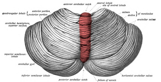
The cerebellar vermis is located in the medial, cortico-nuclear zone of the cerebellum, which is in the posterior fossa of the cranium. The primary fissure in the vermis curves ventrolaterally to the superior surface of the cerebellum, dividing it into anterior and posterior lobes. Functionally, the vermis is associated with bodily posture and locomotion. The vermis is included within the spinocerebellum and receives somatic sensory input from the head and proximal body parts via ascending spinal pathways.

<span class="mw-page-title-main">Basket cell</span>

Basket cells are inhibitory GABAergic interneurons of the brain, found throughout different regions of the cortex and cerebellum.

<span class="mw-page-title-main">Climbing fiber</span> Series of neuronal projections from the inferior olivary nucleus located in the medulla oblongata

Climbing fibers are the name given to a series of neuronal projections from the inferior olivary nucleus located in the medulla oblongata.

<span class="mw-page-title-main">Purkinje cell</span> Specialized neuron in the cerebellum

Purkinje cells, or Purkinje neurons, are a class of GABAergic inhibitory neurons located in the cerebellum. They are named after their discoverer, Czech anatomist Jan Evangelista Purkyně, who characterized the cells in 1839.

<span class="mw-page-title-main">Deep cerebellar nuclei</span>

There are four deep cerebellar nuclei embedded in the white matter of the medullary centre. The nuclei are the fastigial, globose, emboliform, and dentate nuclei.

<span class="mw-page-title-main">Dentate nucleus</span> Nucleus in the centre of each cerebellar hemisphere

The dentate nucleus is a cluster of neurons, or nerve cells, in the central nervous system that has a dentate – tooth-like or serrated – edge. It is located within the deep white matter of each cerebellar hemisphere, and it is the largest single structure linking the cerebellum to the rest of the brain. It is the largest and most lateral, or farthest from the midline, of the four pairs of deep cerebellar nuclei, the others being the globose and emboliform nuclei, which together are referred to as the interposed nucleus, and the fastigial nucleus. The dentate nucleus is responsible for the planning, initiation and control of voluntary movements. The dorsal region of the dentate nucleus contains output channels involved in motor function, which is the movement of skeletal muscle, while the ventral region contains output channels involved in nonmotor function, such as conscious thought and visuospatial function.

<span class="mw-page-title-main">Globose nucleus</span> Part of the interposed nucleus, a structure in the cerebellum.

The globose nucleus is one of the deep cerebellar nuclei. It is located medial to the emboliform nucleus, and lateral to the fastigial nucleus. The globose nucleus and emboliform nucleus are known collectively as the interposed nuclei.

<span class="mw-page-title-main">Fastigial nucleus</span> Grey matter nucleus in the cerebellum

The fastigial nucleus is located in the cerebellum. It is one of the four deep cerebellar nuclei, and is grey matter embedded in the white matter of the cerebellum.

Dysmetria is a lack of coordination of movement typified by the undershoot or overshoot of intended position with the hand, arm, leg, or eye. It is a type of ataxia. It can also include an inability to judge distance or scale.

<span class="mw-page-title-main">Flocculus</span>

The flocculus is a small lobe of the cerebellum at the posterior border of the middle cerebellar peduncle anterior to the biventer lobule. Like other parts of the cerebellum, the flocculus is involved in motor control. It is an essential part of the vestibulo-ocular reflex, and aids in the learning of basic motor skills in the brain.

Cerebellar ataxia is a form of ataxia originating in the cerebellum. Non-progressive congenital ataxia (NPCA) is a classical presentation of cerebral ataxias.

<span class="mw-page-title-main">Mossy fiber (cerebellum)</span> Major input to cerebellum

Mossy fibers are one of the major inputs to cerebellum. There are many sources of this pathway, the largest of which is the cerebral cortex, which sends input to the cerebellum via the pontocerebellar pathway. Other contributors include the vestibular nerve and nuclei, the spinal cord, the reticular formation, and feedback from deep cerebellar nuclei. Axons enter the cerebellum via the middle and inferior cerebellar peduncles, where some branch to make contact with deep cerebellar nuclei. They ascend into the white matter of the cerebellum, where each axon branches to innervate granule cells in several cerebellar folia.

The cerebellothalamic tract or the tractus cerebellothalamicus, is part of the superior cerebellar peduncle. It originates in the cerebellar nuclei, crosses completely in the decussation of the superior cerebellar peduncle, bypasses the red nucleus, and terminates in posterior division of ventral lateral nucleus of thalamus. The ventrolateral nucleus has different divisions and distinct connections, mostly with frontal and parietal lobes. The primary motor cortex and premotor cortex get information from the ventrolateral nucleus projections originating in the interposed nucleus and dentate nuclei. Other dentate nucleus projections via thalamic pathway transmit information to prefrontal cortex and posterior parietal cortex. The cerebellum sends thalamocortical projections and in addition may also send connections from the thalamus to association areas serving cognitive and affective functions.

<span class="mw-page-title-main">Anatomy of the cerebellum</span> Structures in the cerebellum, a part of the brain

The anatomy of the cerebellum can be viewed at three levels. At the level of gross anatomy, the cerebellum consists of a tightly folded and crumpled layer of cortex, with white matter underneath, several deep nuclei embedded in the white matter, and a fluid-filled ventricle in the middle. At the intermediate level, the cerebellum and its auxiliary structures can be broken down into several hundred or thousand independently functioning modules or compartments known as microzones. At the microscopic level, each module consists of the same small set of neuronal elements, laid out with a highly stereotyped geometry.

<span class="mw-page-title-main">Granule cell</span> Type of neuron with a very small cell body

The name granule cell has been used for a number of different types of neurons whose only common feature is that they all have very small cell bodies. Granule cells are found within the granular layer of the cerebellum, the dentate gyrus of the hippocampus, the superficial layer of the dorsal cochlear nucleus, the olfactory bulb, and the cerebral cortex.

<span class="mw-page-title-main">Rhombic lip</span> Posterior section of the developing metencephalon

The rhombic lip is a posterior section of the developing metencephalon which can be recognized transiently within the vertebrate embryo. It extends posteriorly from the roof of the fourth ventricle to dorsal neuroepithelial cells. The rhombic lip can be divided into eight structural units based on rhombomeres 1-8 (r1-r8), which can be recognized at early stages of hindbrain development. Producing granule cells and five brainstem nuclei, the rhombic lip plays an important role in developing a complex cerebellar neural system.

Cerebellar cognitive affective syndrome (CCAS), also called Schmahmann's syndrome is a condition that follows from lesions (damage) to the cerebellum of the brain. It refers to a constellation of deficits in the cognitive domains of executive function, spatial cognition, language, and affect resulting from damage to the cerebellum. Impairments of executive function include problems with planning, set-shifting, abstract reasoning, verbal fluency, and working memory, and there is often perseveration, distractibility and inattention. Language problems include dysprosodia, agrammatism and mild anomia. Deficits in spatial cognition produce visual–spatial disorganization and impaired visual–spatial memory. Personality changes manifest as blunting of affect or disinhibited and inappropriate behavior. These cognitive impairments result in an overall lowering of intellectual function. CCAS challenges the traditional view of the cerebellum being responsible solely for regulation of motor functions. It is now thought that the cerebellum is responsible for monitoring both motor and nonmotor functions. The nonmotor deficits described in CCAS are believed to be caused by dysfunction in cerebellar connections to the cerebral cortex and limbic system.

<span class="mw-page-title-main">AGTPBP1 (gene)</span> Human protein-coding gene

ATP/GTP binding protein 1 is gene that encodes the protein known as cytosolic carboxypeptidase 1 (CCP1), originally named NNA1. Mice with a naturally occurring mutation of the Agtpbp1 gene are known as pcd mice.

References

  1. Bracha, Vlastislav (September 1, 2009). "The cerebellum and eyeblink conditioning: learning vs. network performance hypotheses". Neuroscience. 3 (162): 787–96. doi:10.1016/j.neuroscience.2008.12.042. PMC   2822538 . PMID   19162131.
  2. Kjell, Katarina (December 3, 2018). "A Longer Interstimulus Interval Yields Better Learning in Adults and Young Adolescents". Front Behav Neurosci. 12: 299. doi: 10.3389/fnbeh.2018.00299 . PMC   6286956 . PMID   30559655.
  3. Takehara, K (2016). "The Anatomy and Physiology of Eyeblink Classical Conditioning". The Anatomy and Physiology of Eyeblink Classical Conditioning in: Behavioral Neuroscience of Learning and Memory. Current Topics in Behavioral Neurosciences. Vol. 37. Springer, Cham. pp. 297–323. doi:10.1007/7854_2016_455. ISBN   978-3-319-78755-8. PMID   28025812.
  4. Takehara, K., Kawahara, S., & Kirino, Y. (2003). Time-dependent reorganization of the brain components underlying memory retention in trace eyeblink conditioning. J. Neurosci., 23:9896–9905.
  5. Squire, L. R., Stark, C. E. L., & R. E. Clark (2004). The medial temporal lobe. Annu. Rev. Neurosci.27:279–306.
  6. Bracha, Vlastislav (September 1, 2009). "The cerebellum and eyeblink conditioning: learning vs. network performance hypotheses". Neuroscience. 3 (162): 787–96. doi:10.1016/j.neuroscience.2008.12.042. PMC   2822538 . PMID   19162131.
  7. McCormick DA, Thompson RF (1984) Cerebellum: essential involvement in the classically conditioned eyelid response. Science 223: 296-299.
  8. Hesslow G, Ivarsson M (1994) Suppression of cerebellar Purkinje cells during conditioned responses in ferrets. NeuroReport 5:649-652.
  9. Jirenhed D-A, Bengtsson F, & Hesslow G (2007). Acquisition, Extinction, and Reacquisition of a Cerebellar Cortical Memory Trace. Journal of Neuroscience 27: 2493-2502
  10. Bengtsson, F. and Hesslow, G. (2006). Cerebellar Control of the Inferior Olive. Cerebellum 5: 7-14
  11. Rasmussen, A, Jirenhed, D.-A, Hesslow, G. (2008). Simple and Complex Spike Firing Patterns in Purkinje cells During Classical Conditioning. Cerebellum. 7:563-566