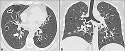
CT scan of the lung showing bullae in the lower lung lobes of a subject with type alpha-1-antitrypsin deficiency. There is also increased lung density in areas with compression of lung tissue by the bullae.
A focal lung pneumatosis is an enclosed pocket of air or gas in the lung and includes blebs, bullae, pulmonary cysts, and lung cavities. Blebs and bullae can be classified by their wall thickness.[1]
A bleb has a wall thickness of less than 1 mm.[2] By radiology definition, it is up to 1 cm in total size.[3] By pathology definition, it originates in the pleurae (rather than in the lung parenchyma).[4]
A bulla has a wall thickness of less than 1 mm.[2] By radiology definition, it has a total size of greater than 1 cm.[3] By pathology definition, it originates in the lung parenchyma (rather than in the pleurae).[4]
A lung cyst has a wall thickness of up to 4 mm.[2] A minimum wall thickness of 1 mm has been suggested,[2] but thin-walled pockets may be included in the definition as well.[5]
A cavity has a wall thickness of more than 4 mm.[2]
The terms above, when referring to sites other than the lungs, often imply fluid content.
Lung cysts are seen in about 8% of the general population, with an increased prevalence in older people, and are not associated with emphysema.[5] They may be part of the aging changes of the lungs, and cause a slight decrease in their diffusing capacity.[5] The presence of multiple pulmonary cysts may indicate a need to evaluate the possibility of bullous or cystic lung diseases.[5] Cavitation indicates workup for serious infection or lung cancer.
A pulmonary cyst is not necessarily the same type of cyst seen in many cystic lung diseases. The cyst for example in pneumocystis pneumonia is not the same as the pulmonary cyst.[citation needed]
Lung metastases rarely cause multiple cystic lung lesions. This form of presentation has been described in metastatic sarcomas.[7]
Incidental blebs and cysts
A focal lung pneumatosis that is an incidental imaging finding such as on a CT scan, without suspicious findings (such as findings indicating any of the diseases listed above), generally does not indicate further follow-up.[8]
1 2 Hansell, DM.; Bankier, AA.; MacMahon, H.; McLoud, TC.; Müller, NL.; Remy, J. (March 2008). "Fleischner Society: glossary of terms for thoracic imaging". Radiology. 246 (3): 697–722. doi:10.1148/radiol.2462070712. PMID18195376.
1 2 Katzenstein (2016). Diagnostic atlas of non-neoplastic lung disease: a practical guide for surgical pathologists. New York, NY: Demos Medical Publishing, LLC/Springer Publishing Company. ISBN978-1-61705-229-3. OCLC951217791.
This page is based on this Wikipedia article Text is available under the CC BY-SA 4.0 license; additional terms may apply. Images, videos and audio are available under their respective licenses.