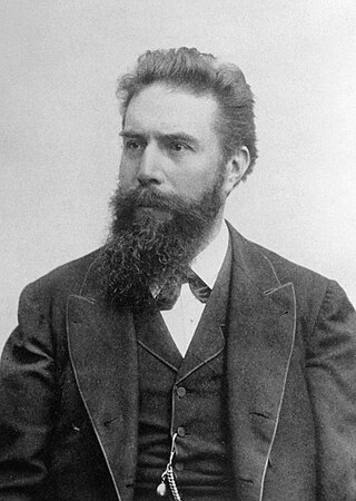
Wilhelm Conrad Röntgen was a German mechanical engineer and physicist, who, on 8 November 1895, produced and detected electromagnetic radiation in a wavelength range known as X-rays or Röntgen rays, an achievement that earned him the inaugural Nobel Prize in Physics in 1901. In honour of Röntgen's accomplishments, in 2004 the International Union of Pure and Applied Chemistry (IUPAC) named element 111, roentgenium, a radioactive element with multiple unstable isotopes, after him. The unit of measurement roentgen was also named after him.
Digital Imaging and Communications in Medicine (DICOM) is the standard for the communication and management of medical imaging information and related data. DICOM is most commonly used for storing and transmitting medical images enabling the integration of medical imaging devices such as scanners, servers, workstations, printers, network hardware, and picture archiving and communication systems (PACS) from multiple manufacturers. It has been widely adopted by hospitals and is making inroads into smaller applications such as dentists' and doctors' offices.
The General Electric Company (GEC) was a major British industrial conglomerate involved in consumer and defence electronics, communications, and engineering. The company was founded in 1886, was Britain's largest private employer with over 250,000 employees in the 1980s, and at its peak in the 1990s, made profits of over £1 billion a year.

Radiography is an imaging technique using X-rays, gamma rays, or similar ionizing radiation and non-ionizing radiation to view the internal form of an object. Applications of radiography include medical radiography and industrial radiography. Similar techniques are used in airport security. To create an image in conventional radiography, a beam of X-rays is produced by an X-ray generator and is projected toward the object. A certain amount of the X-rays or other radiation is absorbed by the object, dependent on the object's density and structural composition. The X-rays that pass through the object are captured behind the object by a detector. The generation of flat two dimensional images by this technique is called projectional radiography. In computed tomography an X-ray source and its associated detectors rotate around the subject which itself moves through the conical X-ray beam produced. Any given point within the subject is crossed from many directions by many different beams at different times. Information regarding attenuation of these beams is collated and subjected to computation to generate two dimensional images in three planes which can be further processed to produce a three dimensional image.

Medical imaging is the technique and process of imaging the interior of a body for clinical analysis and medical intervention, as well as visual representation of the function of some organs or tissues (physiology). Medical imaging seeks to reveal internal structures hidden by the skin and bones, as well as to diagnose and treat disease. Medical imaging also establishes a database of normal anatomy and physiology to make it possible to identify abnormalities. Although imaging of removed organs and tissues can be performed for medical reasons, such procedures are usually considered part of pathology instead of medical imaging.

An X-ray generator is a device that produces X-rays. Together with an X-ray detector, it is commonly used in a variety of applications including medicine, X-ray fluorescence, electronic assembly inspection, and measurement of material thickness in manufacturing operations. In medical applications, X-ray generators are used by radiographers to acquire x-ray images of the internal structures of living organisms, and also in sterilization.
GE HealthCare Technologies Inc., trading as GE HealthCare, is a global medical technology company headquartered in Chicago, Illinois. It was spun-off from GE on January 4, 2023, with GE retaining 20%. As of 2017, it is a manufacturer and distributor of diagnostic imaging agents and radiopharmaceuticals for imaging modalities used in medical imaging procedures. It offers dyes used in magnetic-resonance-imaging procedures; manufactures medical diagnostic equipment, including CT image machines; MRI, XRAY; Ultrasound; Cath Labs; Mammogram; Nuclear Medicine Cameras; and develops Health technology for medical imaging and information technologies, medical diagnostics, patient monitoring systems, disease research, drug discovery, and biopharmaceutical manufacturing. It was incorporated in 1994 and operates in more than 100 countries.
Technicare, formerly known as Ohio Nuclear, made CT, DR and MRI scanners and other medical imaging equipment. Its headquarters was in Solon, Ohio. Originally an independent company which became publicly traded, it was later purchased by Johnson & Johnson. At the time, Invacare was also owned by Technicare. A Harvard Business Case was written about the challenges that precipitated the transition. The company did not do well under Johnson & Johnson and in 1986, under economic pressure following unrelated losses from two Tylenol product tampering cases, J&J folded the company, selling the intellectual property and profitable service business to General Electric, a competitor.
Siemens Healthineers is a German medical device company. It is the parent company for several medical technology companies and is headquartered in Erlangen, Germany. The company dates its early beginnings in 1847 to a small family business in Berlin, co-founded by Werner von Siemens. Siemens Healthineers is 75% owned by Siemens. The name Siemens Medical Solutions was adopted in 2001, and the change to Siemens Healthcare was made in 2008. In 2015, Siemens named Bernd Montag as its new global CEO. In May 2016, the business operations of Siemens Healthcare were rebranded "Siemens Healthineers."

Atkinson Morley Hospital (AMH) was located at Copse Hill near Wimbledon, South-West London, England from 1869 until 2003. Initially a convalescent hospital, it became one of the most advanced brain surgery centres in the world, and was involved in the development of the CT scanner. Following its closure, neuroscience services were relocated to the new Atkinson Morley Wing of St George's Hospital, Tooting.

Positron emission tomography–computed tomography is a nuclear medicine technique which combines, in a single gantry, a positron emission tomography (PET) scanner and an x-ray computed tomography (CT) scanner, to acquire sequential images from both devices in the same session, which are combined into a single superposed (co-registered) image. Thus, functional imaging obtained by PET, which depicts the spatial distribution of metabolic or biochemical activity in the body can be more precisely aligned or correlated with anatomic imaging obtained by CT scanning. Two- and three-dimensional image reconstruction may be rendered as a function of a common software and control system.
Physician self-referral is a term describing the practice of a physician ordering tests on a patient that are performed by either the referring physician himself or a fellow faculty member from whom he receives financial compensation in return for the referral. Examples of self-referral include an internist performing an EKG, a surgeon suggesting an operation that he himself would perform, and a physician ordering imaging tests that would be done at a facility he owns or leases.
Pacemaker failure is the inability of an implanted artificial pacemaker to perform its intended function of regulating the beating of the heart. A pacemaker uses electrical impulses delivered by electrodes in order to contract the heart muscles. Failure of a pacemaker is defined by the requirement of repeat surgical pacemaker-related procedures after the initial implantation. Most implanted pacemakers are dual chambered and have two leads, causing the implantation time to take longer because of this more complicated pacemaker system. These factors can contribute to an increased rate of complications which can lead to pacemaker failure.

The physics of magnetic resonance imaging (MRI) concerns fundamental physical considerations of MRI techniques and technological aspects of MRI devices. MRI is a medical imaging technique mostly used in radiology and nuclear medicine in order to investigate the anatomy and physiology of the body, and to detect pathologies including tumors, inflammation, neurological conditions such as stroke, disorders of muscles and joints, and abnormalities in the heart and blood vessels among others. Contrast agents may be injected intravenously or into a joint to enhance the image and facilitate diagnosis. Unlike CT and X-ray, MRI uses no ionizing radiation and is, therefore, a safe procedure suitable for diagnosis in children and repeated runs. Patients with specific non-ferromagnetic metal implants, cochlear implants, and cardiac pacemakers nowadays may also have an MRI in spite of effects of the strong magnetic fields. This does not apply on older devices, and details for medical professionals are provided by the device's manufacturer.
A shim is a device used to adjust the homogeneity of a magnetic field. Shims received their name from the purely mechanical shims used to adjust position and parallelity of the pole faces of an electromagnet. Coils used to adjust the homogeneity of a magnetic field by changing the current flowing through it were called "electrical current shims" because of their similar function.
Paul Bottomley pioneered the development of magnetic resonance imaging (MRI) leading to modern commercial clinical 1.5 Tesla MRI scanners and techniques for localized magnetic resonance spectroscopy (MRS). Currently, he is Russel H. Morgan Professor of Radiology and Director of the Division of MR Research at Johns Hopkins University, with about 200 peer-reviewed journal articles, over 50 U.S patents and is a Founder and past member of the Board of Directors of ClearPoint Neuro Inc, formerly known as MRI Interventions Inc and SurgiVision Inc, a Johns Hopkins start-up.

In radiography, focal plane tomography is tomography by simultaneously moving the X-ray generator and X-ray detector so as to keep a consistent exposure of only the plane of interest during image acquisition. This was the main method of obtaining tomographs in medical imaging until the late-1970s. It has since been largely replaced by more advanced imaging techniques such as CT and MRI. It remains in use today in a few specialized applications, such as for acquiring orthopantomographs of the jaw in dental radiography.

In a medical facility, such as a hospital or clinic, a gantry holds radiation detectors and/or a radiation source used to diagnose or treat a patient's illness. Radiation sources may produce gamma radiation, x-rays, electromagnetic radiation, or magnetic fields depending on the purpose of the device.

Magnetic resonance imaging (MRI) is in general a safe technique, although injuries may occur as a result of failed safety procedures or human error. During the last 150 years, thousands of papers focusing on the effects or side effects of magnetic or radiofrequency fields have been published. They can be categorized as incidental and physiological. Contraindications to MRI include most cochlear implants and cardiac pacemakers, shrapnel and metallic foreign bodies in the eyes. The safety of MRI during the first trimester of pregnancy is uncertain, but it may be preferable to other options. Since MRI does not use any ionizing radiation, its use generally is favored in preference to CT when either modality could yield the same information.
The history of X-ray computed tomography dates back to at least 1917 with the mathematical theory of the Radon transform In October 1963, William H. Oldendorf received a U.S. patent for a "radiant energy apparatus for investigating selected areas of interior objects obscured by dense material". The first clinical CT scan was performed in 1971 using a scanner invented by Sir Godfrey Hounsfield.