Hamiltosporidium

Last updated

Hamiltosporidium
Daphnia magna with Hamiltosporidium tvaerminensis.JPG
Daphnia magna infected with Hamiltosporidium tvaerminnensis
Scientific classification
Kingdom:
Division:
Order:
Family:
Genus:
Hamiltosporidium

Haag, 2010
Species

Hamiltosporidium tvaerminnensis
Hamiltosporidium magnivora

Contents

Hamiltosporidium is a genus of Microsporidia, which are intracellular and unicellular parasites. [1] The genus, proposed by Haag et al. in 2010, contains two species; Hamiltosporidium tvaerminnensis, and Hamiltosporidium magnivora. [2] Both species infect only the crustacean Daphnia magna (Waterflea).

D. magna and H. tvaerminnensis are a frequently used model organism to study coevolution and local adaptation. [3] [4]

Classification

In 2010 these two species were moved to the new genus Hamiltosporidium. [2] Previously H. tvaerminnensis was wrongly identified as Octosporea bayeri Jírovec, 1936, a parasite of D. magna described in Czech Republic which was never found again. H. magnivora was formerly named Flabelliforma magnivora Larsson, 1998 [5] and belonged to the Flabelliforma genus, before its close phylogenetic relation to H. tvaerminnensis was discovered.

Distribution

Infection of Daphnia populations by Hamiltosporidium have been recorded in the United Kingdom, Russia, Belgium and Israel (H. magnivora), [6] as well as Sweden, Finland and Israel (H. tvaerminnensis). [7] [8] While its host D. magna is found all over the northern hemisphere, the two Hamiltosporidium species seem to have a limited geographic distribution. Genetic differences in host susceptibility have been suggested to contribute to the geographic distribution of H. tvaerminnensis. [8]

Morphology and life cycle

Mostly pyriform spores of Hamiltosporidium tvaerminnensis Hamiltosporidium tvaerminnensis spores2.JPG
Mostly pyriform spores of Hamiltosporidium tvaerminnensis
Spores of Hamiltosporidium magnivora Hamiltosporidium magnivora.JPG
Spores of Hamiltosporidium magnivora

H. magnivora reproduces sexually, while H. tvaerminnensis has an obligatory asexual status. [9] All stages of vegetative reproduction (merogony) are enclosed by a thick plasma membrane, which is in direct contact with the cytoplasma of the host cell. In all stages the nuclei is isolated and clearly visible. The onset of the sporogony is the production of a sporophorous vesicle that is connected to the plasma membrane by tubules. Then fabelliform budding of mostly eight sporoblasts into the vesicle is initiated. Initially the wall of the encysted spore (sporont) has two layers. During the maturating of the sporont two generations of tubules are present in the episporontal space, being reduced at maturity. Mature spores of the genus Hamiltosporidium are polymorph, though pyriform and elongated rod-like spores dominate in H. tvaerminnensis [2] while H. magnivora has lightly pyriform spores only. [5] Pyriform spores measure 4·9–5·6 x 2·2–2·3 μm while elongated spores measure 6·8–12·0 x 1·6–2·1 μm in H. tvaerminnensis. [10] Lightly pyriform spores in H. magnivora measure 2.34-3.03 x 4.07-4.93 μm. [5] The polar filaments in the mature spore are more or less isofilar and the polaroplast has 2 – 3 distinct lamellar regions. [2]

Infection

The unicellular parasites infect the crustacean Daphnia magna. Both species H. magnivora and H. tvaerminnensis infect the fat body, the hypodermis and the ovaries of the crustacean. [5] [10] Infection can be detected by examining crushed animals under a phase contrast microscope (400x). In advanced stages of the infection spores are found in the whole body cavity of host animals, which leads to a clearly visible whitish coloration. The infection leads to reduction in the host fitness by reducing life expectancy, fecundity and competitive ability for resources among D. magna. [11] [12]

Transmission

In H. tvaerminnensis horizontal transmission occurs after the host's death, when spores from decaying cadaver suspend in the water. Spores can persist in the environment, being able to infect new hosts after extended periods of time. [2] Horizontal transmission could not be observed in the lab for H. magnivora but it is suggested to happen in nature as witnessed in H. tvaerminnensis. [6] Both parasite species are vertically transmitted to asexual host offspring with a transmission rate of up to 100% and a somewhat lower transmission rate to sexual offspring (transmission rates have only been reported for H. tvaerminnensis). [13] This means that the parasite is hardly lost in a clone lineage and also survives inside the resting eggs of its host. Thereby it can be kept in a population even though the host is resting while the habitat is dried up or frozen. Because of its combined vertical and horizontal transmission strategies, H. tvaerminnensis can reach prevalence of up to 100% in asexual populations in nature as well as in the laboratory. [12] [14]

Genome

In 2009 a first draft of H. tvaerminnensis genome was sequenced (under the name of Octosporea bayeri). [15] The whole genome has approximately the size of 24.2 Mega-bases (Mb). This stands in contrast to other sequenced microsporidia genomes (for example the compact 2.9 Mb genome of the obligate parasite Encephalitozoon cuniculi [16] ) which are reduced in size. It is suggested that H. tvaerminnensis genome is the largest known microsporidian genome. Almost half of the genome sequence was found to be unique for H. tvaerminnensis. Compared to smaller microsporidian genome, Gene density is rather low in H. tvaerminnensis but variable. [15]

The H. tvaerminnensis population in the Baltic Sea is genetically very homogeneous. So far there were only two main haplotype found in the whole population. Comparison of the genetic diversity to its sister species H. magnivora revealed H. tvaerminnensis asexuality. [9]

Related Research Articles

<i>Daphnia</i> Genus of crustaceans

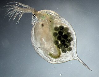
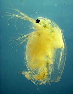
Daphnia is a genus of small planktonic crustaceans, 0.2–6.0 mm (0.01–0.24 in) in length. Daphnia are members of the order Anomopoda, and are one of the several small aquatic crustaceans commonly called water fleas because their saltatory swimming style resembles the movements of fleas. Daphnia spp. live in various aquatic environments ranging from acidic swamps to freshwater lakes and ponds.

Nosema apis is a microsporidian, a small, unicellular parasite recently reclassified as a fungus that mainly affects honey bees. It causes nosemosis, also called nosema, which is the most common and widespread of adult honey bee diseases. The dormant stage of N. apis is a long-lived spore which is resistant to temperature extremes and dehydration, and cannot be killed by freezing the contaminated comb. Nosemosis is a listed disease with the Office International des Epizooties (OIE).

<span class="mw-page-title-main">Microsporidia</span> Phylum of fungi

Microsporidia are a group of spore-forming unicellular parasites. These spores contain an extrusion apparatus that has a coiled polar tube ending in an anchoring disc at the apical part of the spore. They were once considered protozoans or protists, but are now known to be fungi, or a sister group to fungi. These fungal microbes are obligate eukaryotic parasites that use a unique mechanism to infect host cells. They have recently been discovered in a 2017 Cornell study to infect Coleoptera on a large scale. So far, about 1500 of the probably more than one million species are named. Microsporidia are restricted to animal hosts, and all major groups of animals host microsporidia. Most infect insects, but they are also responsible for common diseases of crustaceans and fish. The named species of microsporidia usually infect one host species or a group of closely related taxa. Approximately 10 percent of the species are parasites of vertebrates —several species, most of which are opportunistic, can infect humans, in whom they can cause microsporidiosis.

Microsporidiosis is an opportunistic intestinal infection that causes diarrhea and wasting in immunocompromised individuals. It results from different species of microsporidia, a group of microbial (unicellular) fungi.

<span class="mw-page-title-main">Thelytoky</span> Type of parthenogenesis in which females are produced from unfertilized eggs

Thelytoky is a type of parthenogenesis and is the absence of mating and subsequent production of all female diploid offspring as for example in aphids. Thelytokous parthenogenesis is rare among animals and reported in about 1,500 species, about 1 in 1000 of described animal species, according to a 1984 study. It is more common in invertebrates, like arthropods, but it can occur in vertebrates, including salamanders, fish, and reptiles such as some whiptail lizards.

<span class="mw-page-title-main">Xenoma</span> Growth caused by various species of protists and fungi

A xenoma is a growth caused by various protists and fungi, most notably microsporidia. It can occur on numerous organisms; however is predominantly found on fish.

Drosophila C virus (DCV) belongs to the genus Cripavirus and was previously thought to be a member of the virus family Picornaviridae; it has since been classified as belonging to the Dicistroviridae. It is a single stranded positive sense RNA virus of approximately 9300 nucleotides and it contains two open reading frames. The virus particles are 30 nm in diameter and are made up of approximately 30% of RNA and 70% protein. The virus capsid is composed of three major polypeptides and two minor polypeptides.

Encephalitozoon intestinalis is a parasite. It can cause microsporidiosis.

<i>Encephalitozoon cuniculi</i> Microsporidial pathogen

Encephalitozoon cuniculi is a microsporidial parasite of mammals with world-wide distribution. An important cause of neurologic and renal disease in rabbits, E. cuniculi can also cause disease in immunocompromised people.

<i>Daphnia magna</i> Species of small freshwater animal

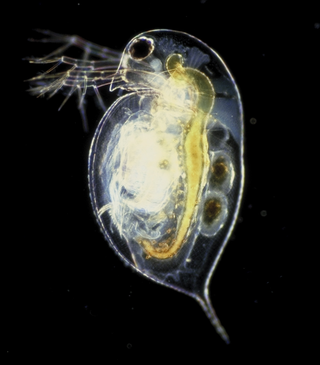
Daphnia magna is a small planktonic crustacean that belongs to the subclass Phyllopoda.

Host–parasite coevolution is a special case of coevolution, where a host and a parasite continually adapt to each other. This can create an evolutionary arms race between them. A more benign possibility is of an evolutionary trade-off between transmission and virulence in the parasite, as if it kills its host too quickly, the parasite will not be able to reproduce either. Another theory, the Red Queen hypothesis, proposes that since both host and parasite have to keep on evolving to keep up with each other, and since sexual reproduction continually creates new combinations of genes, parasitism favours sexual reproduction in the host.

Nosema bombi is a microsporidian, a small, unicellular parasite recently reclassified as a fungus that mainly affects bumble bees. It was reclassified as Vairimorpha bombi in 2020. The parasite infects numerous Bombus spp. at variable rates, and has been found to have a range of deleterious effects on its hosts.

Pasteuria is a genus of mycelial and endospore-forming, nonmotile gram-positive bacteria that are obligate parasites of some nematodes and crustaceans. The genus of Pasteuria was previously classified within the family Alicyclobacillaceae, but has since been moved to the family Pasteuriaceae.

<i>Ordospora colligata</i> Intracellular parasite

Ordospora colligata is an intracellular parasite belonging to the Microsporidia. It is an obligatory gut parasite with the crustacean Daphnia magna as its only host. So far it has been reported from Europe and Asia.

<span class="mw-page-title-main">Pasteuria ramosa</span> Species of bacterium

Pasteuria ramosa is a gram-positive, endospore-forming bacterium in the Bacillus/Clostridia clade within Bacillota. It is an obligate pathogen of cladoceran crustaceans from the genus Daphnia. Daphnia is a genus of small planktonic crustaceans including D. magna, P. ramosa's most popular host target. Other hosts include D. pulex, D. longispina, D. dentifera, and Moina rectirostris. An established and widely used coevolutionary model of host-pathogen interactions exists with P. ramosa and D. magna.

Nematocida parisii is a parasitic species of Microsporidia fungi found in wild isolates of the common nematode, Caenorhabditis elegans. The fungus forms spores and replicates in the intestines before leaving the host.

<span class="mw-page-title-main">Dieter Ebert</span>

Dieter Ebert is professor for Zoology and Evolutionary Biology at the Zoological Institute at the University of Basel in Basel, Switzerland. He is an evolutionary ecologist and geneticist, known for his research on host–pathogen interaction and coevolution, mainly using the model system Daphnia and its parasites.

<i>Enterospora nucleophila</i> Species of parasitic protist

Enterospora nucleophila is a microsporidian infecting the gilt-head bream. It develops primarily within the nuclei of rodlet cells and enterocytes, at the intestinal epithelium. It can also be found in cytoplasmic position within other cell types, including phagocytes, at subepithelial layers. It is the causative agent of emaciative microsporidiosis of gilthead sea bream, a chronic condition manifested as a severe growth arrestment, normally accompanied by trickling mortality.

Hazardia is a genus of microsporidians that parasite insects, with the type host being Culex pipiens. It is currently classified as incertae sedis within the order Amblyosporida of phylum Rozellomycota.

References

  1. "Hamiltosporidium". www.uniprot.org. Retrieved 2016-03-05.
  2. 1 2 3 4 5 Haag, Karen Luisa; Larsson, J. I. Ronny; Refardt, Dominik; Ebert, Dieter (2011-04-01). "Cytological and molecular description of Hamiltosporidium tvaerminnensis gen. et sp. nov., a microsporidian parasite of Daphnia magna, and establishment of Hamiltosporidium magnivora comb. nov" (PDF). Parasitology. 138 (4): 447–462. doi:10.1017/S0031182010001393. hdl: 20.500.11850/33393 . ISSN   1469-8161. PMID   20946698. S2CID   6811605.
  3. Altermatt, F.; Hottinger, F.W. & D. Ebert (2007). "Parasites promote host gene flow in a metapopulation" (PDF). Evolution Ecology. 21 (4): 561–575. doi:10.1007/s10682-006-9136-6. S2CID   4089977.
  4. Zbinden, M.; Haag, C. R.; Ebert, D. (2008-07-01). "Experimental evolution of field populations of Daphnia magna in response to parasite treatment". Journal of Evolutionary Biology. 21 (4): 1068–1078. doi: 10.1111/j.1420-9101.2008.01541.x . ISSN   1420-9101. PMID   18462312.
  5. 1 2 3 4 Larsson J.I.R.; D. Ebert; K.L. Mangin; J. Vavra (1998). "Ultrastructural study and description of Flabelliforma magnivora sp. n. (Microspora: Duboscqiidae), a microsporidian parasite of Daphnia magna (Crustacea: Cladocera: Daphniidae)". Acta Protozoologica. 37: 41–52.
  6. 1 2 Mangin, K. L.; M. Lipsitch & D. Ebert (1995). "Virulence and transmission modes of two microsporidia in Daphnia magna". Parasitology. 111 (2): 133–142. doi:10.1017/s0031182000064878.
  7. Haag, K. L.; Sheikh-Jabbari, E.; Ben-Ami, F.; Ebert, D. (2013-05-01). "Microsatellite and single-nucleotide polymorphisms indicate recurrent transitions to asexuality in a microsporidian parasite". Journal of Evolutionary Biology. 26 (5): 1117–1128. doi: 10.1111/jeb.12125 . ISSN   1420-9101. PMID   23530861.
  8. 1 2 Lange, Benjamin; Kaufmann, Andrea Patricia; Ebert, Dieter (2015-11-01). "Genetic, ecological and geographic covariables explaining host range and specificity of a microsporidian parasite". The Journal of Animal Ecology. 84 (6): 1711–1719. doi: 10.1111/1365-2656.12421 . ISSN   1365-2656. PMID   26147623.
  9. 1 2 Haag, Karen L.; Traunecker, Emmanuel; Ebert, Dieter (2013-01-01). "Single-nucleotide polymorphisms of two closely related microsporidian parasites suggest a clonal population expansion after the last glaciation". Molecular Ecology. 22 (2): 314–326. doi:10.1111/mec.12126. ISSN   1365-294X. PMID   23163569. S2CID   206179470.
  10. 1 2 Vizoso, D. B.; Ebert, D. (2005-07-01). "Phenotypic plasticity of host-parasite interactions in response to the route of infection" (PDF). Journal of Evolutionary Biology. 18 (4): 911–921. doi:10.1111/j.1420-9101.2005.00920.x. ISSN   1010-061X. PMID   16033563. S2CID   12563642.
  11. Ben-Ami, F.; Rigaud, T.; Ebert, D. (2011-06-01). "The expression of virulence during double infections by different parasites with conflicting host exploitation and transmission strategies". Journal of Evolutionary Biology. 24 (6): 1307–1316. doi: 10.1111/j.1420-9101.2011.02264.x . ISSN   1420-9101. PMID   21481055.
  12. 1 2 Bieger, Annette; Ebert, Dieter (2009-05-01). "Expression of parasite virulence at different host population densities under natural conditions" (PDF). Oecologia. 160 (2): 247–255. Bibcode:2009Oecol.160..247B. doi:10.1007/s00442-009-1297-x. ISSN   1432-1939. PMID   19219457. S2CID   2683196.
  13. Vizoso, D. B.; Lass, S.; Ebert, D. (2005-05-01). "Different mechanisms of transmission of the microsporidium Octosporea bayeri: a cocktail of solutions for the problem of parasite permanence" (PDF). Parasitology. 130 (Pt 5): 501–509. doi:10.1017/s0031182004006699. ISSN   0031-1820. PMID   15991493. S2CID   35702689.
  14. Ebert, D. (2005). Ecology, epidemiology and evolution of parasitism in Daphnia. Bethesda (MD): National Library of Medicine (US), National Center for Biotechnology Information.
  15. 1 2 Corradi, Nicolas; Haag, Karen L.; Pombert, Jean-François; Ebert, Dieter; Keeling, Patrick J. (2009-01-01). "Draft genome sequence of the Daphnia pathogen Octosporea bayeri: insights into the gene content of a large microsporidian genome and a model for host-parasite interactions". Genome Biology. 10 (10): R106. doi: 10.1186/gb-2009-10-10-r106 . ISSN   1474-760X. PMC   2784321 . PMID   19807911.
  16. Katinka, Michaël D.; Duprat, Simone; Cornillot, Emmanuel; Méténier, Guy; Thomarat, Fabienne; Prensier, Gérard; Barbe, Valérie; Peyretaillade, Eric; Brottier, Philippe (2001-11-22). "Genome sequence and gene compaction of the eukaryote parasite Encephalitozoon cuniculi". Nature. 414 (6862): 450–453. Bibcode:2001Natur.414..450K. doi: 10.1038/35106579 . ISSN   0028-0836. PMID   11719806.

Further reading