
Arthritis is a general medical term used to describe a disorder that affects joints. Symptoms generally include joint pain and stiffness. Other symptoms may include redness, warmth, swelling, and decreased range of motion of the affected joints. In certain types of arthritis, other organs such as the skin are also affected. Onset can be gradual or sudden.

A finger is a prominent digit on the forelimbs of most tetrapod vertebrate animals, especially those with prehensile extremities such as humans and other primates. Most tetrapods have five digits (pentadactyly), and short digits are typically referred to as toes, while those that are notably elongated are called fingers. In humans, the fingers are flexibly articulated and opposable, serving as an important organ of tactile sensation and fine movements, which are crucial to the dexterity of the hands and the ability to grasp and manipulate objects.
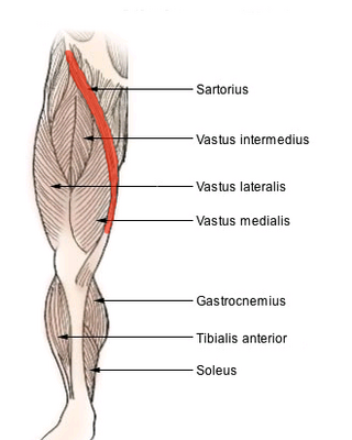
The sartorius muscle is the longest muscle in the human body. It is a long, thin, superficial muscle that runs down the length of the thigh in the anterior compartment.

Osteoarthritis (OA) is a type of degenerative joint disease that results from breakdown of joint cartilage and underlying bone. It is believed to be the fourth leading cause of disability in the world, affecting 1 in 7 adults in the United States alone. The most common symptoms are joint pain and stiffness. Usually the symptoms progress slowly over years. Other symptoms may include joint swelling, decreased range of motion, and, when the back is affected, weakness or numbness of the arms and legs. The most commonly involved joints are the two near the ends of the fingers and the joint at the base of the thumbs, the knee and hip joints, and the joints of the neck and lower back. The symptoms can interfere with work and normal daily activities. Unlike some other types of arthritis, only the joints, not internal organs, are affected.

Psoriatic arthritis (PsA) is a long-term inflammatory arthritis that occurs in people affected by the autoimmune disease psoriasis. The classic features of psoriatic arthritis include dactylitis, skin lesions, and nail lesions. Damage to the nails can include small depressions in the nail (pitting), thickening of the nails, and detachment of the nail from the nailbed. Skin damage consistent with psoriasis frequently occur before the onset of psoriatic arthritis but psoriatic arthritis can precede the rash in 15% of affected individuals. It is classified as a type of seronegative spondyloarthropathy.
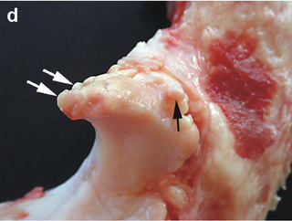
Osteophytes are exostoses that form along joint margins. They should not be confused with enthesophytes, which are bony projections that form at the attachment of a tendon or ligament. Osteophytes are not always distinguished from exostoses in any definite way, although in many cases there are a number of differences. Osteophytes are typically intra-articular.

Bouchard's nodes are hard, bony outgrowths or gelatinous cysts on the proximal interphalangeal joints. They are seen in osteoarthritis, where they are caused by the formation of calcific spurs of the articular (joint) cartilage. Much less commonly, they may be seen in rheumatoid arthritis, where nodes are caused by antibody deposition to the synovium.
Nursing theory is defined as "a creative and conscientious structuring of ideas that project a tentative, purposeful, and systematic view of phenomena". Through systematic inquiry, whether in nursing research or practice, nurses are able to develop knowledge relevant to improving the care of patients. Theory refers to "a coherent group of general propositions used as principles of explanation".
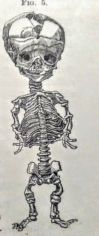
Parrot's sign, refers to at least two medical signs; one relating to a large skull and another to a pupil reaction.
The extensor digitorum muscle is a muscle of the posterior forearm present in humans and other animals. It extends the medial four digits of the hand. Extensor digitorum is innervated by the posterior interosseous nerve, which is a branch of the radial nerve.

Subacute bacterial endocarditis, abbreviated SBE, is a type of endocarditis. Subacute bacterial endocarditis can be considered a form of type III hypersensitivity.

A mallet finger, also known as hammer finger or PLF finger or Hannan finger, is an extensor tendon injury at the farthest away finger joint. This results in the inability to extend the finger tip without pushing it. There is generally pain and bruising at the back side of the farthest away finger joint.

In human anatomy, the abductor digiti minimi is a skeletal muscle situated on the ulnar border of the palm of the hand. It forms the ulnar border of the palm and its spindle-like shape defines the hypothenar eminence of the palm together with the skin, connective tissue, and fat surrounding it. Its main function is to pull the little finger away from the other fingers.

The interphalangeal joints of the hand are the hinge joints between the phalanges of the fingers that provide flexion towards the palm of the hand.

Charles Jacques Bouchard was a French pathologist and an esperantist born in Montier-en-Der, a commune the department of Haute-Marne.

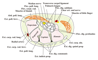
In the human hand, palmar or volar plates are found in the metacarpophalangeal (MCP) and interphalangeal (IP) joints, where they reinforce the joint capsules, enhance joint stability, and limit hyperextension. The plates of the MCP and IP joints are structurally and functionally similar, except that in the MCP joints they are interconnected by a deep transverse ligament. In the MCP joints, they also indirectly provide stability to the longitudinal palmar arches of the hand. The volar plate of the thumb MCP joint has a transverse longitudinal rectangular shape, shorter than those in the fingers.
An adjustable bed is a bed which has a multi-hinged lying surface which can be profiled to a number of different positions. Common adjustments include inclining the upper body and raising the lower body independently of each other. Other common features include height adjustment and tilting the bed to raise the upper body or the lower body into the Trendelenburg or reverse Trendelenburg positions.
Eburnation is a degenerative process of bone commonly found in patients with osteoarthritis or non-union of fractures. Friction in the joint causes the reactive conversion of the sub-chondral bone to an ivory-like surface at the site of the cartilage erosion. The word derives from Latin eburneus, which means "of ivory".

A broken toe is a type of bone fracture. Symptoms include pain when the toe is touched near the break point, or compressed along its length. There may be bruising, swelling, stiffness, or displacement of the broken bone ends from their normal position.
Acquired hand deformity refers to the structural or functional abnormalities that develop in the hand. There are multiple varying causes of acquired hand deformity, triggering significant consequences and complications. Trauma, including blunt force, penetrating injuries, burns, and sports-related incidents, is a primary cause of acquired hand deformities. Inflammatory conditions such as rheumatoid arthritis, gouty arthritis, and systemic lupus erythematosus can also contribute to hand deformities by affecting the joints. Degenerative arthritis, specifically osteoarthritis, functions to evoke impaired hand function due to the gradual deterioration of cartilage. Neurological disorders like cerebral palsy can result in hand contractures due to increased muscle tone and stiffness. There are different types of acquired hand deformities, each with distinct characteristics and underlying causes, such as boutonnière deformity, Dupuytren's contracture, gamekeeper's thumb, hand osteoarthritis deformity, mallet finger, swan-neck deformity, ulnar claw hand, among many others.
