Histopathology of colorectal adenocarcinoma

Last updated
Relative incidence of colorectal cancer types. The vast majority of colorectal cancers are adenocarcinomas. Relative incidence of colorectal cancers.svg
Relative incidence of colorectal cancer types. The vast majority of colorectal cancers are adenocarcinomas.

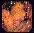
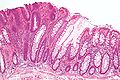
The histopathology of colorectal cancer of the adenocarcinoma type involves analysis of tissue taken from a biopsy or surgery. A pathology report contains a description of the microscopical characteristics of the tumor tissue, including both tumor cells and how the tumor invades into healthy tissues and finally if the tumor appears to be completely removed. The most common form of colon cancer is adenocarcinoma, constituting between 95% [2] and 98% [3] of all cases of colorectal cancer. Other, rarer types include lymphoma, adenosquamous and squamous cell carcinoma. Some subtypes have been found to be more aggressive. [4]

Contents

Macroscopy

Cancers on the right side of the large intestine (ascending colon and cecum) tend to be exophytic, that is, the tumor grows outwards from one location in the bowel wall. This very rarely causes obstruction of feces, and presents with symptoms such as anemia. Left-sided tumors tend to be circumferential, and can obstruct the bowel lumen, much like a napkin ring, and results in thinner caliber stools.

Microscopy

Adenocarcinoma is a malignant epithelial tumor, originating from superficial glandular epithelial cells lining the colon and rectum. It invades the wall, infiltrating the muscularis mucosae layer, the submucosa, and then the muscularis propria. Tumor cells describe irregular tubular structures, harboring pluristratification, multiple lumens, reduced stroma ("back to back" aspect). Sometimes, tumor cells are discohesive and secrete mucus, which invades the interstitium producing large pools of mucus. This occurs in mucinous adenocarcinoma, in which cells are poorly differentiated. If the mucus remains inside the tumor cell, it pushes the nucleus at the periphery, this occurs in "signet-ring cell." Depending on glandular architecture, cellular pleomorphism, and mucosecretion of the predominant pattern, adenocarcinoma may present three degrees of differentiation: well, moderately, and poorly differentiated. [5]

Micrographs (H&E stain)

Microscopic criteria

Colorectal adenocarcinoma, not otherwise specified Colorectal adenocarcinoma, not otherwise specified.jpg
Colorectal adenocarcinoma, not otherwise specified
  • This should be distinguished from cases where piles of well-differentiated mucin-producing cells appear cribriform. In such piles, nuclei show regular polarity with apical mucin, and their nuclei are not markedly enlarged. [6]
  • Varying degrees of gland formation with tall columnar cells
  • Frequently desmoplasia
  • Dirty necrosis, consisting of extensive central necrosis with granular eosinophilic karyorrhectic cell detritus. [6] [7] It is located within the glandular lumina, [7] or often with a garland of cribriform glands in their vicinity. [6]

Subtyping

Pie chart of relative rates of histopathologic subtypes of colorectal carcinoma. Pie chart of colorectal carcinoma etiologies.svg
Pie chart of relative rates of histopathologic subtypes of colorectal carcinoma.

Determining the specific histopathologic subtype of colorectal adenocardinoma is not as important as its staging (see #Staging section below), and about half cases do not have any specific subtype. Still, it is customary to specify it where applicable.

Differential diagnosis

Colorectal adenocarcinoma is distinguished from a colorectal adenoma (mainly tubular and ⁄or villous adenomas) mainly by invasion through the muscularis mucosae. [10]

In carcinoma in situ (Tis), cancer cells invade into the lamina propria, and may involve but not penetrating the muscularis mucosae. This can be classified as an adenoma with "high-grade dysplasia", because prognosis and management are essentially the same. [11]

Grading

Conventional adenocarcinoma may be graded as follows [12]

Gland forming volume or surface
>95%50-95%<50%
Well differentiatedModerately differentiatedLow differentiated
Low gradeHigh grade

Staging

The T stages of bowel cancer. Diagram showing T stages of bowel cancer CRUK 276.svg
The T stages of bowel cancer.

Staging is typically made according to the TNM staging system from the WHO organization, the UICC and the AJCC. The Astler-Coller classification (1954) and the Dukes classification (1932) are now less used. T stands for tumor stage and ranges from 0, no evidence of primary tumor, to T4 when the tumor penetrates the surface of the peritoneum or directly invades other organs or structures. The N stage reflects the number of metastatic lymph nodes and ranges from 0 (no lymph node metastasis) to 2 (four or more lymph node metastasis), and the M stage gives information about distant metastasis (M0 stands for no distant metastasis, and M1 for the presence of distant metastasis). A clinical classification (cTNM) is done at diagnosis and is based on MRI and CT, and a pathological TNM (pTNM) classification is performed after surgery. [13]

The most common metastasis sites for colorectal cancer are the liver, the lung and the peritoneum. [14]

Tumor budding

Tumor budding in colorectal cancer is loosely defined by the presence of individual cells and small clusters of tumor cells at the invasive front of carcinomas. It has been postulated to represent an epithelial–mesenchymal transition (EMT). Tumor budding is a well-established independent marker of a potentially poor outcome in colorectal carcinoma that may allow for dividing people into risk categories more meaningful than those defined by TNM staging, and also potentially guide treatment decisions, especially in T1 and T3 N0 (Stage II, Dukes’ B) colorectal carcinoma. Unfortunately, its universal acceptance as a reportable factor has been held back by a lack of definitional uniformity with respect to both qualitative and quantitative aspects of tumor budding. [15]

Immunohistochemistry

In cases where a metastasis from colorectal cancer is suspected, immunohistochemistry is used to ascertain correct diagnosis. [16] Some proteins are more specifically expressed in colorectal cancer and can be used as diagnostic markers such as CK20 and MUC2. [16] Immunohistochemistry can also be used to screen for Lynch syndrome, a genetic disorder with increased risk of colorectal and other cancers. The diagnosis of Lynch syndrome is made by looking for specific genetic mutations in genes MLH1, MSH2, MSH6, and PMS2. [17] Immunohistochemical testing can also be used to guide treatment and assist in determining the prognosis. Certain markers isolated from the tumor can indicate specific cancer types or susceptibility to different treatments. [18]

Related Research Articles

<span class="mw-page-title-main">Thyroid neoplasm</span> Medical condition

Thyroid neoplasm is a neoplasm or tumor of the thyroid. It can be a benign tumor such as thyroid adenoma, or it can be a malignant neoplasm, such as papillary, follicular, medullary or anaplastic thyroid cancer. Most patients are 25 to 65 years of age when first diagnosed; women are more affected than men. The estimated number of new cases of thyroid cancer in the United States in 2010 is 44,670 compared to only 1,690 deaths. Of all thyroid nodules discovered, only about 5 percent are cancerous, and under 3 percent of those result in fatalities.

<span class="mw-page-title-main">Adenocarcinoma</span> Medical condition

Adenocarcinoma is a type of cancerous tumor that can occur in several parts of the body. It is defined as neoplasia of epithelial tissue that has glandular origin, glandular characteristics, or both. Adenocarcinomas are part of the larger grouping of carcinomas, but are also sometimes called by more precise terms omitting the word, where these exist. Thus invasive ductal carcinoma, the most common form of breast cancer, is adenocarcinoma but does not use the term in its name—however, esophageal adenocarcinoma does to distinguish it from the other common type of esophageal cancer, esophageal squamous cell carcinoma. Several of the most common forms of cancer are adenocarcinomas, and the various sorts of adenocarcinoma vary greatly in all their aspects, so that few useful generalizations can be made about them.

<span class="mw-page-title-main">Carcinoma</span> Malignancy that develops from epithelial cells

Carcinoma is a malignancy that develops from epithelial cells. Specifically, a carcinoma is a cancer that begins in a tissue that lines the inner or outer surfaces of the body, and that arises from cells originating in the endodermal, mesodermal or ectodermal germ layer during embryogenesis.

<span class="mw-page-title-main">Adenoma</span> Type of benign tumor

An adenoma is a benign tumor of epithelial tissue with glandular origin, glandular characteristics, or both. Adenomas can grow from many glandular organs, including the adrenal glands, pituitary gland, thyroid, prostate, and others. Some adenomas grow from epithelial tissue in nonglandular areas but express glandular tissue structure. Although adenomas are benign, they should be treated as pre-cancerous. Over time adenomas may transform to become malignant, at which point they are called adenocarcinomas. Most adenomas do not transform. However, even though benign, they have the potential to cause serious health complications by compressing other structures and by producing large amounts of hormones in an unregulated, non-feedback-dependent manner. Some adenomas are too small to be seen macroscopically but can still cause clinical symptoms.

<span class="mw-page-title-main">Pseudomyxoma peritonei</span> Medical condition

Pseudomyxoma peritonei (PMP) is a clinical condition caused by cancerous cells that produce abundant mucin or gelatinous ascites. The tumors cause fibrosis of tissues and impede digestion or organ function, and if left untreated, the tumors and mucin they produce will fill the abdominal cavity. This will result in compression of organs and will destroy the function of the colon, small intestine, stomach, or other organs. Prognosis with treatment in many cases is optimistic, but the disease is lethal if untreated, with death occurring via cachexia, bowel obstruction, or other types of complications.

An oncocytoma is a tumor made up of oncocytes, epithelial cells characterized by an excessive amount of mitochondria, resulting in an abundant acidophilic, granular cytoplasm. The cells and the tumor that they compose are often benign but sometimes may be premalignant or malignant.

The International Classification of Diseases for Oncology (ICD-O) is a domain-specific extension of the International Statistical Classification of Diseases and Related Health Problems for tumor diseases. This classification is widely used by cancer registries.

<span class="mw-page-title-main">Hürthle cell</span> Medical condition

A Hürthle cell is a cell in the thyroid that is often associated with Hashimoto's thyroiditis as well as benign and malignant tumors. This version is a relatively rare form of differentiated thyroid cancer, accounting for only 3-10% of all differentiated thyroid cancers. Oncocytes in the thyroid are often called Hürthle cells. Although the terms oncocyte, oxyphilic cell, and Hürthle cell are used interchangeably, Hürthle cell is used only to indicate cells of thyroid follicular origin.

<span class="mw-page-title-main">Gleason grading system</span> Score given to a prostate cancer based on its microscopic appearance

The Gleason grading system is used to help evaluate the prognosis of men with prostate cancer using samples from a prostate biopsy. Together with other parameters, it is incorporated into a strategy of prostate cancer staging which predicts prognosis and helps guide therapy. A Gleason score is given to prostate cancer based upon its microscopic appearance. Cancers with a higher Gleason score are more aggressive and have a worse prognosis. Pathological scores range from 2 to 10, with higher numbers indicating greater risks and higher mortality. The system is widely accepted and used for clinical decision making even as it is recognised that certain biomarkers, like ACP1 expression, might yield higher predictive value for future disease course.

<span class="mw-page-title-main">Papillary thyroid cancer</span> Medical condition

Papillary thyroid cancer is the most common type of thyroid cancer, representing 75 percent to 85 percent of all thyroid cancer cases. It occurs more frequently in women and presents in the 20–55 year age group. It is also the predominant cancer type in children with thyroid cancer, and in patients with thyroid cancer who have had previous radiation to the head and neck. It is often well-differentiated, slow-growing, and localized, although it can metastasize.

<span class="mw-page-title-main">Colorectal polyp</span> Growth found in bowel wall

A colorectal polyp is a polyp occurring on the lining of the colon or rectum. Untreated colorectal polyps can develop into colorectal cancer.

<span class="mw-page-title-main">Signet ring cell</span> Cell with a large vacuole

In histology, a signet ring cell is a cell with a large vacuole. The malignant type is seen predominantly in carcinomas. Signet ring cells are most frequently associated with stomach cancer, but can arise from any number of tissues including the prostate, bladder, gallbladder, breast, colon, ovarian stroma and testis.

<span class="mw-page-title-main">Signet ring cell carcinoma</span> Medical condition

Signet ring cell carcinoma (SRCC) is a rare form of highly malignant adenocarcinoma that produces mucin. It is an epithelial malignancy characterized by the histologic appearance of signet ring cells.

<span class="mw-page-title-main">Sebaceous carcinoma</span> Medical condition

Sebaceous carcinoma, also known as sebaceous gland carcinoma (SGc), sebaceous cell carcinoma, and meibomian gland carcinoma is an uncommon malignant cutaneous tumor. Most are typically about 1.4 cm at presentation. SGc originates from sebaceous glands in the skin and, therefore, may originate anywhere in the body where these glands are found. SGc can be divided into 2 types: periocular and extraocular. The periocular region is rich in sebaceous glands making it a common site of origin. The cause of these lesions in the vast majority of cases is unknown. Occasional cases may be associated with Muir-Torre syndrome. SGc accounts for approximately 0.7% of all skin cancers, and the incidence of SGc is highest in Caucasian, Asian, and Indian populations. Due to the rarity of this tumor and variability in clinical and histological presentation, SGc is often misdiagnosed as an inflammatory condition or a more common neoplasm. SGc is commonly treated with wide local excision or Mohs micrographic surgery, and the relative survival rates at 5 and 10 years are 92.72 and 86.98%, respectively.

<span class="mw-page-title-main">Hyperplastic polyp</span> Type of colorectal polyp

A hyperplastic polyp is a type of colorectal polyp.

Colon cancer staging is an estimate of the amount of penetration of a particular cancer. It is performed for diagnostic and research purposes, and to determine the best method of treatment. The systems for staging colorectal cancers depend on the extent of local invasion, the degree of lymph node involvement and whether there is distant metastasis.

<span class="mw-page-title-main">Ceruminous adenocarcinoma</span> Medical condition

Ceruminous adenocarcinoma is a malignant neoplasm derived from ceruminous glands of the external auditory canal. This tumor is rare, with several names used in the past. Synonyms have included cylindroma, ceruminoma, ceruminous adenocarcinoma, not otherwise specified (NOS), ceruminous adenoid cystic carcinoma (ACC), and ceruminous mucoepidermoid carcinoma.

<span class="mw-page-title-main">Colorectal adenoma</span> Medical condition

The colorectal adenoma is a benign glandular tumor of the colon and the rectum. It is a precursor lesion of the colorectal adenocarcinoma. They often manifest as colorectal polyps.

A histopathologic diagnosis of prostate cancer is the discernment of whether there is a cancer in the prostate, as well as specifying any subdiagnosis of prostate cancer if possible. The histopathologic subdiagnosis of prostate cancer has implications for the possibility and methodology of any subsequent Gleason scoring. The most common histopathological subdiagnosis of prostate cancer is acinar adenocarcinoma, constituting 93% of prostate cancers. The most common form of acinar adenocarcinoma, in turn, is "adenocarcinoma, not otherwise specified", also termed conventional, or usual acinar adenocarcinoma.

<span class="mw-page-title-main">Invasive cribriform carcinoma of the breast</span> Medical condition

Invasive cribriform carcinoma of the breast (ICCB), also termed invasive cribriform carcinoma, is a rare type of breast cancer that accounts for 0.3% to 0.6% of all carcinomas in the breast. It originates in a lactiferous duct as opposed to the lobules that form the alveoli in the breasts' mammary glands. ICCB was first described by Dixon and colleagues in 1983 as a tumor that on microscopic histopathological inspection had a cribriform pattern, i.e. a tissue pattern consisting of numerous "Swiss cheese"-like open spaces and/or sieve-like small holes. The latest edition (2019) of the World Health Organization (2019) termed these lesions invasive cribriform carcinomas indicating that by definition they must have a component that invades out of their ducts of origin into adjacent tissues. In situ ductal cancers that have a cribriform histopathology are regarded as belonging to the group of ductal carcinoma in situ tumors.

References

  1. Kang, Hakjung; O’Connell, Jessica B.; Leonardi, Michael J.; Maggard, Melinda A.; McGory, Marcia L.; Ko, Clifford Y. (2006). "Rare tumors of the colon and rectum: a national review". International Journal of Colorectal Disease. 22 (2): 183–189. doi:10.1007/s00384-006-0145-2. ISSN   0179-1958. PMID   16845516. S2CID   34693873.
  2. "Colon, Rectosigmoid, and Rectum Equivalent Terms and Definitions C180-C189, C199, C209, (Excludes lymphoma and leukemia M9590 – M9992 and Kaposi sarcoma M9140) - Colon Solid Tumor Rules 2018. July 2019 Update" (PDF). National Cancer Institute .
  3. "Colorectal cancer types". Cancer Treatment Centers of America . Retrieved 2020-01-16.
  4. Di Como, Joseph A. (October 2015). "Adenosquamous carcinoma of the colon and rectum: a population based clinical outcomes study involving 578 patients from the Surveillance Epidemiology and End Result (SEER) database (1973–2010)". Journal of the American College of Surgeons. 221 (4): 56. doi:10.1016/j.jamcollsurg.2015.08.044.
  5. "Moderately differentiated adenocarcinoma (colon)". pathologyatlas.ro. Archived from the original on January 26, 2016.
  6. 1 2 3 4 5 6 Robert V Rouse. "Adenocarcinoma of the Colon and Rectum". Stanford University School of Medicine. Original posting/updates: 1/31/10, 7/15/11, 11/12/11
  7. 1 2 Li, Lianhuang; Jiang, Weizhong; Yang, Yinghong; Chen, Zhifen; Feng, Changyin; Li, Hongsheng; Guan, Guoxian; Chen, Jianxin (2014). "Identification of dirty necrosis in colorectal carcinoma based on multiphoton microscopy". Journal of Biomedical Optics. 19 (6): 066008. Bibcode:2014JBO....19f6008L. doi: 10.1117/1.JBO.19.6.066008 . ISSN   1083-3668. PMID   24967914. S2CID   8053157.
  8. Remo, Andrea; Fassan, Matteo; Vanoli, Alessandro; Bonetti, Luca Reggiani; Barresi, Valeria; Tatangelo, Fabiana; Gafà, Roberta; Giordano, Guido; Pancione, Massimo; Grillo, Federica; Mastracci, Luca (2019). "Morphology and Molecular Features of Rare Colorectal Carcinoma Histotypes". Cancers. 11 (7): 1036. doi: 10.3390/cancers11071036 . ISSN   2072-6694. PMC   6678907 . PMID   31340478.
  9. 1 2 3 4 Initially copied from: Remo, Andrea; Fassan, Matteo; Vanoli, Alessandro; Bonetti, Luca Reggiani; Barresi, Valeria; Tatangelo, Fabiana; Gafà, Roberta; Giordano, Guido; Pancione, Massimo; Grillo, Federica; Mastracci, Luca (2019). "Morphology and Molecular Features of Rare Colorectal Carcinoma Histotypes". Cancers. 11 (7): 1036. doi: 10.3390/cancers11071036 . ISSN   2072-6694. PMC   6678907 . PMID   31340478. Attribution 4.0 International (CC BY 4.0) license
  10. Robert V Rouse. "Colorectal Adenoma Containing Invasive Adenocarcinoma". Stanford University School of Medicine.
  11. Finlay A Macrae. "Overview of colon polyps". UpToDate. This topic last updated: Dec 10, 2018.
  12. Fleming M, Ravula S, Tatishchev SF, Wang HL (September 2012). "Colorectal carcinoma: Pathologic aspects". J Gastrointest Oncol. 3 (3): 153–73. doi:10.3978/j.issn.2078-6891.2012.030. PMC   3418538 . PMID   22943008.
  13. "Colon and Rectum Cancer Staging, American Joint Committee on Cancer, 7th Edition" (PDF). Retrieved 27 Feb 2019.
  14. "Metastatic Cancer". National Cancer Institute . February 6, 2017. Archived from the original on April 4, 2017. Retrieved April 5, 2017.
  15. Mitrovic B, Schaeffer DF, Riddell RH, Kirsch R (October 2012). "Tumor budding in colorectal carcinoma: time to take notice". Modern Pathology. 25 (10): 1315–1325. doi: 10.1038/modpathol.2012.94 . PMID   22790014.
  16. 1 2 Taliano RJ, LeGolvan M, Resnick MB (February 2013). "Immunohistochemistry of colorectal carcinoma: current practice and evolving applications". Human Pathology. 44 (2): 151–163. doi:10.1016/j.humpath.2012.04.017. PMID   22939578.
  17. Bui QM, Lin D, Ho W (February 2017). "Approach to Lynch Syndrome for the Gastroenterologist". Digestive Diseases and Sciences. 62 (2): 299–304. doi:10.1007/s10620-016-4346-4. PMID   27990589. S2CID   32833106.
  18. Vakiani E (December 2017). "Molecular Testing of Colorectal Cancer in the Modern Era: What Are We Doing and Why?". Surgical Pathology Clinics. 10 (4): 1009–1020. doi:10.1016/j.path.2017.07.013. PMID   29103530.