Related Research Articles

Neurosurgery or neurological surgery, known in common parlance as brain surgery, is the medical specialty concerned with the surgical treatment of disorders which affect any portion of the nervous system including the brain, spinal cord and peripheral nervous system.
Lars Leksell (1907–1986) was a Swedish physician and Professor of Neurosurgery at the Karolinska Institute in Stockholm, Sweden. He was the inventor of radiosurgery.

Radiosurgery is surgery using radiation, that is, the destruction of precisely selected areas of tissue using ionizing radiation rather than excision with a blade. Like other forms of radiation therapy, it is usually used to treat cancer. Radiosurgery was originally defined by the Swedish neurosurgeon Lars Leksell as "a single high dose fraction of radiation, stereotactically directed to an intracranial region of interest".

Minimally invasive procedures encompass surgical techniques that limit the size of incisions needed, thereby reducing wound healing time, associated pain, and risk of infection. Surgery by definition is invasive and many operations requiring incisions of some size are referred to as open surgery. Incisions made during open surgery can sometimes leave large wounds that may be painful and take a long time to heal. Advancements in medical technologies have enabled the development and regular use of minimally invasive procedures. For example, endovascular aneurysm repair, a minimally invasive surgery, has become the most common method of repairing abdominal aortic aneurysms in the US as of 2003. The procedure involves much smaller incisions than the corresponding open surgery procedure of open aortic surgery.

Stereotactic surgery is a minimally invasive form of surgical intervention that makes use of a three-dimensional coordinate system to locate small targets inside the body and to perform on them some action such as ablation, biopsy, lesion, injection, stimulation, implantation, radiosurgery (SRS), etc.
Thalamotomy is a surgical procedure in which a functional lesion is made into the thalamus to improve the overall brain function in patients. First introduced in the 1950s, it is primarily effective for tremors such as those associated with Parkinson's disease, where a selected portion of the thalamus is surgically destroyed (ablated). Neurosurgeons use specialized equipment to precisely locate an area of the thalamus, usually choosing to work on only one side. Bilateral procedures are poorly tolerated because of increased complications and risk, including vision and speech problems. The positive effects on tremors are immediate. Other less destructive procedures are sometimes preferred, such as subthalamic deep brain stimulation, since this procedure can also improve tremors and other symptoms of PD.

Neuroimaging is the use of quantitative (computational) techniques to study the structure and function of the central nervous system, developed as an objective way of scientifically studying the healthy human brain in a non-invasive manner. Increasingly it is also being used for quantitative research studies of brain disease and psychiatric illness. Neuroimaging is highly multidisciplinary involving neuroscience, computer science, psychology and statistics, and is not a medical specialty. Neuroimaging is sometimes confused with neuroradiology.
A fiducial marker or fiducial is an object placed in the field of view of an imaging system that appears in the image produced, for use as a point of reference or a measure. It may be either something placed into or on the imaging subject, or a mark or set of marks in the reticle of an optical instrument.
NeuroArm is an engineering research surgical robot specifically designed for neurosurgery. It is the first image-guided, MR-compatible surgical robot that has the capability to perform both microsurgery and stereotaxy.
Image-guided radiation therapy is the process of frequent imaging, during a course of radiation treatment, used to direct the treatment, position the patient, and compare to the pre-therapy imaging from the treatment plan. Immediately prior to, or during, a treatment fraction, the patient is localized in the treatment room in the same position as planned from the reference imaging dataset. An example of IGRT would include comparison of a cone beam computed tomography (CBCT) dataset, acquired on the treatment machine, with the computed tomography (CT) dataset from planning. IGRT would also include matching planar kilovoltage (kV) radiographs or megavoltage (MV) images with digital reconstructed radiographs (DRRs) from the planning CT.
Neuronavigation is the set of computer-assisted technologies used by neurosurgeons to guide or "navigate” within the confines of the skull or vertebral column during surgery, and used by psychiatrists to accurately target rTMS. The set of hardware for these purposes is referred to as a neuronavigator.
The Anne Arundel Medical Center (AAMC) is a regional health system headquartered in Annapolis, Maryland. In addition to the main campus in Annapolis, the group has outpatient pavilions in Bowie, Kent Island, Odenton, Easton and Waugh Chapel.
Patient registration is used to correlate the reference position of a virtual 3D dataset gathered by computer medical imaging with the reference position of the patient. This procedure is crucial in computer assisted surgery, in order to insure the reproducitibility of the preoperative registration and the clinical situation during surgery. The use of the term "patient registration" out of this context can lead to a confusion with the procedure of registering a patient into the files of a medical institution.
Computer-assisted surgery (CAS) represents a surgical concept and set of methods, that use computer technology for surgical planning, and for guiding or performing surgical interventions. CAS is also known as computer-aided surgery, computer-assisted intervention, image-guided surgery, digital surgery and surgical navigation, but these are terms that are more or less synonymous with CAS. CAS has been a leading factor in the development of robotic surgery.

Surgical planning is the preoperative method of pre-visualising a surgical intervention, in order to predefine the surgical steps and furthermore the bone segment navigation in the context of computer-assisted surgery. The surgical planning is most important in neurosurgery and oral and maxillofacial surgery. The transfer of the surgical planning to the patient is generally made using a medical navigation system.

Russell A. Brown, an American physician and computer scientist, is the inventor of the N-localizer technology that enables guidance of stereotactic surgery or radiosurgery using medical images that are obtained via computed tomography (CT), magnetic resonance imaging (MRI), or positron emission tomography (PET).
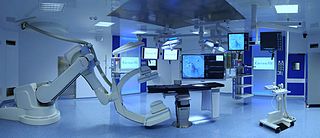
A hybrid operating room is a surgical theatre that is equipped with advanced medical imaging devices such as fixed C-Arms, X-ray computed tomography (CT) scanners or magnetic resonance imaging (MRI) scanners. These imaging devices enable minimally-invasive surgery. Minimally-invasive surgery is intended to be less traumatic for the patient and minimize incisions on the patient and perform surgery procedure through one or several small cuts.

Ferenc Andras Jolesz was a Hungarian-American physician and scientist best known for his research on image guided therapy, the process by which information derived from diagnostic imaging is used to improve the localization and targeting of diseased tissue to monitor and control treatment during surgical and interventional procedures. He pioneered the field of Magnetic Resonance Imaging-guided interventions and introduced of a variety of new medical procedures based on novel combinations of imaging and therapy delivery.
Augmented reality-assisted surgery (ARAS) is a surgical tool utilizing technology that superimposes a computer-generated image on a surgeon's view of the operative field, thus providing a composite view for the surgeon of the patient with a computer generated overlay enhancing the operative experience. It can be used for training, preparation for an operation, or performance of an operation. ARAS can be performed using a wide array of technology, including an optical head-mounted display (OHMD)—such as the Google Glass XE 22.1 or Vuzix STAR 1200 XL—and a digital overlay from robotic and laparoscopic surgery feeds. The technique has been primarily been tested in the urological and cardiovascular domains.
Bone malrotation refers to the situation that results when a bone heals out of rotational alignment from another bone, or part of bone. It often occurs as the result of a surgical complication after a fracture where intramedullary nailing (IMN) occurs, especially in the femur and tibial bones, but can also occur genetically at birth. The severity of this complication is often neglected due to its complexity to detect and treat, yet if left untreated, bone malrotation can significantly impact regular bodily functioning, and even lead to severe arthritis. Detection throughout history has become more advanced and accurate, ranging from clinical assessment to ultrasounds to CT scans. Treatment can include an osteotomy, a major surgical procedure where bones are cut and realigned correctly, or compensatory methods, where individuals learn to externally or internally rotate their limb to compensate for the rotation. Further research is currently being examined in this area to reduce occurrences of malrotation, including detailed computer navigation to improve visual accuracy during surgery.
References
- ↑ "ClarifEye". Philips.
- ↑ "Surgical Navigation Technology Based on Augmented Reality and Integrated 3D Intraoperative Imaging: A Spine Cadaveric Feasibility and Accuracy Study".
- ↑ "10 Augmented Reality Surgery Companies". 24 October 2019.
- ↑ "Surgery and Treatment -". Dedicated Computing. Retrieved 2018-03-14.
- ↑ Grace J, Wang Y, Robinson D, Tahuil C, Xu R (2018). Retrospective Analysis: Collateral nerve damage and local tissue trauma associated with endovenous laser ablation therapy. Ultrasound Guided Endovenous Laser Ablation Union International de Phlebology World Congress. Melbourne Australia.
- 1 2 Mezger U, Jendrewski C, Bartels M (April 2013). "Navigation in surgery". Langenbeck's Archives of Surgery. 398 (4): 501–14. doi:10.1007/s00423-013-1059-4. PMC 3627858 . PMID 23430289.
- 1 2 3 4 5 6 7 8 Abedin-Nasab, Mohammad (2019), Handbook of Robotic and Image-Guided Surgery (1 ed.), Elsevier, ISBN 9780128142455
- ↑ "Image-Guided Surgery". care.american-rhinologic.org. Retrieved 2018-03-14.
- ↑ Jakubovic R, Guha D, Gupta S, Lu M, Jivraj J, Standish BA, et al. (October 2018). "High Speed, High Density Intraoperative 3D Optical Topographical Imaging with Efficient Registration to MRI and CT for Craniospinal Surgical Navigation". Scientific Reports. 8 (1): 14894. Bibcode:2018NatSR...814894J. doi:10.1038/s41598-018-32424-z. PMC 6173775 . PMID 30291261.
- ↑ Kelly PJ (January 2000). "Stereotactic surgery: what is past is prologue". Neurosurgery. 46 (1): 16–27. doi: 10.1093/neurosurgery/46.1.16 . PMID 10626931.
- ↑ Galloway, RL Jr. (2015). "Introduction and Historical Perspectives on Image-Guided Surgery". In Golby, AJ (ed.). Image-Guided Neurosurgery. Amsterdam: Elsevier. pp. 2–4. doi:10.1016/B978-0-12-800870-6.00001-7. ISBN 978-0-12-800870-6.
- ↑ Sturm V, Pastyr O, Schlegel W, Scharfenberg H, Zabel HJ, Netzeband G, Schabbert S, Berberich W (1983). "Stereotactic computer tomography with a modified Riechert-Mundinger device as the basis for integrated stereotactic neuroradiological investigations". Acta Neurochirurgica. 68 (1–2): 11–17. doi:10.1007/BF01406197. PMID 6344559. S2CID 38864553.