Digital painting
Ploem started painting as a small boy and was educated in drawing and painting in Maastricht, the Netherlands. While still at secondary school he attended an evening course in drawing and painting at the Kunstnijverheidsschool Maastricht. Ploem's presence in Paris was important for his knowledge and interest in art since he could regularly visit his cousins in Paris, the painter Frits Klein and his son Yves Klein. He visited the Kleins when Yves was making his first monochromes.[ citation needed ]
In the last years of his activities at the faculty of medicine at Leiden University, he concentrated on research in image analysis. He was asked to participate in a European project with the aim of automating cancer cell recognition using computer analysis. It concerned a collaborative project with the German optical company Leitz/Leica Microsystems, and the Institute for Mathematical Morphology in Fontainebleau, France. Together with a team, Professor Jean Serra at this institute had developed an image analysis method, now internationally known as mathematical morphology. With his experience as an analogue painter, Ploem saw the possibility of also applying the methods of mathematical morphology to the creation of digital art.
At the International Symposium on Mathematical Morphology in Amsterdam (1998), Ploem presented a paper on the creation of computer graphics with Mathematical Morphology, using for the first time, the transforming algorithms from the Fontainebleau group for the creation of digital art. He wrote about it in the chapter of a book ( ISBN 978-0-7923-5133-7) published on that occasion. [15]
His first digital graphics of nature scenes were shown in his exposition at a regional art centre in the Pyrenees (Ossega, June 1997).[ citation needed ]
He was invited for a symposium on Art et Science at the University of Caen, France (April 2001). At the art exposition connected with this symposium, he presented 6 digital graphics that were dominated by chaotic transformations of rock art themes. A similar invitation was made by the University of Basel in Switzerland (April 2002).[ citation needed ]
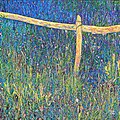 Meadow near Sauto, Pyrenees, France
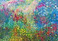 In the valley of the Eyne, Pyrenees
 David and Goliath (multiple image transformations of a romanesque painting in the Sant Climent church in Taull)


