Laminoplasty

Last updated
Laminoplasty
Laminoplastie HWK 6 - CT axial 001 - cropped Annotation.jpg
CT scan of Laminoplasty of cervical vertebra
ICD-9-CM 03.09

Laminoplasty is an orthopaedic/neurosurgical surgical procedure for treating spinal stenosis by relieving pressure on the spinal cord. The main purpose of this procedure is to provide relief to patients who may have symptoms of numbness, pain, or weakness in arm movement. The procedure involves cutting the lamina on both sides of the affected vertebrae (cutting through on one side and merely cutting a groove on the other) and then "swinging" the freed flap of bone open thus relieving the pressure on the spinal cord. The spinous process may be removed to allow the lamina bone flap to be swung open. The bone flap is then propped open using small wedges or pieces of bone such that the enlarged spinal canal will remain in place.[ citation needed ]

Contents

This technique contrasts with vertebral laminectomy in the amount of bone and muscle tissue that has to be removed, displaced, or dissected in the procedure.

History

Laminoplasty is a surgical procedure that has been developed as an alternative to cervical laminectomy, which is used to treat cervical myelopathy. Laminoplasty reconstructs the vertebral lamina to decompress the spinal cord. The term laminoplasty means, "to create a hinge to lift the lamina." [1]

To treat myelopathy and ossified posterior longitudinal ligament (OPLL), there are two approaches that can expand the spinal canal. These approaches are the anterior approach which is a direct removal of the cord compressing lesion, or a posterior approach which is an indirect decompression of the spinal cord. Laminectomy was one of the main methods for the posterior approach, however, the creation of laminoplasty was able to avoid several problems associated with the laminectomy procedure. Some risks of the laminectomy procedure include postoperative segmental instability, kyphosis, perineural adhesions, and late neurological deterioration. [1]

The laminoplasty procedure was created by Japanese orthopedic surgeons during the 1970s to 1980s. Over the years, laminoplasty has evolved its technique. The first laminoplasty technique developed was from modifying the Miyazaki and Kirita's technique for laminectomy. [1] This method was described by Oyama as Z-shaped laminoplasty. The name was given due to the z-shape formed when cutting the laminae. The next method is called, en bloc laminoplasty, and it was a modification of the en bloc laminectomy, which was developed by Tsuji. En bloc laminoplasty decompresses the spine by making the laminae act as a flap, and this flap hovered over the cord without sutures or bone grafts. Later in 1977, Hirabayashi and his colleagues introduced the open-door laminoplasty, which was inspired by the en bloc laminoplasty. This method uses sutures on the facet capsule to leave the flap open. After this method, Kurokawa and his team developed the double door laminoplasty. This procedure involves cutting the laminae midline, and hinges are made bilaterally.[ citation needed ]

Not only are there many other methods of laminoplasty being created, these new methods falling under the open door or double door laminoplasty category, but also, other techniques are being developed in order to preserve the cervical muscle attachment on the spinous processes.

Anatomy of the spine

Spinal column curvature-en Spinal column curvature-en.svg
Spinal column curvature-en

The spine is one of the main components of the central nervous system (CNS). This structure's function is to provide the body with support and to protect the spinal cord. The spinal cord serves 3 main functions for the body. It provides sensation, autonomic and motor control for all bodily functions and parts. The spinal cord is the most complex yet organized part of the CNS. The entire spinal structure may be divided into 4 sections that create an overall S-shaped curve. These sections include the cervical, thoracic, lumbar, and sacral regions. Intervertebral discs stacked on top of one another make up the structure of the overall spine. These discs are separate and cushioned in between them, and with age, these discs become brittle and flat. Sensory stimulation is recognized and processed through the spinal cord, these include pain and temperature, touch, and proprioception. Our body's entire neural network sends any of this sensory information to the spinal cord to process. In order to stabilize spinal movement, there are many ligaments throughout the spine to hold the vertebrae and intervertebral discs together. These elements all working together allow for spinal movement and overall bodily stability and support. [2]

Laminoplasty purpose

The main purpose of this procedure is to provide relief to patients who may develop symptoms of numbness, pain, or weakness in arm movement. Patients may also experience difficulty with hand and finger movement, along with balance and walking difficulty. This surgical procedure is also commonly performed in order to remove pressure from the spinal cord in the neck, which may be due to various reasons. These include: tumors, fractures, arthritis, bone spurs, disc herniations, or degenerative problems. [3]

Method

Z-Shaped Laminoplasty - Adapted From R. Kurokawa et al. Z-Shaped Laminoplasty.png
Z-Shaped Laminoplasty - Adapted From R. Kurokawa et al.
Open Door Laminoplasty - Adapted From R. Kurokawa et al. Open Door Laminoplasty.png
Open Door Laminoplasty - Adapted From R. Kurokawa et al.
Double Door Laminoplasty - Adapted From R. Kurokawa et al. Double Door Laminoplasty.png
Double Door Laminoplasty - Adapted From R. Kurokawa et al.

Positioning

In the positioning of the patient, neuromonitoring is performed. The two forms of neuromonitoring implemented are somatosensory evoked potentials (SSEP) and transcranial motor evoked potentials (tcMEP). After monitoring the patient for some time, anesthetic induction and positioning are done. An intubation tube is added to ensure an oral pathway. Individuals with severe myelopathy will need a fiberoptic intubation scope to prevent the risk of extension of the cervical spine during the intubation process. The patient is then placed on a Jackson table with a Mayfield tong. The chest, iliac crests, arms, and knees all have gel padding and mats placed for support. The patient's head is flexed while the neutral alignment of the cervical spine is established. The final step is checking the SSEP and tcMEP signals. [4]

=== Exposure couple centimeters just inferior to the C7 vertebra. To maintain hemostasis, a monopolar electrocautery is used during the incision process. The incision is usually performed in the region of C3 to C7. The paraspinal muscles are then pushed off the laminae to the medial edge. A lateral radiograph is done with a radiopaque marker, which is placed on the dorsal osseous region. This allows the regions of interest to be determined. [4]

Decompression

There are several types of laminoplasty techniques for the decompression of the spinal cord. Depending on the laminoplasty technique, the surgeon may repair the vertebral lamina plane with rigid or semi-rigid fixations. Autograft and allograft bone blocks also may be used, but are dependent on the technique. The four most common techniques are Z-shaped, en bloc, open door, and double door laminoplasty. [4]

Z-shaped laminoplasty

The C2 to C7 laminae are thinned out with a drill. It is important to decompress more than one region of the laminae. A z-shape is cut on the laminae, and the laminae are lifted and connected with a suture. [1]

Open door laminoplasty

The C2 to C7 laminae are drilled at the lateral borders of the laminae. One side of the bone is completely cut, while the other side acts as a hinge. The laminae are then lifted to increase space in the spinal canal. The flap of the laminae is kept open with a suture that is fixed to the facet capsule. [1] [5]

Double door laminoplasty

At the midline of the vertebral laminae, a drill is used to cut the bone. The lateral sides of the laminae serve as a hinge that allows expansion of the spinal canal. Specifically, the inner cortex of the lateral portion was part of the hinge. An artificial spacer is used to keep the opening fixed. One common spacer that is used is hydroxyapatite. [1]

Complications and success rates

Complications of this surgical procedure can include nerve damage to either the nerve roots or the spinal cord, which would result in limb weakness or paralysis respectively. Other complications can include infection, spinal fluid leak, or unsuccessful relief of compression. [6] Upon review of over 60 studies, there has been evidence showing that these complications are greater in patients of older age, while it is less common that factors such as body mass index, smoking status, the duration of symptoms as well as baseline severity score contribute to perioperative complications. These studies have also indicated that neck pain is more prevalent in patients undergoing laminoplasty compared to that of other surgical techniques, whereas C-5 palsy occurs less in laminoplasty. [7] There is also a slower progression rate of OPLL in laminoplasty compared to laminectomy. Laminoplasty has a progression rate of only 45.4%, while 52.5% is associated with laminectomy. Other miscellaneous complications that were reported include hematoma, progressive kyphosis, and incomplete decompression. The probability of these complications range from 5-20% after this procedure. [8]

While the success rate of the laminoplasty procedure is dependent on the underlying condition that causes the need of surgery, the vast majority of patients that undergo this procedure see significant relief of pain and approximately 75% of patients see improvement. [7]

Rehabilitation

It is encouraged and sometimes required that patients partake in rehabilitative therapy after undergoing a laminoplasty in order to regain the strength and flexibility of the operated area. While factors such as the age of patients as well as the duration of symptoms prior to surgery influence recovery time, a study assessing the recovery process in patients showed evidence that the preoperative values of all parameters significantly improved 5 years after the surgery. [9]

Related Research Articles

Neurosurgery Medical specialty of disorders which affect any portion of the nervous system

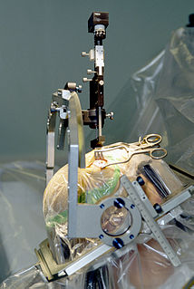
Neurosurgery or neurological surgery, known in common parlance as brain surgery, is the medical specialty concerned with the surgical treatment of disorders which affect any portion of the nervous system including the brain, spinal cord and peripheral nervous system.

Lumbar spinal stenosis Medical condition of the spine

Lumbar spinal stenosis (LSS) is a medical condition in which the spinal canal narrows and compresses the nerves and blood vessels at the level of the lumbar vertebrae. Spinal stenosis may also affect the cervical or thoracic region, in which case it is known as cervical spinal stenosis or thoracic spinal stenosis. Lumbar spinal stenosis can cause pain in the low back or buttocks, abnormal sensations, and the absence of sensation (numbness) in the legs, thighs, feet, or buttocks, or loss of bladder and bowel control.

Laminectomy

A laminectomy is a surgical procedure that removes a portion of a vertebra called the lamina, which is the roof of the spinal canal. It is a major spine operation with residual scar tissue and may result in postlaminectomy syndrome. Depending on the problem, more conservative treatments may be viable.

Spondylosis Degeneration of the vertebral column.

Spondylosis is the degeneration of the vertebral column from any cause. In the more narrow sense it refers to spinal osteoarthritis, the age-related wear and tear of the spinal column, which is the most common cause of spondylosis. The degenerative process in osteoarthritis chiefly affects the vertebral bodies, the neural foramina and the facet joints. If severe, it may cause pressure on the spinal cord or nerve roots with subsequent sensory or motor disturbances, such as pain, paresthesia, imbalance, and muscle weakness in the limbs.

Myelopathy describes any neurologic deficit related to the spinal cord. When due to trauma, it is known as (acute) spinal cord injury. When inflammatory, it is known as myelitis. Disease that is vascular in nature is known as vascular myelopathy. The most common form of myelopathy in humans, cervical spondylotic myelopathy (CSM), is caused by arthritic changes (spondylosis) of the cervical spine, which result in narrowing of the spinal canal ultimately causing compression of the spinal cord. In Asian populations, spinal cord compression often occurs due to a different, inflammatory process affecting the posterior longitudinal ligament.

Degenerative disc disease Medical condition

Degenerative disc disease (DDD) is a medical condition typically brought on by the normal aging process in which there are anatomic changes and possibly a loss of function of one or more intervertebral discs of the spine. DDD can take place with or without symptoms, but is typically identified once symptoms arise. The root cause is thought to be loss of soluble proteins within the fluid contained in the disc with resultant reduction of the oncotic pressure, which in turn causes loss of fluid volume. Normal downward forces cause the affected disc to lose height, and the distance between vertebrae is reduced. The anulus fibrosus, the tough outer layers of a disc, also weakens. This loss of height causes laxity of the longitudinal ligaments, which may allow anterior, posterior, or lateral shifting of the vertebral bodies, causing facet joint malalignment and arthritis; scoliosis; cervical hyperlordosis; thoracic hyperkyphosis; lumbar hyperlordosis; narrowing of the space available for the spinal tract within the vertebra ; or narrowing of the space through which a spinal nerve exits with resultant inflammation and impingement of a spinal nerve, causing a radiculopathy.

Spinal fusion Immobilization or ankylosis of two or more vertebrae by fusion of the vertebral bodies

Spinal fusion, also called spondylodesis or spondylosyndesis, is a neurosurgical or orthopedic surgical technique that joins two or more vertebrae. This procedure can be performed at any level in the spine and prevents any movement between the fused vertebrae. There are many types of spinal fusion and each technique involves using bone grafting—either from the patient (autograft), donor (allograft), or artificial bone substitutes—to help the bones heal together. Additional hardware is often used to hold the bones in place while the graft fuses the two vertebrae together. The placement of hardware can be guided by fluoroscopy, navigation systems, or robotics.

Wobbler disease is a catchall term referring to several possible malformations of the cervical vertebrae that cause an unsteady (wobbly) gait and weakness in dogs and horses. A number of different conditions of the cervical (neck) spinal column cause similar clinical signs. These conditions may include malformation of the vertebrae, intervertebral disc protrusion, and disease of the interspinal ligaments, ligamenta flava, and articular facets of the vertebrae. Wobbler disease is also known as cervical vertebral instability, cervical spondylomyelopathy (CSM), and cervical vertebral malformation (CVM). In dogs, the disease is most common in large breeds, especially Great Danes and Doberman Pinschers. In horses, it is not linked to a particular breed, though it is most often seen in tall, race-bred horses of Thoroughbred or Standardbred ancestry. It is most likely inherited to at least some extent in dogs and horses.

Ligamenta flava Ligaments connecting the laminae of adjacent vertebrae

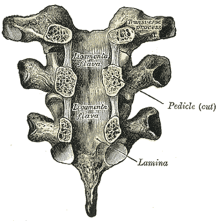
The ligamenta flava are a series of ligaments that connect the ventral parts of the laminae of adjacent vertebrae. They help to preserve upright posture, preventing hyperflexion, and ensuring that the vertebral column straightens after flexion. Hypertrophy can cause spinal stenosis.

Microsurgical lumbar laminoplasty is a minimally invasive technique for decompressing pinched nerves in the lumbar spine. Pinched or compressed nerves may result from herniated discs, lumbar spinal stenosis, or spondylolisthesis.

Spinal disc herniation Injury to the connective tissue between spinal vertebrae

Spinal disc herniation is an injury to the cushioning and connective tissue between vertebrae, usually caused by excessive strain or trauma to the spine. It may result in back pain, pain or sensation in different parts of the body, and physical disability. The most conclusive diagnostic tool for disc herniation is MRI, and treatment may range from painkillers to surgery. Protection from disc herniation is best provided by core strength and an awareness of body mechanics including posture.

Anterior cervical discectomy and fusion

Anterior cervical discectomy and fusion (ACDF) is a surgical procedure to treat nerve root or spinal cord compression by decompressing the spinal cord and nerve roots of the cervical spine with a discectomy, followed by inter-vertebral fusion to stabilize the corresponding vertebrae. This procedure is used when other non-surgical treatments have failed.

Cervical spinal stenosis Medical condition

Cervical spinal stenosis is a bone disease involving the narrowing of the spinal canal at the level of the neck. It is frequently due to chronic degeneration, but may also be congenital. Treatment is frequently surgical.

Neurogenic claudication Medical condition

Neurogenic claudication (NC), also known as pseudoclaudication, is the most common symptom of lumbar spinal stenosis (LSS) and describes intermittent leg pain from impingement of the nerves emanating from the spinal cord. Neurogenic means that the problem originates within the nervous system. Claudication, from the Latin word for to limp, refers to painful cramping or weakness in the legs. NC should therefore be distinguished from vascular claudication, which stems from a circulatory problem rather than a neural one.

Laminotomy

A laminotomy is an orthopaedic neurosurgical procedure that removes part of the lamina of a vertebral arch in order to relieve pressure in the vertebral canal. A laminotomy is less invasive than conventional vertebral column surgery techniques, such as laminectomy because it leaves more ligaments and muscles attached to the vertebral column intact and it requires removing less bone from the vertebra. As a result, laminotomies typically have a faster recovery time and result in fewer postoperative complications. Nevertheless, possible risks can occur during or after the procedure like infection, hematomas, and dural tears. Laminotomies are commonly performed as treatment for lumbar spinal stenosis and herniated disks. MRI and CT scans are often used pre- and post surgery to determine if the procedure was successful.

Spinal decompression

Spinal decompression is the relief of pressure on the spinal cord or on one or more compressed nerve roots passing through or exiting the spinal column. Decompression of the spinal neural elements is a key component in treating spinal radiculopathy, myelopathy and claudication.

Spinal stenosis Disease of the bony spine that results in narrowing of the spinal canal

Spinal stenosis is an abnormal narrowing of the spinal canal or neural foramen that results in pressure on the spinal cord or nerve roots. Symptoms may include pain, numbness, or weakness in the arms or legs. Symptoms are typically gradual in onset and improve with bending forwards. Severe symptoms may include loss of bladder control, loss of bowel control, or sexual dysfunction.

The ventral slot technique is a procedure that allows the surgeon to reach and decompress the spinal cord and associated nerve roots from a ventral route in veterinary medicine. There are also alternative ways to open the spinal canal from dorsal by performing a hemilaminectomy, but this often gives only limited access. Even when the main pathological changes evolve from the midline, it is necessary to choose a ventral approach.

Vertebra Bone in the vertebral column

The spinal column, characteristic of each vertebrate species, is a moderately flexible series of vertebrae, each constituting a characteristic irregular bone whose complex structure is composed primarily of bone, and secondarily of hyaline cartilage. They show variation in the proportion contributed by these two tissue types; such variations correlate on one hand with the cerebral/caudal rank, and on the other with phylogenetic differences among the vertebrate taxa.

Ossification of the posterior longitudinal ligament Medical condition

Ossification of the posterior longitudinal ligament (OPLL) is a process of fibrosis, calcification, and ossification of the posterior longitudinal ligament of the spine, that may involve the spinal dura. Once considered a disorder unique to people of Asian heritage, it is now recognized as an uncommon disorder in a variety of patients with myelopathy.

References

  1. 1 2 3 4 5 6 Hirano, Yoshitaka; Ohara, Yukoh; Mizuno, Junichi; Itoh, Yasunobu (January 2018). "History and Evolution of Laminoplasty". Neurosurgery Clinics of North America. 29 (1): 107–113. doi:10.1016/j.nec.2017.09.019. ISSN   1558-1349. PMID   29173422.
  2. Nógrádi, Antal; Vrbová, Gerta (2013). Anatomy and Physiology of the Spinal Cord. Landes Bioscience.
  3. Bakhsheshian, Joshua; Mehta, Vivek A.; Liu, John C. (2017). "Current Diagnosis and Management of Cervical Spondylotic Myelopathy". Global Spine Journal. 7 (6): 572–586. doi:10.1177/2192568217699208. PMC   5582708 . PMID   28894688.
  4. 1 2 3 Tobert, Daniel (2017). Operative Techniques in Orthopedics. pp. 242–247. ISBN   978-1451193145.
  5. , Jenis L. Cervical laminaplasty. J Med Ins. 2014;2014(6). doi:https://doi.org/10.24296/jomi/6.
  6. Button, Gavin. "Cervical Laminectomy and Laminoplasty Risks and Success Rate in Portland, Oregon | Gavin Button, MD". spineportland.com.
  7. 1 2 Tetreault, Lindsay; Ibrahim, Ahmed; Côté, Pierre; Singh, Anoushka; Fehlings, Michael G. (January 2016). "A systematic review of clinical and surgical predictors of complications following surgery for degenerative cervical myelopathy". Journal of Neurosurgery. Spine. 24 (1): 77–99. doi: 10.3171/2015.3.SPINE14971 . ISSN   1547-5646. PMID   26407090.
  8. Singhatanadgige, Weerasak; Limthongkul, Worawat; Valone, Frank; Yingsakmongkol, Wicharn; Riew, K. Daniel (2016). "Outcomes following Laminoplasty or Laminectomy and Fusion in Patients with Myelopathy Caused by Ossification of the Posterior Longitudinal Ligament: A Systematic Review". Global Spine Journal. 6 (7): 702–709. doi:10.1055/s-0036-1578805. PMC   5077712 . PMID   27781191.
  9. Suzuki, Akinobu; Misawa, Hiromichi; Simogata, Mitsuhiko; Tsutsumimoto, Takahiro; Takaoka, Kunio; Nakamura, Hiroaki (2009-12-15). "Recovery Process Following Cervical Laminoplasty in Patients With Cervical Compression Myelopathy: Prospective Cohort Study". Spine. 34 (26): 2874–9. doi:10.1097/BRS.0b013e3181bb0e33. ISSN   0362-2436. PMID   19949366. S2CID   205513164.