
Shoulder problems including pain, are one of the more common reasons for physician visits for musculoskeletal symptoms. The shoulder is the most movable joint in the body. However, it is an unstable joint because of the range of motion allowed. This instability increases the likelihood of joint injury, often leading to a degenerative process in which tissues break down and no longer function well.

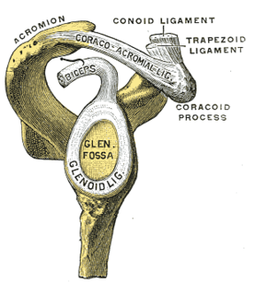
The human shoulder is made up of three bones: the clavicle (collarbone), the scapula, and the humerus as well as associated muscles, ligaments and tendons. The articulations between the bones of the shoulder make up the shoulder joints. The shoulder joint, also known as the glenohumeral joint, is the major joint of the shoulder, but can more broadly include the acromioclavicular joint. In human anatomy, the shoulder joint comprises the part of the body where the humerus attaches to the scapula, and the head sits in the glenoid cavity. The shoulder is the group of structures in the region of the joint.
A joint dislocation, also called luxation, occurs when there is an abnormal separation in the joint, where two or more bones meet. A partial dislocation is referred to as a subluxation. Dislocations are often caused by sudden trauma on the joint like an impact or fall. A joint dislocation can cause damage to the surrounding ligaments, tendons, muscles, and nerves. Dislocations can occur in any joint major or minor. The most common joint dislocation is a shoulder dislocation.
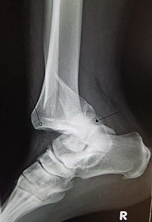
Dysbaric osteonecrosis or DON is a form of avascular necrosis where there is death of a portion of the bone that is thought to be caused by nitrogen embolism in divers. Although the definitive pathologic process is poorly understood, there are several hypotheses:

Avascular necrosis (AVN), also called osteonecrosis or bone infarction, is death of bone tissue due to interruption of the blood supply. Early on there may be no symptoms. Gradually joint pain may develop which may limit the ability to move. Complications may include collapse of the bone or nearby joint surface.

The shoulder joint is structurally classified as a synovial ball and socket joint and functionally as a diarthrosis and multiaxial joint. It involves articulation between the glenoid cavity of the scapula and the head of the humerus.

The shoulder girdle or pectoral girdle is the set of bones in the appendicular skeleton which connects to the arm on each side. In humans it consists of the clavicle and scapula; in those species with three bones in the shoulder, it consists of the clavicle, scapula, and coracoid. Some mammalian species have only the scapula.
Dead arm syndrome starts with repetitive motion and forces on the posterior capsule of the shoulder. The posterior capsule is a band of fibrous tissue that interconnects with tendons of the rotator cuff of the shoulder. Four muscles and their tendons make up the rotator cuff. They cover the outside of the shoulder to hold, protect and move the joint.
A SLAP tear or SLAP lesion is an injury to the glenoid labrum. SLAP is an acronym for "superior labral tear from anterior to posterior".

A dislocated shoulder is when the head of the humerus is out of the shoulder joint. Symptoms include shoulder pain and instability. Complications may include a Bankart lesion, Hill-Sachs lesion, rotator cuff tear, or injury to the axillary nerve.
Shoulder surgery is a means of treating injured shoulders. Many surgeries have been developed to repair the muscles, connective tissue, or damaged joints that can arise from traumatic or overuse injuries to the shoulder.

A Hill–Sachs lesion, or Hill–Sachs fracture, is a cortical depression in the posterolateral head of the humerus. It results from forceful impaction of the humeral head against the anteroinferior glenoid rim when the shoulder is dislocated anteriorly.

An ALPSAlesion is an injury at the front of the shoulder associated with shoulder dislocation.

Perthes lesion is variant of Bankart lesion, presenting as an anterior glenohumeral injury that occurs when the scapular periosteum remains intact but is stripped medially and the anterior labrum is avulsed from the glenoid but remains partially attached to the scapula by intact periosteum.
Humeral avulsion of the glenohumeral ligament (HAGL) is defined as an avulsion of the inferior glenohumeral ligament from the anatomic neck of the humerus. In other words, it occurs when we have disruption of the ligaments that join the humerus to the glenoid. HAGL tends to occur in 7.5-9.3% of cases of anterior shoulder instability. Making it an uncommon cause of anterior shoulder instability. Avulsion of this ligamentous complex may occur in three sites: glenoid insertion (40%), the midsubstance (35%) and the humeral insertion (25%). Bony humeral avulsion of the glenohumeral ligament (BHAGL) refers when we have HAGL with bony fracture.
The Latarjet operation, also known as the Latarjet-Bristow procedure, is a surgical procedure used to treat recurrent shoulder dislocations, typically caused by bone loss or a fracture of the glenoid. The procedure was first described by French surgeon Dr. Michel Latarjet in 1954.
Orthopedic surgery is the branch of surgery concerned with conditions involving the musculoskeletal system. Orthopedic surgeons use both surgical and nonsurgical means to treat musculoskeletal injuries, sports injuries, degenerative diseases, infections, bone tumours, and congenital limb deformities. Trauma surgery and traumatology is a sub-specialty dealing with the operative management of fractures, major trauma and the multiply-injured patient.

