Bankart lesion

Last updated
Bankart lesion
Gray328.png
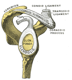
The glenoid labrum, labeled glenoid ligament, is damaged in a Bankart lesion. Lateral view demonstrating the articular surface of the right scapula is shown.
Symptoms Shoulder instability and widespread shoulder discomfort, and catching, locking, or popping feelings in shoulders. [1]
Risk factors Anterior shoulder dislocation and/or repeated anterior shoulder subluxations. [2]
Diagnostic method X-ray and MRI.
Differential diagnosis Anterior labroligamentous periosteal sleeve avulsion, Rotator Cuff Tears, SLAP Lesion, Impingement, Perthes lesion, Glenolabral articular disruption, Humeral avulsion of the glenohumeral ligament. [2]

A Bankart lesion is a type of shoulder injury that occurs following a dislocated shoulder. [3] It is an injury of the anterior (inferior) glenoid labrum of the shoulder. [4] When this happens, a pocket at the front of the glenoid forms that allows the humeral head to dislocate into it. It is an indication for surgery and often accompanied by a Hill-Sachs lesion, damage to the posterior humeral head. [5]

Contents

A bony Bankart is a Bankart lesion that includes a fracture of the anterior-inferior glenoid cavity of the scapula bone. [6]

The Bankart lesion is named after English orthopedic surgeon Arthur Sydney Blundell Bankart (1879–1951). [7]

Signs and symptoms

Bankart lesions are characterized by recurrent shoulder instability and widespread shoulder discomfort. Some individuals may experience catching, locking, or popping feelings in their shoulders. The majority of Bankart lesion patients have primary or recurrent anterior shoulder dislocation. [1]

Diagnosis

The diagnosis is usually initially made by a combination of physical exam and medical imaging, where the latter may be projectional radiography (in cases of bony Bankart) and/or MRI of the shoulder. The presence of intra-articular contrast allows for better evaluation of the glenoid labrum. [8] Type V SLAP tears extends into the Bankart defect. [9]

Treatment

Arthroscopic repair of Bankart injuries have good success rates, though nearly one-third of patients require further surgery for continued instability after the initial procedure in a study of young adults, with higher re-operation rates in those less than 20 years of age. [10] Options for repair include an arthroscopic technique or a more invasive open Latarjet procedure, [11] with the open technique tending to have a lower incidence of recurrent dislocation, but also a reduced range of motion following surgery. [12]

See also

Related Research Articles

<span class="mw-page-title-main">Shoulder problem</span> Medical condition

Shoulder problems including pain, are one of the more common reasons for physician visits for musculoskeletal symptoms. The shoulder is the most movable joint in the body. However, it is an unstable joint because of the range of motion allowed. This instability increases the likelihood of joint injury, often leading to a degenerative process in which tissues break down and no longer function well.

<span class="mw-page-title-main">Shoulder</span> Part of the body

The human shoulder is made up of three bones: the clavicle (collarbone), the scapula, and the humerus as well as associated muscles, ligaments and tendons.

<span class="mw-page-title-main">Joint dislocation</span> Medical injury

A joint dislocation, also called luxation, occurs when there is an abnormal separation in the joint, where two or more bones meet. A partial dislocation is referred to as a subluxation. Dislocations are often caused by sudden trauma on the joint like an impact or fall. A joint dislocation can cause damage to the surrounding ligaments, tendons, muscles, and nerves. Dislocations can occur in any major joint or minor joint. The most common joint dislocation is a shoulder dislocation.

<span class="mw-page-title-main">Rotator cuff tear</span> Medical condition

Rotator cuff tendinopathy is a process of senescence. The pathophysiology is mucoid degeneration. Most people develop rotator cuff tendinopathy within their lifetime.

<span class="mw-page-title-main">Shoulder joint</span> Synovial ball and socket joint in the shoulder

The shoulder joint is structurally classified as a synovial ball-and-socket joint and functionally as a diarthrosis and multiaxial joint. It involves an articulation between the glenoid fossa of the scapula and the head of the humerus. Due to the very loose joint capsule that gives a limited interface of the humerus and scapula, it is the most mobile joint of the human body.

Dead arm syndrome starts with repetitive motion and forces on the posterior capsule of the shoulder. The posterior capsule is a band of fibrous tissue that interconnects with tendons of the rotator cuff of the shoulder. Four muscles and their tendons make up the rotator cuff. They cover the outside of the shoulder to hold, protect and move the joint.

<span class="mw-page-title-main">SLAP tear</span> Medical condition

A SLAP tear or SLAP lesion is an injury to the superior glenoid labrum that initiates in the back of the labrum and stretches toward the front into the attachment point of the long head of the biceps tendon. SLAP is an acronym for "Superior Labrum Anterior and Posterior". SLAP lesions are commonly seen in overhead throwing athletes but middle-aged labor workers can also be affected, and they can be caused by chronic overuse or an acute stretch injury of the shoulder.

<span class="mw-page-title-main">Glenoid labrum</span> Ligament in the shoulder blade

The glenoid labrum is a fibrocartilaginous structure rim attached around the margin of the glenoid cavity in the shoulder blade. The shoulder joint is considered a ball and socket joint. However, in bony terms the 'socket' is quite shallow and small, covering at most only a third of the 'ball'. The socket is deepened by the glenoid labrum, stabilizing the shoulder joint.

<span class="mw-page-title-main">Dislocated shoulder</span> Injury

A dislocated shoulder is a condition in which the head of the humerus is detached from the shoulder joint. Symptoms include shoulder pain and instability. Complications may include a Bankart lesion, Hill-Sachs lesion, rotator cuff tear, or injury to the axillary nerve.

Shoulder surgery is a means of treating injured shoulders. Many surgeries have been developed to repair the muscles, connective tissue, or damaged joints that can arise from traumatic or overuse injuries to the shoulder.

<span class="mw-page-title-main">Hill–Sachs lesion</span> Cortical depression in the posterolateral head of the humerus

A Hill–Sachs lesion, or Hill–Sachs fracture, is a cortical depression in the posterolateral head of the humerus. It results from forceful impaction of the humeral head against the anteroinferior glenoid rim when the shoulder is dislocated anteriorly.

<span class="mw-page-title-main">Shoulder replacement</span>

Shoulder replacement is a surgical procedure in which all or part of the glenohumeral joint is replaced by a prosthetic implant. Such joint replacement surgery generally is conducted to relieve arthritis pain or fix severe physical joint damage.

<span class="mw-page-title-main">ALPSA lesion</span> Type of shoulder injury

An ALPSAlesion is an injury at the front of the shoulder associated with shoulder dislocation.

A Bankart repair is an operation for habitual anterior shoulder dislocation. The joint capsule is sewed to the detached glenoid labrum, without duplication of the subscapularis tendon.

<span class="mw-page-title-main">Hip arthroscopy</span>

Hip arthroscopy refers to the viewing of the interior of the acetabulofemoral (hip) joint through an arthroscope and the treatment of hip pathology through a minimally invasive approach. This technique is sometimes used to help in the treatment of various joint disorders and has gained popularity because of the small incisions used and shorter recovery times when compared with conventional surgical techniques. Hip arthroscopy was not feasible until recently, new technology in both the tools used and the ability to distract the hip joint has led to a recent surge in the ability to do hip arthroscopy and the popularity of it.

<span class="mw-page-title-main">Perthes lesion</span> Medical condition

Perthes lesion is a variant of Bankart lesion, presenting as an anterior glenohumeral injury that occurs when the scapular periosteum remains intact but is stripped medially and the anterior labrum is avulsed from the glenoid but remains partially attached to the scapula by intact periosteum.

Humeral avulsion of the glenohumeral ligament (HAGL) is defined as an avulsion of the inferior glenohumeral ligament from the anatomic neck of the humerus. In other words, it occurs when we have disruption of the ligaments that join the humerus to the glenoid. HAGL tends to occur in 7.5-9.3% of cases of anterior shoulder instability. Making it an uncommon cause of anterior shoulder instability. Avulsion of this ligamentous complex may occur in three sites: glenoid insertion (40%), the midsubstance (35%) and the humeral insertion (25%). Bony humeral avulsion of the glenohumeral ligament (BHAGL) refers when we have HAGL with bony fracture.

The Latarjet operation, also known as the Latarjet-Bristow procedure, is a surgical procedure used to treat recurrent shoulder dislocations, typically caused by bone loss or a fracture of the glenoid. The procedure was first described by French surgeon Dr. Michel Latarjet in 1954.

Bryan L. Reuss is an American orthopaedic surgeon. He received a Bachelor of Arts Degree in Biology with distinction from the University of Kansas in 1996 and graduated with honors with an M.D. from the University of Nebraska College of Medicine in 2000. From 2000 to 2005, Dr. Reuss was an Orthopaedic Surgery Resident Physician in the Orlando Regional Healthcare System. In 2004-2005 he was awarded the "Resident of the Year" for Orlando Regional Healthcare chosen from all the hospital system's Resident Doctors. He completed an Orthopaedic Fellowship at the University of Cincinnati/Wellington Orthopaedics and Sports Medicine Fellowship in 2006. In 2010, Dr. Reuss was awarded the Sports Medicine Person of the Year from the Athletic Trainers Association of Florida.

<span class="mw-page-title-main">Labral reconstruction</span> Medical procedure

Labral reconstruction is a type of hip arthroscopy in which the patient's native labrum is partially or completely removed and reconstructed using either autograft or allograft tissue. Originally described in 2009 using the ligamentum teres capitis, arthroscopic labral reconstruction using a variety of graft tissue has demonstrated promising short and mid-term clinical outcomes. Most importantly, labral reconstruction has demonstrated utility when the patient's native labral tissue is far too damaged for debridement or repair.

References

  1. 1 2 Tupe, Rishikesh N.; Tiwari, Vivek (August 3, 2023). "Anteroinferior Glenoid Labrum Lesion (Bankart Lesion)". StatPearls Publishing. PMID   36508533 . Retrieved October 30, 2023.
  2. 1 2 "Bankart lesion". Physiopedia. Retrieved October 30, 2023.
  3. Major, Nancy M.; Anderson, Mark W. (2020). "10. Shoulder". Musculoskeletal MRI. Philadelphia: Elsevier. pp. 218–219. ISBN   978-0-323-415606.
  4. Widjaja A, Tran A, Bailey M, Proper S (2006). "Correlation between Bankart and Hill-Sachs lesions in anterior shoulder dislocation". ANZ Journal of Surgery. 76 (6): 436–8. doi:10.1111/j.1445-2197.2006.03760.x. PMID   16768763. S2CID   42257934.
  5. Porcellini, Giuseppe; Campi, Fabrizio; Paladini, Paolo (2002). "Arthroscopic approach to acute bony Bankart lesion". Arthroscopy: The Journal of Arthroscopic and Related Surgery. 18 (7): 764–769. doi:10.1053/jars.2002.35266. ISSN   0749-8063. PMID   12209435.
  6. "bony Bankart at The Steadman Clinic Vail, CO. © 2001 by LeadingMD". Archived from the original on 2011-07-26. Retrieved 2011-05-16.
  7. Who Named It.com - Bankart's Lesion
  8. Jana, M; Srivastava, DN; Sharma, R; Gamanagatti, S; Nag, H; Mittal, R; Upadhyay, AD (April 2011). "Spectrum of magnetic resonance imaging findings in clinical glenohumeral instability". The Indian Journal of Radiology & Imaging. 21 (2): 98–106. doi: 10.4103/0971-3026.82284 . PMC   3137866 . PMID   21799591.
  9. Chang, D; Mohana-Borges, A; Borso, M; Chung, CB (October 2008). "SLAP lesions: anatomy, clinical presentation, MR imaging diagnosis and characterization". European Journal of Radiology. 68 (1): 72–87. doi:10.1016/j.ejrad.2008.02.026. PMID   18499376.
  10. Flinkkilä, T; Knape, R; Sirniö, K; Ohtonen, P; Leppilahti, J (16 March 2017). "Long-term results of arthroscopic Bankart repair: Minimum 10 years of follow-up". Knee Surgery, Sports Traumatology, Arthroscopy. 26 (1): 94–99. doi:10.1007/s00167-017-4504-z. PMID   28303281. S2CID   6692528.
  11. Zimmermann, SM; Scheyerer, MJ; Farshad, M; Catanzaro, S; Rahm, S; Gerber, C (7 December 2016). "Long-Term Restoration of Anterior Shoulder Stability: A Retrospective Analysis of Arthroscopic Bankart Repair Versus Open Latarjet Procedure" (PDF). The Journal of Bone and Joint Surgery. American Volume. 98 (23): 1954–1961. doi:10.2106/jbjs.15.01398. PMID   27926676. S2CID   24940288.
  12. Wang, L; Liu, Y; Su, X; Liu, S (8 October 2015). "A Meta-Analysis of Arthroscopic versus Open Repair for Treatment of Bankart Lesions in the Shoulder". Medical Science Monitor. 21: 3028–35. doi:10.12659/msm.894346. PMC   4603609 . PMID   26446430.