Pulled elbow

Last updated
Pulled elbow
Other namesRadial head subluxation, annular ligament displacement, [1] nursemaid's elbow, [2] babysitter's elbow, subluxatio radii
Gray331.png
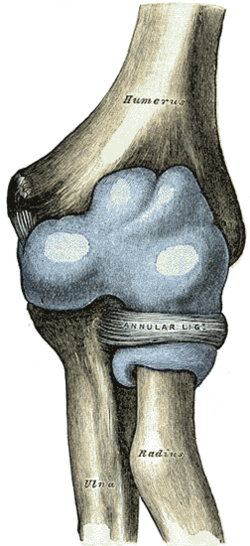
Capsule of elbow-joint (distended). Anterior aspect. (Nursemaid's elbow involves the head of radius slipping out from the anular ligament of radius.)
Specialty Emergency medicine
Symptoms Unwilling to move the arm [2]
Usual onset1 to 4 years old [2]
CausesSudden pull on an extended arm [2]
Diagnostic method Based on symptoms, Xrays [2]
Differential diagnosis Elbow fracture [3]
Treatment Reduction (forearm into a palms down position with straightening at the elbow) [1] [2]
Prognosis Recovery within minutes of reduction [1]
FrequencyCommon [2]

A pulled elbow, also known as a radial head subluxation, [4] is when the ligament that wraps around the radial head slips off. [1] Often a child will hold their arm against their body with the elbow slightly bent. [1] They will not move the arm as this results in pain. [2] Touching the arm, without moving the elbow, is usually not painful. [1]

Contents

A pulled elbow typically results from a sudden pull on an extended arm. [2] This may occur when lifting or swinging a child by the arms. [2] The underlying mechanism involves slippage of the annular ligament off of the head of the radius followed by the ligament getting stuck between the radius and humerus. [1] Diagnosis is often based on symptoms. [2] X-rays may be done to rule out other problems. [2]

Prevention is by avoiding potential causes. [2] Treatment is by reduction. [2] Moving the forearm into a palms down position with straightening at the elbow appears to be more effective than moving it into a palms up position followed by bending at the elbow. [1] [4] [5] Following a successful reduction the child should return to normal within a few minutes. [1] A pulled elbow is common. [2] It generally occurs in children between the ages of 1 and 4 years old, though it can happen up to 7 years old. [2]

Signs and symptoms

Symptoms include:

Cause

This injury has also been reported in babies younger than six months and in older children up to the preteen years. There is a slight predilection for this injury to occur in girls and in the left arm. The classic mechanism of injury is longitudinal traction on the arm with the wrist in pronation, as occurs when the child is lifted up by the wrist. There is no support for the common assumption that a relatively small head of the radius as compared to the neck of the radius predisposes the young to this injury.[ citation needed ]

Pathophysiology

The distal attachment of the annular ligament covering the radial head is weaker in children than in adults, allowing it to be more easily torn. The older child will usually point to the dorsal aspect of the proximal forearm when asked where it hurts. This may mislead one to suspect a buckle fracture of the proximal radius. [7] There is no tear in the soft tissue (probably due to the pliability of young connective tissues). [7]

The forearm contains two bones: the radius and the ulna. These bones are attached to each other both at the proximal, or elbow, end and also at the distal, or wrist, end. Among other movements, the forearm is capable of pronation and supination, which is to say rotation about the long axis of the forearm. In this movement the ulna, which is connected to the humerus by a simple hinge-joint, remains stationary, while the radius rotates, carrying the wrist and hand with it. To allow this rotation, the proximal (elbow) end of the radius is held in proximity to the ulna by a ligament known as the annular ligament. This is a circular ligamentous structure within which the radius is free, with constraints existing elsewhere in the forearm, to rotate. The proximal end of the radius in young children is conical, with the wider end of the cone nearest the elbow. With the passage of time the shape of this bone changes, becoming more cylindrical but with the proximal end being widened.[ citation needed ]

If the forearm of a young child is pulled, it is possible for this traction to pull the radius into the annular ligament with enough force to cause it to be jammed therein. This causes significant pain, partial limitation of flexion/extension of the elbow and total loss of pronation/supination in the affected arm. The situation is rare in adults, or in older children, because the changing shape of the radius associated with growth prevents it.[ citation needed ]

Diagnosis

Diagnosis is often based on symptoms. [2] X-rays may be done to rule out other problems. [2]

Treatment

To resolve the problem, the affected arm is moved in a way that causes the joint to move back into a normal position. The two main methods are hyperpronation and a combination of supination and flexion. Hyperpronation has a higher success rate and is less painful than a supination-flexion maneuver. [4] [8]

Related Research Articles

Carpal bones Eight small bones that make up the wrist (or carpus) that connects the hand to the forearm

The carpal bones are the eight small bones that make up the wrist that connects the hand to the forearm. The term "carpus" is derived from the Latin carpus and the Greek καρπός (karpós), meaning "wrist". In human anatomy, the main role of the wrist is to facilitate effective positioning of the hand and powerful use of the extensors and flexors of the forearm, and the mobility of individual carpal bones increase the freedom of movements at the wrist.

Ulna

The ulna is a long bone found in the forearm that stretches from the elbow to the smallest finger, and when in anatomical position, is found on the medial side of the forearm. It runs parallel to the radius, the other long bone in the forearm. The ulna is usually slightly longer than the radius, but the radius is thicker. Therefore, the radius is considered to be the larger of the two.

Biceps

The biceps or biceps brachii is a large muscle that lies on the front of the upper arm between the shoulder and the elbow. Both heads of the muscle arise on the scapula and join to form a single muscle belly which is attached to the upper forearm. While the biceps crosses both the shoulder and elbow joints, its main function is at the elbow where it flexes the forearm and supinates the forearm. Both these movements are used when opening a bottle with a corkscrew: first biceps screws in the cork (supination), then it pulls the cork out (flexion).

The forearm is the region of the upper limb between the elbow and the wrist. The term forearm is used in anatomy to distinguish it from the arm, a word which is most often used to describe the entire appendage of the upper limb, but which in anatomy, technically, means only the region of the upper arm, whereas the lower "arm" is called the forearm. It is homologous to the region of the leg that lies between the knee and the ankle joints, the crus.

Radius (bone) One of the two long bones of the forearm

The radius or radial bone is one of the two large bones of the forearm, the other being the ulna. It extends from the lateral side of the elbow to the thumb side of the wrist and runs parallel to the ulna. The ulna is usually slightly longer than the radius, but the radius is thicker. Therefore the radius is considered to be the larger of the two. It is a long bone, prism-shaped and slightly curved longitudinally.

Upper limb

The upper limbs or upper extremities are the forelimbs of an upright-postured tetrapod vertebrate, extending from the scapulae and clavicles down to and including the digits, including all the musculatures and ligaments involved with the shoulder, elbow, wrist and knuckle joints. In humans, each upper limb is divided into the arm, forearm and hand, and is primarily used for climbing, lifting and manipulating objects.

Supinator muscle

In human anatomy, the supinator is a broad muscle in the posterior compartment of the forearm, curved around the upper third of the radius. Its function is to supinate the forearm.

Galeazzi fracture Medical condition

The Galeazzi fracture is a fracture of the distal third of the radius with dislocation of the distal radioulnar joint. It classically involves an isolated fracture of the junction of the distal third and middle third of the radius with associated subluxation or dislocation of the distal radio-ulnar joint; the injury disrupts the forearm axis joint.

Madelungs deformity Medical condition

Madelung's deformity is usually characterized by malformed wrists and wrist bones and is often associated with Léri-Weill dyschondrosteosis. It can be bilateral or just in the one wrist. It has only been recognized within the past hundred years. Named after Otto Wilhelm Madelung (1846–1926), a German surgeon, who described it in detail, it was noted by others. Guillaume Dupuytren mentioned it in 1834, Auguste Nélaton in 1847, and Joseph-François Malgaigne in 1855.

Annular ligament of radius

The annular ligament is a strong band of fibers that encircles the head of the radius, and retains it in contact with the radial notch of the ulna.

Proximal radioulnar articulation

The proximal radioulnar joint is a synovial pivot joint between the circumference of the head of the radius and the ring formed by the radial notch of the ulna and the annular ligament.

Distal radioulnar articulation

The distal radioulnar articulation is a synovial pivot-type joint between the two bones in the forearm; the radius and ulna. It is one of two joints between the radius and ulna, the other being the proximal radioulnar articulation. The distal radioulnar articulation is the one of the two closest to the wrist and hand.

Triangular fibrocartilage

The Triangular fibrocartilage complex (TFCC) is formed by the triangular fibrocartilage discus (TFC), the radioulnar ligaments (RULs) and the ulnocarpal ligaments (UCLs).

Humeroradial joint

The humeroradial joint is the joint between the head of the radius and the capitulum of the humerus, is a limited ball-and-socket joint, hinge type of synovial joint.

Head of radius A bone of the arm

The head of the radius has a cylindrical form, and on its upper surface is a shallow cup or fovea for articulation with the capitulum of the humerus. The circumference of the head is smooth; it is broad medially where it articulates with the radial notch of the ulna, narrow in the rest of its extent, which is embraced by the annular ligament.

Elbow Joint between the upper and lower parts of the arm

The elbow is the visible joint between the upper and lower parts of the arm. It includes prominent landmarks such as the olecranon, the elbow pit, the lateral and medial epicondyles, and the elbow joint. The elbow joint is the synovial hinge joint between the humerus in the upper arm and the radius and ulna in the forearm which allows the forearm and hand to be moved towards and away from the body.

Supracondylar humerus fracture Medical condition

A supracondylar humerus fracture is a fracture of the distal humerus just above the elbow joint. The fracture is usually transverse or oblique and above the medial and lateral condyles and epicondyles. This fracture pattern is relatively rare in adults, but is the most common type of elbow fracture in children. In children, many of these fractures are non-displaced and can be treated with casting. Some are angulated or displaced and are best treated with surgery. In children, most of these fractures can be treated effectively with expectation for full recovery. Some of these injuries can be complicated by poor healing or by associated blood vessel or nerve injuries with serious complications.

The Essex-Lopresti fracture is a fracture of the radial head with concomitant dislocation of the distal radio-ulnar joint and disruption of the interosseous membrane. The injury is named after Peter Essex-Lopresti who described it in 1951.

The Hume fracture is an injury of the elbow comprising a fracture of the olecranon with an associated anterior dislocation of the radial head which occurs in children. It was originally described as an undisplaced olecranon fracture, but more recently includes displaced fractures and can be considered a variant of the Monteggia fracture.

Quadrate ligament

In human anatomy, the quadrate ligament or ligament of Denucé is one of the ligaments of the proximal radioulnar joint in the upper forearm.

References

  1. 1 2 3 4 5 6 7 8 9 Browner, EA (August 2013). "Nursemaid's Elbow (Annular Ligament Displacement)". Pediatrics in Review. 34 (8): 366–7, discussion 367. doi:10.1542/pir.34-8-366. PMID   23908364.
  2. 1 2 3 4 5 6 7 8 9 10 11 12 13 14 15 16 17 18 "Nursemaid's Elbow". OrthoInfo - AAOS. February 2014. Retrieved 4 December 2017.
  3. Cohen-Rosenblum, A; Bielski, RJ (1 June 2016). "Elbow Pain After a Fall: Nursemaid's Elbow or Fracture?". Pediatric Annals. 45 (6): e214–7. doi:10.3928/00904481-20160506-01. PMID   27294496.
  4. 1 2 3 Krul, M; van der Wouden, JC; Kruithof, EJ; van Suijlekom-Smit, LW; Koes, BW (28 July 2017). "Manipulative interventions for reducing pulled elbow in young children". The Cochrane Database of Systematic Reviews. 7: CD007759. doi:10.1002/14651858.CD007759.pub4. PMC   6483272 . PMID   28753234.
  5. Bexkens, R; Washburn, FJ; Eygendaal, D; van den Bekerom, MP; Oh, LS (January 2017). "Effectiveness of reduction maneuvers in the treatment of nursemaid's elbow: A systematic review and meta-analysis". The American Journal of Emergency Medicine. 35 (1): 159–163. doi:10.1016/j.ajem.2016.10.059. PMID   27836316. S2CID   2315716.
  6. Radial Head Subluxation Joint Reduction at eMedicine
  7. 1 2 Nursemaid Elbow at eMedicine
  8. Bexkens, R; Washburn, FJ; Eygendaal, D; van den Bekerom, MP; Oh, LS (2 November 2016). "Effectiveness of reduction maneuvers in the treatment of nursemaid's elbow: A systematic review and meta-analysis". The American Journal of Emergency Medicine. 35 (1): 159–163. doi:10.1016/j.ajem.2016.10.059. PMID   27836316. S2CID   2315716.
Classification
D
External resources