Shin splints

Last updated

Shin splints
Other namesMedial tibial stress syndrome (MTSS), [1] soleus syndrome, [2] tibial stress syndrome, [2] periostitis [2]
Tibia - frontal view2.png
Red area represents the tibia. Pain is generally in the inner and lower 2/3rds of tibia.
Specialty Sports medicine
Symptoms Pain along the inside edge of the shinbone [1]
Complications Stress fracture [2]
Risk factors Runners, dancers, military personnel [2]
Diagnostic method Based on symptoms, medical imaging [2]
Differential diagnosis Stress fracture, tendinitis, exertional compartment syndrome [1]
TreatmentRest with gradual return to exercise [1] [2]
Prognosis Good [2]
Frequency4% to 35% (at-risk groups) [2]

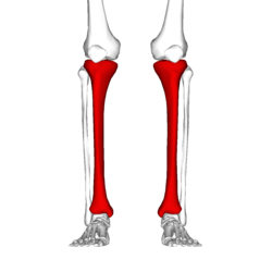
A shin splint, also known as medial tibial stress syndrome, is pain along the inside edge of the shinbone (tibia) due to inflammation of tissue in the area. [1] Generally this is between the middle of the lower leg and the ankle. [2] The pain may be dull or sharp, and is generally brought on by high-impact exercise that overloads the tibia. [1] It generally resolves during periods of rest. [3] Complications may include stress fractures. [2]

Contents

Shin splints typically occur due to excessive physical activity. [1] Groups that are commonly affected include runners, dancers, gymnasts, and military personnel. [2] The underlying mechanism is not entirely clear. [2] Diagnosis is generally based on the symptoms, with medical imaging done to rule out other possible causes. [2]

Shin splints are generally treated by rest followed by a gradual return to exercise over a period of weeks. [1] [2] [3] Other measures such as nonsteroidal anti-inflammatory drugs (NSAIDs), cold packs, physical therapy, and compression may be used. [1] [2] Shoe insoles may help some people. [1] Surgery is rarely required, but may be done if other measures are not effective. [2] Rates of shin splints in at-risk groups range from 4% to 35%. [2] The condition occurs more often in women. [2] It was first described in 1958. [2]

Signs and symptoms

Shin splint pain is described as a recurring dull ache, sometimes becoming an intense pain, along the inner part of the lower two-thirds of the tibia. [4] The pain increases during exercise, and some individuals experience swelling in the pain area. [5] In contrast, stress fracture pain is localized to the fracture site. [6]

Women are several times more likely to progress to stress fractures from shin splints. [7] [8] [9] This is due in part to women having a higher incidence of diminished bone density and osteoporosis. [10] [ citation needed ]

Causes

Shin splints typically occur due to excessive physical activity. [1] Groups that are commonly affected include runners, dancers, and military personnel. [2]

Risk factors for developing shin splints include:

People who have previously had shin splints are more likely to have them again. [12]

Pathophysiology

While the exact mechanism is unknown, shin splints can be attributed to the overloading of the lower leg due to biomechanical irregularities resulting in an increase in stress exerted on the tibia. A sudden increase in intensity or frequency in activity level fatigues muscles too quickly to help shock absorption properly, forcing the tibia to absorb most of the impact. Lack of cushioning footwear, especially on hard surfaces, does not absorb transmitting forces while running or jumping. [13] This stress is associated with the onset of shin splints. [14] Muscle imbalance, including weak core muscles, inflexibility and tightness of lower leg muscles, including the gastrocnemius, soleus, and plantar muscles (commonly the flexor digitorum longus) can increase the possibility of shin splints. [15] The pain associated with shin splints is caused from a disruption of Sharpey's fibres that connect the medial soleus fascia through the periosteum of the tibia where it inserts into the bone. [14] With repetitive stress, the impact forces eccentrically fatigue the soleus and create repeated tibial bending or bowing, contributing to shin splints. The impact is made worse by running uphill, downhill, on uneven terrain, or on hard surfaces. Improper footwear, including worn-out shoes, can also contribute to shin splints. [16] [17]

Diagnosis

Magnetic resonance image of the lower leg in the coronal plane showing high signal (bright) areas around the tibia as signs of shin splints. Shinsplint-mri.jpg
Magnetic resonance image of the lower leg in the coronal plane showing high signal (bright) areas around the tibia as signs of shin splints.

Shin splints are generally diagnosed from a history and physical examination. [3] The important factors on history are the location of pain, what triggers the pain, and the absence of cramping or numbness. [3]

On physical examination, gentle pressure over the tibia will recreate the type of pain experienced. [12] [18] Generally more than a 5 cm length of tibia is involved. [12] Swelling, redness, or poor pulses in addition to the symptoms of shin splints indicate a different underlying cause. [3]

Differential diagnosis

Other potential causes include stress fractures, compartment syndrome, nerve entrapment, and popliteal artery entrapment syndrome. [18] If the cause is unclear, medical imaging such as a bone scan or magnetic resonance imaging (MRI) may be performed. [3] Bone scans and MRI can differentiate between stress fractures and shin splints. [12]

Treatment

Treatments include rest, ice, and gradually returning to activity. [15] Rest and ice help the tibia to recover from sudden, high levels of stress and reduce inflammation and pain levels. It is important to reduce significantly any pain or swelling before returning to activity. Strengthening exercises should be performed after pain has subsided, on calves, quadriceps and gluteals. [15] Cross training (e.g., cycling, swimming, boxing) is recommended in order to maintain aerobic fitness. [19] Individuals should return to activity gradually, beginning with a short and low intensity level. Over multiple weeks, they can slowly work up to normal activity level. It is important to decrease activity level if any pain returns. Individuals should consider running on other surfaces besides asphalt, such as grass, to decrease the amount of force the lower leg must absorb. [7]

Orthoses and insoles help to offset biomechanical irregularities, like pronation, and help to support the arch of the foot. [20] Other conservative interventions include improving form during exercise, footwear refitting, orthotics, manual therapy, balance training (e.g., using a balance board), cortisone injections, and calcium and vitamin D supplementation. [15]

Deep tissue massage is one of the massage techniques that may be useful. A technique such as deep transverse friction to relieve muscle tightness will help stop the build-up of scar tissue. This can overall release tension in the calf muscle area, relieving pressure that is causing pain. [13]

Less-common forms of treatment for more-severe cases of shin splints include extracorporeal shockwave therapy (ESWT) and surgery. [21] Surgery does not guarantee 100% recovery, and is only performed in extreme cases where non-surgical options have been tried for at least a year. [22]

Epidemiology

Rates of shin splints in at-risk groups are 4% to 35%. [2] Women are affected more often than men. [23] [24]

Related Research Articles

<span class="mw-page-title-main">Running</span> Method of terrestrial locomotion allowing rapid movement on foot

Running is a method of terrestrial locomotion by which humans and other animals move rapidly on foot. Running is a gait with an aerial phase in which all feet are above the ground. This is in contrast to walking, where one foot is always in contact with the ground, the legs are kept mostly straight, and the center of gravity vaults over the stance leg or legs in an inverted pendulum fashion. A feature of a running body from the viewpoint of spring-mass mechanics is that changes in kinetic and potential energy within a stride co-occur, with energy storage accomplished by springy tendons and passive muscle elasticity. The term "running" can refer to a variety of speeds ranging from jogging to sprinting.

<span class="mw-page-title-main">Human leg</span> Lower extremity or limb of the human body (foot, lower leg, thigh and hip)

The leg is the entire lower limb of the human body, including the foot, thigh or sometimes even the hip or buttock region. The major bones of the leg are the femur, tibia, and adjacent fibula. The thigh is between the hip and knee, while the calf (rear) and shin (front) are between the knee and foot.

<span class="mw-page-title-main">Knee</span> Leg joint in primates

In humans and other primates, the knee joins the thigh with the leg and consists of two joints: one between the femur and tibia, and one between the femur and patella. It is the largest joint in the human body. The knee is a modified hinge joint, which permits flexion and extension as well as slight internal and external rotation. The knee is vulnerable to injury and to the development of osteoarthritis.

<span class="mw-page-title-main">Compartment syndrome</span> Condition in which increased pressure results in insufficient blood supply

Compartment syndrome is a condition in which increased pressure within one of the body's anatomical compartments results in insufficient blood supply to tissue within that space. There are two main types: acute and chronic. Compartments of the leg or arm are most commonly involved.

<span class="mw-page-title-main">Ankle</span> Region where the foot and the leg meet

The ankle, the talocrural region or the jumping bone (informal) is the area where the foot and the leg meet. The ankle includes three joints: the ankle joint proper or talocrural joint, the subtalar joint, and the inferior tibiofibular joint. The movements produced at this joint are dorsiflexion and plantarflexion of the foot. In common usage, the term ankle refers exclusively to the ankle region. In medical terminology, "ankle" can refer broadly to the region or specifically to the talocrural joint.

<span class="mw-page-title-main">Achilles tendon</span> Tendon at the back of the lower leg

The Achilles tendon or heel cord, also known as the calcaneal tendon, is a tendon at the back of the lower leg, and is the thickest in the human body. It serves to attach the plantaris, gastrocnemius (calf) and soleus muscles to the calcaneus (heel) bone. These muscles, acting via the tendon, cause plantar flexion of the foot at the ankle joint, and flexion at the knee.

<span class="mw-page-title-main">Bone fracture</span> Physical damage to the continuity of a bone

A bone fracture is a medical condition in which there is a partial or complete break in the continuity of any bone in the body. In more severe cases, the bone may be broken into several fragments, known as a comminuted fracture. A bone fracture may be the result of high force impact or stress, or a minimal trauma injury as a result of certain medical conditions that weaken the bones, such as osteoporosis, osteopenia, bone cancer, or osteogenesis imperfecta, where the fracture is then properly termed a pathologic fracture.

<span class="mw-page-title-main">Plantar fasciitis</span> Connective tissue disorder of the heel

Plantar fasciitis or plantar heel pain is a disorder of the plantar fascia, which is the connective tissue which supports the arch of the foot. It results in pain in the heel and bottom of the foot that is usually most severe with the first steps of the day or following a period of rest. Pain is also frequently brought on by bending the foot and toes up towards the shin. The pain typically comes on gradually, and it affects both feet in about one-third of cases.

<span class="mw-page-title-main">Flat feet</span> Deformity in which the foot arches contact the ground

Flat feet, also called pes planus or fallen arches, is a postural deformity in which the arches of the foot collapse, with the entire sole of the foot coming into complete or near-complete contact with the ground. Sometimes children are born with flat feet (congenital). There is a functional relationship between the structure of the arch of the foot and the biomechanics of the lower leg. The arch provides an elastic, springy connection between the forefoot and the hind foot so that a majority of the forces incurred during weight bearing on the foot can be dissipated before the force reaches the long bones of the leg and thigh.

<span class="mw-page-title-main">Stress fracture</span> Medical condition

A stress fracture is a fatigue-induced bone fracture caused by repeated stress over time. Instead of resulting from a single severe impact, stress fractures are the result of accumulated injury from repeated submaximal loading, such as running or jumping. Because of this mechanism, stress fractures are common overuse injuries in athletes.

<span class="mw-page-title-main">Osgood–Schlatter disease</span> Inflammation of the patellar ligament

Osgood–Schlatter disease (OSD) is inflammation of the patellar ligament at the tibial tuberosity (apophysitis) usually affecting adolescents during growth spurts. It is characterized by a painful bump just below the knee that is worse with activity and better with rest. Episodes of pain typically last a few weeks to months. One or both knees may be affected and flares may recur.

<span class="mw-page-title-main">Tibialis posterior muscle</span> Muscle in the most central of all the leg muscles

The tibialis posterior muscle is the most central of all the leg muscles, and is located in the deep posterior compartment of the leg. It is the key stabilizing muscle of the lower leg.

<span class="mw-page-title-main">Soleus muscle</span> Powerful muscle in the back part of the lower leg

In humans and some other mammals, the soleus is a powerful muscle in the back part of the lower leg. It runs from just below the knee to the heel and is involved in standing and walking. It is closely connected to the gastrocnemius muscle, and some anatomists consider this combination to be a single muscle, the triceps surae. Its name is derived from the Latin word "solea", meaning "sandal".

Periostitis, also known as periostalgia, is a medical condition caused by inflammation of the periosteum, a layer of connective tissue that surrounds bone. The condition is generally chronic, and is marked by tenderness and swelling of the bone and pain.

<span class="mw-page-title-main">Ankle fracture</span> Medical condition

An ankle fracture is a break of one or more of the bones that make up the ankle joint. Symptoms may include pain, swelling, bruising, and an inability to walk on the injured leg. Complications may include an associated high ankle sprain, compartment syndrome, stiffness, malunion, and post-traumatic arthritis.

<span class="mw-page-title-main">Popliteal artery entrapment syndrome</span> Abnormal popliteal artery compression by surrounding structures

The popliteal artery entrapment syndrome (PAES) is an uncommon pathology that occurs when the popliteal artery is compressed by the surrounding popliteal fossa myofascial structures. This results in claudication and chronic leg ischemia. This condition mainly occurs more in young athletes than in the elderlies. Elderlies, who present with similar symptoms, are more likely to be diagnosed with peripheral artery disease with associated atherosclerosis. Patients with PAES mainly present with intermittent feet and calf pain associated with exercises and relieved with rest. PAES can be diagnosed with a combination of medical history, physical examination, and advanced imaging modalities such as duplex ultrasound, computer tomography, or magnetic resonance angiography. Management can range from non-intervention to open surgical decompression with a generally good prognosis. Complications of untreated PAES can include stenotic artery degeneration, complete popliteal artery occlusion, distal arterial thromboembolism, or even formation of an aneurysm.

<span class="mw-page-title-main">Patellofemoral pain syndrome</span> Medical condition

Patellofemoral pain syndrome is knee pain as a result of problems between the kneecap and the femur. The pain is generally in the front of the knee and comes on gradually. Pain may worsen with sitting, excessive use, or climbing and descending stairs.

<span class="mw-page-title-main">Patellar dislocation</span> Medical condition

A patellar dislocation is a knee injury in which the patella (kneecap) slips out of its normal position. Often the knee is partly bent, painful and swollen. The patella is also often felt and seen out of place. Complications may include a patella fracture or arthritis.

<span class="mw-page-title-main">Tibial plateau fracture</span> Medical condition

A tibial plateau fracture is a break of the upper part of the tibia (shinbone) that involves the knee joint. This could involve the medial, lateral, central, or bicondylar. Symptoms include pain, swelling, and a decreased ability to move the knee. People are generally unable to walk. Complication may include injury to the artery or nerve, arthritis, and compartment syndrome.

Running injuries affect about half of runners annually. The frequencies of various RRI depend on the type of running, such as speed and mileage. Some injuries are acute, caused by sudden overstress, such as side stitch, strains, and sprains. Many of the common injuries that affect runners are chronic, developing over longer periods as the result of overuse. Common overuse injuries include shin splints, stress fractures, Achilles tendinitis, Iliotibial band syndrome, Patellofemoral pain, and plantar fasciitis.

References

  1. 1 2 3 4 5 6 7 8 9 10 11 12 Alaia, Michael J. (August 2019). "Shin Splints". OrthoInfo. American Academy of Orthopaedic Surgeons. Retrieved 15 November 2020.
  2. 1 2 3 4 5 6 7 8 9 10 11 12 13 14 15 16 17 18 19 20 21 22 Reshef, N; Guelich, DR (April 2012). "Medial tibial stress syndrome". Clinics in Sports Medicine. 31 (2): 273–290. doi:10.1016/j.csm.2011.09.008. PMID   22341017. S2CID   262469440.
  3. 1 2 3 4 5 6 7 McClure, CJ; Oh, R (January 2019). "Medial Tibial Stress Syndrome". NCBI Bookshelf. PMID   30860714. Archived from the original on 29 March 2023.
  4. Carr, K.; Sevetson, E.; Aukerman, D. (2008). "Clinical inquiries. How can you help athletes prevent and treat shin splints?". The Journal of Family Practice. 57 (6): 406–408. PMID   18544325.
  5. Tweed, J.L.; Avil, S.J.; Campbell, J.A.; Barnes, M.R. (2008). "Etiologic factors in the development of medial tibial stress syndrome: A review of the literature". Journal of the American Podiatric Medical Association. 98 (2): 107–111. doi:10.7547/0980436. PMID   18347118.
  6. Edwards, Peter H.; Wright, Michelle L.; Hartman, Jodi F. (2005). "A Practical Approach for the Differential Diagnosis of Chronic Leg Pain in the Athlete". The American Journal of Sports Medicine. 33 (8): 1241–1249. doi:10.1177/0363546505278305. PMID   16061959. S2CID   7828716.
  7. 1 2 3 4 Yates, B.; White, S. (2004). "The incidence and risk factors in the development of medial tibial stress syndrome among naval recruits". American Journal of Sports Medicine. 32 (3): 772–780. doi:10.1177/0095399703258776. PMID   15090396. S2CID   24603853.
  8. Bennett, Jason E.; Reinking, Mark F.; Pluemer, Bridget; Pentel, Adam; Seaton, Marcus; Killian, Clyde (2001). "Factors Contributing to the Development of Medial Tibial Stress Syndrome in High School Runners". Journal of Orthopaedic & Sports Physical Therapy. 31 (9): 504–510. doi:10.2519/jospt.2001.31.9.504. PMID   11570734.
  9. Haycock, Christine E.; Gillette, Joan V. (1976). "Susceptibility of women athletes to injury. Myths vs reality". Journal of the American Medical Association. 236 (2): 163–165. doi:10.1001/jama.1976.03270020033020. PMID   947011.
  10. Healthwise Staff. (7 December 2020). Low bone density. University of Michigan Health. Retrieved 26 June 2021, from https://www.uofmhealth.org/health-library/tp23004spec
  11. Brukner, Peter (2000). "Exercise-related lower leg pain: An overview". Medicine & Science in Sports & Exercise. 32 (3 Suppl): S1–S3. doi: 10.1249/00005768-200003001-00001 . PMID   10730988.
  12. 1 2 3 4 Moen, MH; Tol, JL; Weir, A; Steunebrink, M; De Winter, TC (2009). "Medial tibial stress syndrome: a critical review". Sports Medicine (Auckland, N.Z.). 39 (7): 523–546. doi:10.2165/00007256-200939070-00002. PMID   19530750. S2CID   40720414.
  13. 1 2 De Permentier, Patrick (2014). "An Anatomical and Physiological Evaluation of the Periosteal Layer Surrounding Bone and Its Implication in Massage Therapy". Journal of the Australian Traditional-Medicine Society. 20: 272–277.
  14. 1 2 Craig, Debbie I. (2008). "Medial Tibial Stress Syndrome: Evidence-Based Prevention". Journal of Athletic Training. 43 (3): 316–318. doi:10.4085/1062-6050-43.3.316. PMC   2386425 . PMID   18523568.
  15. 1 2 3 4 Galbraith, R. Michael; Lavallee, Mark E. (7 October 2009). "Medial tibial stress syndrome: conservative treatment options". Current Reviews in Musculoskeletal Medicine. 2 (3): 127–133. doi:10.1007/s12178-009-9055-6. ISSN   1935-973X. PMC   2848339 . PMID   19809896.
  16. Lobby, Mackenzie (9 September 2014). "Running 101: How To Select The Best Pair Of Running Shoes". Competitor.com. Archived from the original on 13 September 2014.
  17. "Shin Splints Symptoms, Treatment, Recovery, and Prevention". WebMD.
  18. 1 2 Patel, Deepak S.; Roth, Matt; Kapil, Neha (2011). "Stress fractures: diagnosis, treatment, and prevention" (PDF). American Family Physician. 83 (1): 39–46. PMID   21888126. S2CID   1736230. Archived from the original (PDF) on 12 February 2019.
  19. Couture, Christopher (2002). "Tibial Stress Injuries: Decisive Diagnosis and Treatment of 'Shin Splints'". The Physician and Sportsmedicine. 30 (6): 29–36. doi:10.3810/psm.2002.06.337. PMID   20086529. S2CID   39133382.
  20. Loudon, Janice K.; Dolphino, Martin R. (2010). "Use of Foot Orthoses and Calf Stretching for Individuals with Medial Tibial Stress Syndrome". Foot & Ankle Specialist. 3 (1): 15–20. doi:10.1177/1938640009355659. PMID   20400435. S2CID   3374384.
  21. Rompe, Jan D.; Cacchio, Angelo; Furia, John P.; Maffulli, Nicola (2010). "Low-Energy Extracorporeal Shock Wave Therapy as a Treatment for Medial Tibial Stress Syndrome". The American Journal of Sports Medicine. 38 (1): 125–132. doi:10.1177/0363546509343804. PMID   19776340. S2CID   21114701.
  22. Yates, Ben; Allen, Mike J.; Barnes, Mike R. (2003). "Outcome of Surgical Treatment of Medial Tibial Stress Syndrome". The Journal of Bone and Joint Surgery. American Volume. 85 (10): 1974–1980. doi:10.2106/00004623-200310000-00017. PMID   14563807.
  23. Newman, Phil; Witchalls, Jeremy; Waddington, Gordon; Adams, Roger (2013). "Risk factors associated with medial tibial stress syndrome in runners: a systematic review and meta-analysis". Open Access Journal of Sports Medicine. 4: 229–241. doi: 10.2147/OAJSM.S39331 . ISSN   1179-1543. PMC   3873798 . PMID   24379729.
  24. Macleod MA, Houston AS, Sanders L, Anagnostopoulos C (1999). "Incidence of trauma related stress fractures and shin splints in male and female army recruits: retrospective case study". BMJ. 318 (7175): 29. doi:10.1136/bmj.318.7175.29. PMC   27673 . PMID   9872880.