The foot is an anatomical structure found in many vertebrates. It is the terminal portion of a limb which bears weight and allows locomotion. In many animals with feet, the foot is a separate organ at the terminal part of the leg made up of one or more segments or bones, generally including claws and/or nails.

The tibia, also known as the shinbone or shankbone, is the larger, stronger, and anterior (frontal) of the two bones in the leg below the knee in vertebrates ; it connects the knee with the ankle. The tibia is found on the medial side of the leg next to the fibula and closer to the median plane. The tibia is connected to the fibula by the interosseous membrane of leg, forming a type of fibrous joint called a syndesmosis with very little movement. The tibia is named for the flute tibia. It is the second largest bone in the human body, after the femur. The leg bones are the strongest long bones as they support the rest of the body.

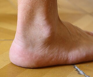
The ankle, the talocrural region or the jumping bone (informal) is the area where the foot and the leg meet. The ankle includes three joints: the ankle joint proper or talocrural joint, the subtalar joint, and the inferior tibiofibular joint. The movements produced at this joint are dorsiflexion and plantarflexion of the foot. In common usage, the term ankle refers exclusively to the ankle region. In medical terminology, "ankle" can refer broadly to the region or specifically to the talocrural joint.

In humans and many other primates, the calcaneus or heel bone is a bone of the tarsus of the foot which constitutes the heel. In some other animals, it is the point of the hock.

Clubfoot is a congenital or acquired defect where one or both feet are rotated inward and downward. Congenital clubfoot is the most common congenital malformation of the foot with an incidence of 1 per 1000 births. In approximately 50% of cases, clubfoot affects both feet, but it can present unilaterally causing one leg or foot to be shorter than the other. Most of the time, it is not associated with other problems. Without appropriate treatment, the foot deformity will persist and lead to pain and impaired ability to walk, which can have a dramatic impact on the quality of life.

Pes cavus, also known as high arch, is an orthopedic condition that presents as a hollow arch underneath the foot with a pronounced high ridge at the top when weight bearing.

The Maisonneuve fracture is a spiral fracture of the proximal third of the fibula associated with a tear of the distal tibiofibular syndesmosis and the interosseous membrane. There is an associated fracture of the medial malleolus or rupture of the deep deltoid ligament of the ankle. This type of injury can be difficult to detect.

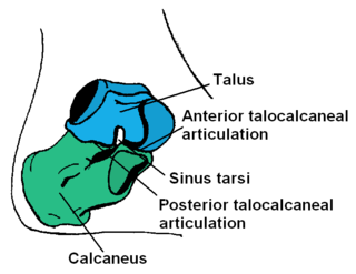
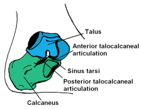
The talus, talus bone, astragalus, or ankle bone is one of the group of foot bones known as the tarsus. The tarsus forms the lower part of the ankle joint. It transmits the entire weight of the body from the lower legs to the foot.

An ankle fracture is a break of one or more of the bones that make up the ankle joint. Symptoms may include pain, swelling, bruising, and an inability to walk on the injured leg. Complications may include an associated high ankle sprain, compartment syndrome, stiffness, malunion, and post-traumatic arthritis.

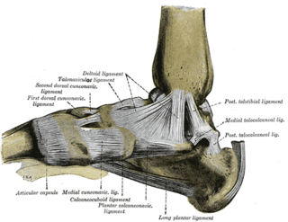
The deltoid ligament is a strong, flat, triangular band, attached, above, to the apex and anterior and posterior borders of the medial malleolus. The deltoid ligament supports the ankle joint and also resists excessive eversion of the foot. The deltoid ligament is composed of 4 fibers:
- Anterior tibiotalar ligament
- Tibiocalcaneal ligament
- Posterior tibiotalar ligament
- Tibionavicular ligament.

The lateral collateral ligament of ankle joint are ligaments of the ankle which attach to the fibula.

A calcaneal fracture is a break of the calcaneus. Symptoms may include pain, bruising, trouble walking, and deformity of the heel. It may be associated with breaks of the hip or back.

A trimalleolar fracture is a fracture of the ankle that involves the lateral malleolus, the medial malleolus, and the distal posterior aspect of the tibia, which can be termed the posterior malleolus. The trauma is sometimes accompanied by ligament damage and dislocation.
Foot and ankle surgery is a sub-specialty of orthopedics and podiatry that deals with the treatment, diagnosis and prevention of disorders of the foot and ankle. Orthopaedic surgeons are medically qualified, having been through four years of college, followed by 4 years of medical school or osteopathic medical school to obtain an M.D. or D.O. followed by specialist training as a resident in orthopaedics, and only then do they sub-specialise in foot and ankle surgery. Training for a podiatric foot and ankle surgeon consists of four years of college, four years of podiatric medical school (D.P.M.), 3–4 years of a surgical residency and an optional 1 year fellowship.
The Ponseti method is a manipulative technique that corrects congenital clubfoot without invasive surgery. It was developed by Ignacio V. Ponseti of the University of Iowa Hospitals and Clinics, US, in the 1950s, and was repopularized in 2000 by John Herzenberg in the US and Europe and in Africa by NHS surgeon Steve Mannion. It is a standard treatment for clubfoot.
Ankle replacement, or ankle arthroplasty, is a surgical procedure to replace the damaged articular surfaces of the human ankle joint with prosthetic components. This procedure is becoming the treatment of choice for patients requiring arthroplasty, replacing the conventional use of arthrodesis, i.e. fusion of the bones. The restoration of range of motion is the key feature in favor of ankle replacement with respect to arthrodesis. However, clinical evidence of the superiority of the former has only been demonstrated for particular isolated implant designs.
Unlike the flexible flat foot that is commonly encountered in young children, congenital vertical talus is characterized by presence of a very rigid foot deformity. The foot deformity in congenital vertical talus consists of various components, namely a prominent calcaneus caused by the ankle equines or plantar flexion, a convex and rounded sole of the foot caused by prominence of the head of the talus, and a dorsiflexion and abduction of the forefoot and midfoot on the hindfoot. It gets its name from the foot's resemblance to the bottom of a rocking chair. There are two subcategories of congenital vertical talus; namely idiopathic or isolated type, and non-idiopathic type, which may be seen in association with arthrogryposis multiplex congenital, genetic syndromes and other neuromuscular disorders.

Tarsal coalition is an abnormal connecting bridge of tissue between two normally-separate tarsal (foot) bones, and is considered a sort of birth defect. The term 'coalition' means a coming together of two or more entities to merge into one mass. The tissue connecting the bones, often referred to as a "bar", may be composed of fibrous or osseous tissue. The two most common types of tarsal coalitions are calcaneo-navicular and talo-calcaneal, comprising 90% of all tarsal coalitions. There are other bone coalition combinations possible, but they are very rare. Symptoms tend to occur in the same location, regardless of the location of coalition: on the lateral foot, just anterior and below the lateral malleolus. This area is called the sinus tarsi.
In February 2021, the U.S. Food and Drug Administration (FDA) approved the Patient Specific Talus Spacer 3D-printed talus implant for humanitarian use. The Patient Specific Talus Spacer is the first in the world and first of its kind implant to replace the talus; the bone in the ankle joint that connects the leg and the foot. The implant is used for the treatment of avascular necrosis (AVN) of the ankle joint, a serious and progressive condition that causes the death of bone tissue stemming from a lack of blood supply to the area. The implant provides a joint sparing alternative to other surgical interventions commonly used in late stage AVN that may disable motion of the ankle joint.
Subtalar arthroereisis is a common treatment for symptomatic pes planus, also known as flatfoot. There are two forms of pes planus: rigid flatfoot (RFF) and flexible flatfoot (FFF). The symptoms of the former typically necessitate surgical intervention. The latter may manifest fatigue or pain, but is typically asymptomatic.


