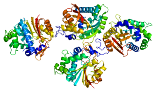
Thioredoxin is a class of small redox proteins known to be present in all organisms. It plays a role in many important biological processes, including redox signaling. In humans, thioredoxins are encoded by TXN and TXN2 genes. Loss-of-function mutation of either of the two human thioredoxin genes is lethal at the four-cell stage of the developing embryo. Although not entirely understood, thioredoxin is linked to medicine through their response to reactive oxygen species (ROS). In plants, thioredoxins regulate a spectrum of critical functions, ranging from photosynthesis to growth, flowering and the development and germination of seeds. Thioredoxins play a role in cell-to-cell communication.

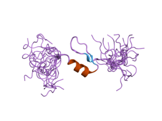
Amylin, or islet amyloid polypeptide (IAPP), is a 37-residue peptide hormone. It is co-secreted with insulin from the pancreatic β-cells in the ratio of approximately 100:1 (insulin:amylin). Amylin plays a role in glycemic regulation by slowing gastric emptying and promoting satiety, thereby preventing post-prandial spikes in blood glucose levels.

In the inner ear, stereocilia are the mechanosensing organelles of hair cells, which respond to fluid motion in numerous types of animals for various functions, including hearing and balance. They are about 10–50 micrometers in length and share some similar features of microvilli. The hair cells turn the fluid pressure and other mechanical stimuli into electric stimuli via the many microvilli that make up stereocilia rods. Stereocilia exist in the auditory and vestibular systems.

Agouti-signaling protein is a protein that in humans is encoded by the ASIP gene. It is responsible for the distribution of melanin pigment in mammals. Agouti interacts with the melanocortin 1 receptor to determine whether the melanocyte produces phaeomelanin, or eumelanin. This interaction is responsible for making distinct light and dark bands in the hairs of animals such as the agouti, which the gene is named after. In other species such as horses, agouti signalling is responsible for determining which parts of the body will be red or black. Mice with wildtype agouti will be grey-brown, with each hair being partly yellow and partly black. Loss of function mutations in mice and other species cause black fur coloration, while mutations causing expression throughout the whole body in mice cause yellow fur and obesity.

Insulin-degrading enzyme, also known as IDE, is an enzyme.

Fatty acid-binding protein 2 (FABP2), also known as Intestinal-type fatty acid-binding protein (I-FABP), is a protein that in humans is encoded by the FABP2 gene.
In enzymology, a peptide-methionine (R)-S-oxide reductase (EC 1.8.4.12) is an enzyme that catalyzes the chemical reaction

Insulin induced gene 1, also known as INSIG1, is a protein which in humans is encoded by the INSIG1 gene.

Nicotinamide N-methyltransferase (NNMT) is an enzyme that in humans is encoded by the NNMT gene. NNMT catalyzes the methylation of nicotinamide and similar compounds using the methyl donor S-adenosyl methionine (SAM-e) to produce S-adenosyl-L-homocysteine (SAH) and 1-methylnicotinamide.

Phosphatidylinositol binding clathrin assembly protein, also known as PICALM, is a protein which in humans is encoded by the PICALM gene.

Stanniocalcin-2 is a protein that in humans is encoded by the STC2 gene.

The LBH gene is a highly conserved human gene that produces the LBH protein, a transcription co-factor in the Wnt/β-catenin pathway. Upon transcriptional activation of β-catenin, LBH goes on to act as a regulator of cell proliferation and differentiation through multiple transcriptional targets. The gene is located on the p arm of chromosome 2 and is roughly 28 kb long. Current ongoing studies are examining its role in developmental and oncological settings.

Aldo-keto reductase family 1 member B10 is an enzyme that in humans is encoded by the AKR1B10 gene.

Methionine-R-sulfoxide reductase B2, mitochondrial is an enzyme that in humans is encoded by the MSRB2 gene. The MRSB2 enzyme catalyzes the reduction of methionine sulfoxide to methionine.

Methionine-R-sulfoxide reductase B1 is an enzyme that in humans is encoded by the SEPX1 gene.

Fibronectin type III domain-containing protein 5, the precursor of irisin, is a type I transmembrane glycoprotein that is encoded by the FNDC5 gene. Irisin is a cleaved version of FNDC5, named after the Greek messenger goddess Iris.

Krüppel-like factor 15 is a protein that in humans is encoded by the KLF15 gene in the Krüppel-like factor family. Its former designation KKLF stands for kidney-enriched Krüppel-like factor.

Methionine sulfoxide is the organic compound with the formula CH3S(O)CH2CH2CH(NH2)CO2H. It is an amino acid that occurs naturally although it is formed post-translationally.
Peptide-methionine (S)-S-oxide reductase (EC 1.8.4.11, MsrA, methionine sulphoxide reductase A, methionine S-oxide reductase (S-form oxidizing), methionine sulfoxide reductase A, peptide methionine sulfoxide reductase, formerly protein-methionine-S-oxide reductase) is an enzyme with systematic name peptide-L-methionine:thioredoxin-disulfide S-oxidoreductase (L-methionine (S)-S-oxide-forming). This enzyme catalyses the following chemical reaction

The mitochondrial theory of ageing has two varieties: free radical and non-free radical. The first is one of the variants of the free radical theory of ageing. It was formulated by J. Miquel and colleagues in 1980 and was developed in the works of Linnane and coworkers (1989). The second was proposed by A. N. Lobachev in 1978.


