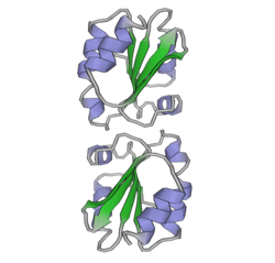
Thioredoxin

Last updated
TXN
Tioredoksin, gomodimer.png
Available structures
PDB Ortholog search: PDBe RCSB
Identifiers
Aliases TXN , TRDX, TRX, TRX1, thioredoxin, Trx80
External IDs OMIM: 187700 MGI: 98874 HomoloGene: 128202 GeneCards: TXN
Orthologs
SpeciesHumanMouse
Entrez
Ensembl
UniProt
RefSeq (mRNA)

NM_003329
NM_001244938

NM_011660

RefSeq (protein)

NP_001231867
NP_003320

NP_035790

Location (UCSC) Chr 9: 110.24 – 110.26 Mb Chr 4: 57.94 – 57.96 Mb
PubMed search [3] [4]
Wikidata
View/Edit Human View/Edit Mouse

Thioredoxin (TRX or TXN) is a class of small redox proteins known to be present in all organisms. It plays a role in many important biological processes, including redox signaling. In humans, thioredoxins are encoded by TXN and TXN2 genes. [5] [6] Loss-of-function mutation of either of the two human thioredoxin genes is lethal at the four-cell stage of the developing embryo. Although not entirely understood, thioredoxin is linked to medicine through their response to reactive oxygen species (ROS). In plants, thioredoxins regulate a spectrum of critical functions, ranging from photosynthesis to growth, flowering and the development and germination of seeds. Thioredoxins play a role in cell-to-cell communication. [7]

Occurrence

They are found in nearly all known organisms and are essential for life in mammals. [8] [9]

Function

The primary function of thioredoxin (Trx) is the reduction of oxidized cysteine residues and the cleavage of disulfide bonds. [10] Multiple in vitro substrates for thioredoxin have been identified, including ribonuclease, choriogonadotropins, coagulation factors, glucocorticoid receptor, and insulin. Reduction of insulin is classically used as an activity test. [11] The thioredoxins are maintained in their reduced state by the flavoenzyme thioredoxin reductase, in a NADPH-dependent reaction. [12] Thioredoxins act as electron donors to peroxidases and ribonucleotide reductase. [13] The related glutaredoxins share many of the functions of thioredoxins, but are reduced by glutathione rather than a specific reductase.

Structure and mechanism

Thioredoxin is a 12-kD oxidoreductase protein. Thioredoxin proteins also have a characteristic tertiary structure termed the thioredoxin fold. The active site contains a dithiols in a CXXC motif. These two cysteines are the key to the ability of thioredoxin to reduce other proteins.

For Trx1, this process begins by attack of Cys32, one of the residues conserved in the thioredoxin CXXC motif, onto the oxidized group of the substrate. [14] Almost immediately after this event Cys35, the other conserved Cys residue in Trx1, forms a disulfide bond with Cys32, thereby transferring 2 electrons to the substrate which is now in its reduced form. Oxidized Trx1 is then reduced by thioredoxin reductase, which in turn is reduced by NADPH as described above. [14]

Mechanism of Trx1 reducing a substrate FigMech.png
Mechanism of Trx1 reducing a substrate

Trx1 can regulate non-redox post-translational modifications. [15] In the mice with cardiac-specific overexpression of Trx1, the proteomics study found that SET and MYND domain-containing protein 1 (SMYD1), a lysine methyltransferase highly expressed in cardiac and other muscle tissues, is also upregulated. This suggests that Trx1 may also play an role in protein methylation via regulating SMYD1 expression, which is independent of its oxidoreductase activity. [15]

Plants have an unusually complex complement of Trx's composed of six well-defined types (Trxs f, m, x, y, h, and o) that reside in diverse cell compartments and function in an array of processes. Thioredoxin proteins move from cell to cell, representing a novel form of cellular communication in plants. [7]

Interactions

Thioredoxin has been shown to interact with:

Effect on cardiac hypertrophy

Trx1 has been shown to downregulate cardiac hypertrophy, the thickening of the walls of the lower heart chambers, by interactions with several different targets. Trx1 upregulates the transcriptional activity of nuclear respiratory factors 1 and 2 (NRF1 and NRF2) and stimulates the expression of peroxisome proliferator-activated receptor γ coactivator 1-α (PGC-1α). [26] [27] Furthermore, Trx1 reduces two cysteine residues in histone deacetylase 4 (HDAC4), which allows HDAC4 to be imported from the cytosol, where the oxidized form resides, [28] into the nucleus. [29] Once in the nucleus, reduced HDAC4 downregulates the activity of transcription factors such as NFAT that mediate cardiac hypertrophy. [14] Trx 1 also controls microRNA levels in the heart and has been found to inhibit cardiac hypertrophy by upregulating miR-98/let-7. [30] Trx1 can regulate the expression level of SMYD1, thus may indirectly modulate protein methylation for purpose of cardiac protection. [15]

Thioredoxin in skin care

Thioredoxin is used in skin care products as an antioxidant in conjunction with glutaredoxin and glutathione.[ citation needed ]

See also

Related Research Articles

<span class="mw-page-title-main">Protein disulfide-isomerase</span> Class of enzymes

Protein disulfide isomerase, or PDI, is an enzyme in the endoplasmic reticulum (ER) in eukaryotes and the periplasm of bacteria that catalyzes the formation and breakage of disulfide bonds between cysteine residues within proteins as they fold. This allows proteins to quickly find the correct arrangement of disulfide bonds in their fully folded state, and therefore the enzyme acts to catalyze protein folding.

Thioredoxin reductases are enzymes that reduce thioredoxin (Trx). Two classes of thioredoxin reductase have been identified: one class in bacteria and some eukaryotes and one in animals. In bacteria TrxR also catalyzes the reduction of glutaredoxin like proteins known as NrdH. Both classes are flavoproteins which function as homodimers. Each monomer contains a FAD prosthetic group, a NADPH binding domain, and an active site containing a redox-active disulfide bond.

Ferredoxins are iron–sulfur proteins that mediate electron transfer in a range of metabolic reactions. The term "ferredoxin" was coined by D.C. Wharton of the DuPont Co. and applied to the "iron protein" first purified in 1962 by Mortenson, Valentine, and Carnahan from the anaerobic bacterium Clostridium pasteurianum.

<span class="mw-page-title-main">Glutathione reductase</span> Enzyme

Glutathione reductase (GR) also known as glutathione-disulfide reductase (GSR) is an enzyme that in humans is encoded by the GSR gene. Glutathione reductase catalyzes the reduction of glutathione disulfide (GSSG) to the sulfhydryl form glutathione (GSH), which is a critical molecule in resisting oxidative stress and maintaining the reducing environment of the cell. Glutathione reductase functions as dimeric disulfide oxidoreductase and utilizes an FAD prosthetic group and NADPH to reduce one molar equivalent of GSSG to two molar equivalents of GSH:

<span class="mw-page-title-main">Vitamin K epoxide reductase</span> Class of enzymes

Vitamin K epoxide reductase (VKOR) is an enzyme that reduces vitamin K after it has been oxidised in the carboxylation of glutamic acid residues in blood coagulation enzymes. VKOR is a member of a large family of predicted enzymes that are present in vertebrates, Drosophila, plants, bacteria and archaea. In some plant and bacterial homologues, the VKOR domain is fused with domains of the thioredoxin family of oxidoreductases.

<span class="mw-page-title-main">Glutaredoxin</span>

Glutaredoxins are small redox enzymes of approximately one hundred amino-acid residues that use glutathione as a cofactor. In humans this oxidation repair enzyme is also known to participate in many cellular functions, including redox signaling and regulation of glucose metabolism. Glutaredoxins are oxidized by substrates, and reduced non-enzymatically by glutathione. In contrast to thioredoxins, which are reduced by thioredoxin reductase, no oxidoreductase exists that specifically reduces glutaredoxins. Instead, glutaredoxins are reduced by the oxidation of glutathione. Reduced glutathione is then regenerated by glutathione reductase. Together these components compose the glutathione system.

<span class="mw-page-title-main">Peroxiredoxin</span> Family of antioxidant enzymes

Peroxiredoxins are a ubiquitous family of antioxidant enzymes that also control cytokine-induced peroxide levels and thereby mediate signal transduction in mammalian cells. The family members in humans are PRDX1, PRDX2, PRDX3, PRDX4, PRDX5, and PRDX6. The physiological importance of peroxiredoxins is indicated by their relative abundance. Their function is the reduction of peroxides, specifically hydrogen peroxide, alkyl hydroperoxides, and peroxynitrite.

<span class="mw-page-title-main">ASK1</span> Protein-coding gene in the species Homo sapiens

Apoptosis signal-regulating kinase 1 (ASK1) also known as mitogen-activated protein kinase 5 (MAP3K5) is a member of MAP kinase family and as such a part of mitogen-activated protein kinase pathway. It activates c-Jun N-terminal kinase (JNK) and p38 mitogen-activated protein kinases in a Raf-independent fashion in response to an array of stresses such as oxidative stress, endoplasmic reticulum stress and calcium influx. ASK1 has been found to be involved in cancer, diabetes, rheumatoid arthritis, cardiovascular and neurodegenerative diseases.

NAD<sup>+</sup> kinase Enzyme

NAD+ kinase (EC 2.7.1.23, NADK) is an enzyme that converts nicotinamide adenine dinucleotide (NAD+) into NADP+ through phosphorylating the NAD+ coenzyme. NADP+ is an essential coenzyme that is reduced to NADPH primarily by the pentose phosphate pathway to provide reducing power in biosynthetic processes such as fatty acid biosynthesis and nucleotide synthesis. The structure of the NADK from the archaean Archaeoglobus fulgidus has been determined.

<span class="mw-page-title-main">Formaldehyde dehydrogenase</span>

In enzymology, a formaldehyde dehydrogenase (EC 1.2.1.46) is an enzyme that catalyzes the chemical reaction

<span class="mw-page-title-main">P4HB</span> Protein-coding gene in the species Homo sapiens

Protein disulfide-isomerase, also known as the beta-subunit of prolyl 4-hydroxylase (P4HB), is an enzyme that in humans encoded by the P4HB gene. The human P4HB gene is localized in chromosome 17q25. Unlike other prolyl 4-hydroxylase family proteins, this protein is multifunctional and acts as an oxidoreductase for disulfide formation, breakage, and isomerization. The activity of P4HB is tightly regulated. Both dimer dissociation and substrate binding are likely to enhance its enzymatic activity during the catalysis process.

<span class="mw-page-title-main">TXNIP</span> Mammalian protein found in Homo sapiens

Thioredoxin-interacting protein is a protein that in humans is encoded by the TXNIP gene.

<span class="mw-page-title-main">PRDX3</span> Protein-coding gene in the species Homo sapiens

Thioredoxin-dependent peroxide reductase, mitochondrial is an enzyme that in humans is encoded by the PRDX3 gene. It is a member of the peroxiredoxin family of antioxidant enzymes.

<span class="mw-page-title-main">GLRX2</span> Protein-coding gene in the species Homo sapiens

Glutaredoxin 2 (GLRX2) is an enzyme that in humans encoded by the GLRX2 gene. GLRX2, also known as GRX2, is a glutaredoxin family protein and a thiol-disulfide oxidoreductase that maintains cellular thiol homeostasis. This gene consists of four exons and three introns, spanned 10 kilobase pairs, and localized to chromosome 1q31.2–31.3.

<span class="mw-page-title-main">NDUFA2</span> Protein-coding gene in the species Homo sapiens

NADH dehydrogenase [ubiquinone] 1 alpha subcomplex subunit 2 is a protein that in humans is encoded by the NDUFA2 gene. The NDUFA2 protein is a subunit of NADH dehydrogenase (ubiquinone), which is located in the mitochondrial inner membrane and is the largest of the five complexes of the electron transport chain. Mutations in the NDUFA2 gene are associated with Leigh's syndrome.

<span class="mw-page-title-main">TXN2</span> Protein-coding gene in the species Homo sapiens

Thioredoxin, mitochondrial also known as thioredoxin-2 is a protein that in humans is encoded by the TXN2 gene on chromosome 22. This nuclear gene encodes a mitochondrial member of the thioredoxin family, a group of small multifunctional redox-active proteins. The encoded protein may play important roles in the regulation of the mitochondrial membrane potential and in protection against oxidant-induced apoptosis.

In biochemistry, S-nitrosylation is the covalent attachment of a nitric oxide group to a cysteine thiol within a protein to form an S-nitrosothiol (SNO). S-Nitrosylation has diverse regulatory roles in bacteria, yeast and plants and in all mammalian cells. It thus operates as a fundamental mechanism for cellular signaling across phylogeny and accounts for the large part of NO bioactivity.

<span class="mw-page-title-main">Ferredoxin-thioredoxin reductase</span>

Ferredoxin-thioredoxin reductase EC 1.8.7.2, systematic name ferredoxin:thioredoxin disulfide oxidoreductase, is a [4Fe-4S] protein that plays an important role in the ferredoxin/thioredoxin regulatory chain. It catalyzes the following reaction:

<span class="mw-page-title-main">Methionine sulfoxide</span> Chemical compound

Methionine sulfoxide is the organic compound with the formula CH3S(O)CH2CH2CH(NH2)CO2H. It is an amino acid that occurs naturally although it is formed post-translationally.

<span class="mw-page-title-main">PDIA2</span> Protein-coding gene in the species Homo sapiens

Protein disulfide isomerase family A member 2 is a protein that in humans is encoded by the PDIA2 gene.

References

  1. 1 2 3 GRCh38: Ensembl release 89: ENSG00000136810 - Ensembl, May 2017
  2. 1 2 3 GRCm38: Ensembl release 89: ENSMUSG00000028367 - Ensembl, May 2017
  3. "Human PubMed Reference:". National Center for Biotechnology Information, U.S. National Library of Medicine.
  4. "Mouse PubMed Reference:". National Center for Biotechnology Information, U.S. National Library of Medicine.
  5. Wollman EE, d'Auriol L, Rimsky L, Shaw A, Jacquot JP, Wingfield P, Graber P, Dessarps F, Robin P, Galibert F (October 1988). "Cloning and expression of a cDNA for human thioredoxin". The Journal of Biological Chemistry. 263 (30): 15506–12. doi: 10.1016/S0021-9258(19)37617-3 . PMID   3170595.
  6. "Entrez Gene: TXN2 thioredoxin 2".
  7. 1 2 Meng L, Wong JH, Feldman LJ, Lemaux PG, Buchanan BB (February 2010). "A membrane-associated thioredoxin required for plant growth moves from cell to cell, suggestive of a role in intercellular communication". Proceedings of the National Academy of Sciences of the United States of America. 107 (8): 3900–5. Bibcode:2010PNAS..107.3900M. doi: 10.1073/pnas.0913759107 . PMC   2840455 . PMID   20133584.
  8. Holmgren A (August 1989). "Thioredoxin and glutaredoxin systems" (PDF). The Journal of Biological Chemistry. 264 (24): 13963–6. doi: 10.1016/S0021-9258(18)71625-6 . PMID   2668278. Archived from the original (PDF) on 2007-09-29. Retrieved 2007-02-23.
  9. Nordberg J, Arnér ES (December 2001). "Reactive oxygen species, antioxidants, and the mammalian thioredoxin system". Free Radical Biology & Medicine. 31 (11): 1287–312. doi:10.1016/S0891-5849(01)00724-9. PMID   11728801.
  10. Nakamura H, Nakamura K, Yodoi J (1997-01-01). "Redox regulation of cellular activation". Annual Review of Immunology. 15 (1): 351–69. doi:10.1146/annurev.immunol.15.1.351. PMID   9143692.
  11. "Entrez Gene: TXN thioredoxin".
  12. Mustacich D, Powis G (February 2000). "Thioredoxin reductase". The Biochemical Journal. 346 (1): 1–8. doi:10.1042/0264-6021:3460001. PMC   1220815 . PMID   10657232.
  13. Arnér ES, Holmgren A (October 2000). "Physiological functions of thioredoxin and thioredoxin reductase". European Journal of Biochemistry. 267 (20): 6102–9. doi: 10.1046/j.1432-1327.2000.01701.x . PMID   11012661.
  14. 1 2 3 Nagarajan N, Oka S, Sadoshima J (December 2016). "Modulation of signaling mechanisms in the heart by thioredoxin 1". Free Radical Biology & Medicine. 109: 125–131. doi:10.1016/j.freeradbiomed.2016.12.020. PMC   5462876 . PMID   27993729.
  15. 1 2 3 Liu T, Wu C, Jain MR, Nagarajan N, Yan L, Dai H, Cui C, Baykal A, Pan S, Ago T, Sadoshima J, Li H (December 2015). "Master redox regulator Trx1 upregulates SMYD1 & modulates lysine methylation". Biochimica et Biophysica Acta (BBA) - Proteins and Proteomics. 1854 (12): 1816–1822. doi:10.1016/j.bbapap.2015.09.006. PMC   4721509 . PMID   26410624.
  16. Liu Y, Min W (June 2002). "Thioredoxin promotes ASK1 ubiquitination and degradation to inhibit ASK1-mediated apoptosis in a redox activity-independent manner". Circulation Research. 90 (12): 1259–66. doi: 10.1161/01.res.0000022160.64355.62 . PMID   12089063.
  17. Morita K, Saitoh M, Tobiume K, Matsuura H, Enomoto S, Nishitoh H, Ichijo H (November 2001). "Negative feedback regulation of ASK1 by protein phosphatase 5 (PP5) in response to oxidative stress". The EMBO Journal. 20 (21): 6028–36. doi:10.1093/emboj/20.21.6028. PMC   125685 . PMID   11689443.
  18. Saitoh M, Nishitoh H, Fujii M, Takeda K, Tobiume K, Sawada Y, Kawabata M, Miyazono K, Ichijo H (May 1998). "Mammalian thioredoxin is a direct inhibitor of apoptosis signal-regulating kinase (ASK) 1". The EMBO Journal. 17 (9): 2596–606. doi:10.1093/emboj/17.9.2596. PMC   1170601 . PMID   9564042.
  19. Matsumoto K, Masutani H, Nishiyama A, Hashimoto S, Gon Y, Horie T, Yodoi J (July 2002). "C-propeptide region of human pro alpha 1 type 1 collagen interacts with thioredoxin". Biochemical and Biophysical Research Communications. 295 (3): 663–7. doi:10.1016/s0006-291x(02)00727-1. PMID   12099690.
  20. Makino Y, Yoshikawa N, Okamoto K, Hirota K, Yodoi J, Makino I, Tanaka H (January 1999). "Direct association with thioredoxin allows redox regulation of glucocorticoid receptor function". The Journal of Biological Chemistry. 274 (5): 3182–8. doi: 10.1074/jbc.274.5.3182 . PMID   9915858.
  21. Li X, Luo Y, Yu L, Lin Y, Luo D, Zhang H, He Y, Kim YO, Kim Y, Tang S, Min W (April 2008). "SENP1 mediates TNF-induced desumoylation and cytoplasmic translocation of HIPK1 to enhance ASK1-dependent apoptosis". Cell Death and Differentiation. 15 (4): 739–50. doi: 10.1038/sj.cdd.4402303 . PMID   18219322.
  22. Nishiyama A, Matsui M, Iwata S, Hirota K, Masutani H, Nakamura H, Takagi Y, Sono H, Gon Y, Yodoi J (July 1999). "Identification of thioredoxin-binding protein-2/vitamin D(3) up-regulated protein 1 as a negative regulator of thioredoxin function and expression". The Journal of Biological Chemistry. 274 (31): 21645–50. doi: 10.1074/jbc.274.31.21645 . PMID   10419473.
  23. Matthews JR, Wakasugi N, Virelizier JL, Yodoi J, Hay RT (August 1992). "Thioredoxin regulates the DNA binding activity of NF-kappa B by reduction of a disulphide bond involving cysteine 62". Nucleic Acids Research. 20 (15): 3821–30. doi:10.1093/nar/20.15.3821. PMC   334054 . PMID   1508666.
  24. Hirota K, Matsui M, Iwata S, Nishiyama A, Mori K, Yodoi J (April 1997). "AP-1 transcriptional activity is regulated by a direct association between thioredoxin and Ref-1". Proceedings of the National Academy of Sciences of the United States of America. 94 (8): 3633–8. Bibcode:1997PNAS...94.3633H. doi: 10.1073/pnas.94.8.3633 . PMC   20492 . PMID   9108029.
  25. Shao D, Oka S, Liu T, Zhai P, Ago T, Sciarretta S, Li H, Sadoshima J (February 2014). "A redox-dependent mechanism for regulation of AMPK activation by Thioredoxin1 during energy starvation". Cell Metabolism. 19 (2): 232–45. doi:10.1016/j.cmet.2013.12.013. PMC   3937768 . PMID   24506865.
  26. Ago T, Yeh I, Yamamoto M, Schinke-Braun M, Brown JA, Tian B, Sadoshima J (2006). "Thioredoxin1 upregulates mitochondrial proteins related to oxidative phosphorylation and TCA cycle in the heart". Antioxidants & Redox Signaling. 8 (9–10): 1635–50. doi:10.1089/ars.2006.8.1635. PMID   16987018.
  27. Yamamoto M, Yang G, Hong C, Liu J, Holle E, Yu X, Wagner T, Vatner SF, Sadoshima J (November 2003). "Inhibition of endogenous thioredoxin in the heart increases oxidative stress and cardiac hypertrophy". The Journal of Clinical Investigation. 112 (9): 1395–406. doi:10.1172/JCI17700. PMC   228400 . PMID   14597765.
  28. Matsushima S, Kuroda J, Ago T, Zhai P, Park JY, Xie LH, Tian B, Sadoshima J (February 2013). "Increased oxidative stress in the nucleus caused by Nox4 mediates oxidation of HDAC4 and cardiac hypertrophy". Circulation Research. 112 (4): 651–63. doi:10.1161/CIRCRESAHA.112.279760. PMC   3574183 . PMID   23271793.
  29. Ago T, Liu T, Zhai P, Chen W, Li H, Molkentin JD, Vatner SF, Sadoshima J (June 2008). "A redox-dependent pathway for regulating class II HDACs and cardiac hypertrophy". Cell. 133 (6): 978–93. doi: 10.1016/j.cell.2008.04.041 . PMID   18555775. S2CID   2678474.
  30. Yang Y, Ago T, Zhai P, Abdellatif M, Sadoshima J (February 2011). "Thioredoxin 1 negatively regulates angiotensin II-induced cardiac hypertrophy through upregulation of miR-98/let-7". Circulation Research. 108 (3): 305–13. doi:10.1161/CIRCRESAHA.110.228437. PMC   3249645 . PMID   21183740.

Further reading