
An electron microscope is a microscope that uses a beam of electrons as a source of illumination. They use electron optics that are analogous to the glass lenses of an optical light microscope to control the electron beam, for instance focusing them to produce magnified images or electron diffraction patterns. As the wavelength of an electron can be up to 100,000 times smaller than that of visible light, electron microscopes have a much higher resolution of about 0.1 nm, which compares to about 200 nm for light microscopes. Electron microscope may refer to:

A scanning electron microscope (SEM) is a type of electron microscope that produces images of a sample by scanning the surface with a focused beam of electrons. The electrons interact with atoms in the sample, producing various signals that contain information about the surface topography and composition of the sample. The electron beam is scanned in a raster scan pattern, and the position of the beam is combined with the intensity of the detected signal to produce an image. In the most common SEM mode, secondary electrons emitted by atoms excited by the electron beam are detected using a secondary electron detector. The number of secondary electrons that can be detected, and thus the signal intensity, depends, among other things, on specimen topography. Some SEMs can achieve resolutions better than 1 nanometer.
Walter Cox McCrone Jr. was an American chemist who worked extensively on applications of polarized light microscopy and is sometimes characterized as the "father of modern microscopy". He was also an expert in electron microscopy, crystallography, ultra-microanalysis, and particle identification. In 1960 he founded the McCrone Research Institute, a non-profit educational and research organization for microscopy based in Chicago.

Monthly Notices of the Royal Astronomical Society (MNRAS) is a peer-reviewed scientific journal in astronomy, astrophysics and related fields. It publishes original research in two formats: papers and letters. MNRAS publishes more articles per year than any other astronomy journal.

Confocal microscopy, most frequently confocal laser scanning microscopy (CLSM) or laser scanning confocal microscopy (LSCM), is an optical imaging technique for increasing optical resolution and contrast of a micrograph by means of using a spatial pinhole to block out-of-focus light in image formation. Capturing multiple two-dimensional images at different depths in a sample enables the reconstruction of three-dimensional structures within an object. This technique is used extensively in the scientific and industrial communities and typical applications are in life sciences, semiconductor inspection and materials science.

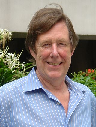
David John Hugh Cockayne FRS FInstP was Professor in the physical examination of materials in the Department of Materials at the University of Oxford and professorial fellow at Linacre College from 2000 to 2009. He was the president of the International Federation of Societies for Microscopy from 2003 till 2007, then vice-president 2007 to 2010.
Joseph Ó Ruanaidh is a scientist and frequently cited author in the field of digital watermarking.

Richard Henderson is a British molecular biologist and biophysicist and pioneer in the field of electron microscopy of biological molecules. Henderson shared the Nobel Prize in Chemistry in 2017 with Jacques Dubochet and Joachim Frank. "Thanks to his work, we can look at individual atoms of living nature, thanks to cryo-electron microscopes we can see details without destroying samples, and for this he won the Nobel Prize in Chemistry."

Bruker Corporation is an American manufacturer of scientific instruments for molecular and materials research, as well as for industrial and applied analysis. It is headquartered in Billerica, Massachusetts, and is the publicly traded parent company of Bruker Scientific Instruments and Bruker Energy & Supercon Technologies (BEST) divisions.
The Microscopy Society of America (MSA) was founded in 1942 as The Electron Microscope Society of America and is a non-profit organization that provides microanalytical facilities for studies within the sciences. Currently, there are approximately 3000 members. The society holds an annual meeting, which is usually held in the beginning of August. It has 30 local affiliates across the United States. The society has a program for examining and certifying technologists of electron microscopes. The organization produces two journals: Microscopy Today, and Microscopy and Microanalysis. As of 2024, the President is Jay Potts.

The Royal Microscopical Society (RMS) is a learned society for the promotion of microscopy. It was founded in 1839 as the Microscopical Society of London making it the oldest organisation of its kind in the world. In 1866, the society gained its royal charter and took its current name. Founded as a society of amateurs, its membership consists of individuals of all skill levels in numerous related fields from throughout the world. Every year since 1841, the society has published its own scientific journal, the Journal of Microscopy, which contains peer-reviewed papers and book reviews. The society is a registered charity that is dedicated to advancing science, developing careers and supporting wider understanding of science and microscopy through its Outreach activities.
The Journal of Cell Science is a peer-reviewed scientific journal in the field of cell biology. The journal is published by The Company of Biologists. The journal is partnered with Publons, is part of the Review Commons initiative and has two-way integration with bioRxiv. Journal of Cell Science is a hybrid journal and publishes 24 issues a year. Content over 6 months old is free to read.
Vernon Ellis Cosslett, FRS was a British microscopist.

The Journal of Cell Biology is a peer-reviewed scientific journal published by Rockefeller University Press.
David Bernard Williams was the dean of the College of Engineering at the Ohio State University from 2011-2021. He was previously the fifth president of the University of Alabama in Huntsville in Huntsville, Alabama from March 2007 until April 2011, and Vice Provost for Research and Harold Chambers Senior Professor of Materials Science and Engineering at Lehigh University in Bethlehem, Pennsylvania.
Colin James Richard Sheppard, usually cited as C. J. R. Sheppard, is senior scientist at the Italian Institute of Technology in Genoa, Italy. His areas of research are in optics, microscopy and imaging, including confocal and multiphoton microscopy, diffraction, 3D imaging and reconstruction, superresolution, beam propagation, and pulse propagation.

Ondrej L. Krivanek is a Czech/British physicist resident in the United States, and a leading developer of electron-optical instrumentation. He won the Kavli Prize for Nanoscience in 2020 for his substantial innovations in atomic resolution electron microscopy.
Peter Duncumb is a British physicist specialising in X-ray microscopy and microanalysis. He is best known for his contribution to the development of the first electron microprobe.

Lena Fitting Kourkoutis was an American physicist working in the field of electron microscopy, and a professor of applied and engineering physics at Cornell University. Her research focused on the use of aberration-corrected scanning transmission electron microscope, providing atomic resolution, at cryogenic temperatures (>77K) to study physical processes such as superconductivity and biological structures such as proteins.
The International Conference on X-Ray Microscopy (XRM) is a biennial international conference on X-ray imaging. The scope includes a range of topics in X-ray imaging, both in the soft and hard X-ray spectrum. Imaging by synchrotron light sources is the dominant topic, but small scale laboratory imaging is also included in many talks and posters. A number of subtopics are covered, including but not limited to X-ray microtomography, Phase-contrast X-ray imaging, Ptychography, and X-ray optics. The conference is typically five days long and held in summer. The conference is organized by an international committee and a local host organization. This local host is in most cases a synchrotron facility or an institute closely connected to a synchrotron. The host of the conference is decided two conferences in advance with a majority vote by all conference attendees. For example, the 14th XRM (2018) was decided during the 12th XRM (2014).
