Related Research Articles

Necrosis is a form of cell injury which results in the premature death of cells in living tissue by autolysis. The term "necrosis" came about in the mid-19th century and is commonly attributed to German pathologist Rudolf Virchow, who is often regarded as one of the founders of modern pathology. Necrosis is caused by factors external to the cell or tissue, such as infection, or trauma which result in the unregulated digestion of cell components. In contrast, apoptosis is a naturally occurring programmed and targeted cause of cellular death. While apoptosis often provides beneficial effects to the organism, necrosis is almost always detrimental and can be fatal.

A gumma is a soft, non-cancerous growth resulting from the tertiary stage of syphilis.

A lipoma is a benign tumor made of fat tissue. They are generally soft to the touch, movable, and painless. They usually occur just under the skin, but occasionally may be deeper. Most are less than 5 cm (2.0 in) in size. Common locations include upper back, shoulders, and abdomen. It is possible to have several lipomas.

A granuloma is an aggregation of macrophages that forms in response to chronic inflammation. This occurs when the immune system attempts to isolate foreign substances that it is otherwise unable to eliminate. Such substances include infectious organisms including bacteria and fungi, as well as other materials such as foreign objects, keratin, and suture fragments.

Dystrophic calcification (DC) is the calcification occurring in degenerated or necrotic tissue, as in hyalinized scars, degenerated foci in leiomyomas, and caseous nodules. This occurs as a reaction to tissue damage, including as a consequence of medical device implantation. Dystrophic calcification can occur even if the amount of calcium in the blood is not elevated, in contrast to metastatic calcification, which is a consequence of a systemic mineral imbalance, including hypercalcemia and/or hyperphosphatemia, that leads to calcium deposition in healthy tissues. In dystrophic calcification, basophilic calcium salt deposits aggregate, first in the mitochondria, then progressively throughout the cell. These calcifications are an indication of previous microscopic cell injury, occurring in areas of cell necrosis when activated phosphatases bind calcium ions to phospholipids in the membrane.
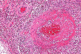
Fibrinoid necrosis is a pathological lesion that affects blood vessels, and is characterized by the occurrence of endothelial damage, followed by leakage of plasma proteins, including fibrinogen, from the vessel lumen; these proteins infiltrate and deposit within the vessel walls, where fibrin polymerization subsequently ensues.

A benign tumor is a mass of cells (tumor) that does not invade neighboring tissue or metastasize. Compared to malignant (cancerous) tumors, benign tumors generally have a slower growth rate. Benign tumors have relatively well differentiated cells. They are often surrounded by an outer surface or stay contained within the epithelium. Common examples of benign tumors include moles and uterine fibroids.

A hamartoma is a mostly benign, local malformation of cells that resembles a neoplasm of local tissue but is usually due to an overgrowth of multiple aberrant cells, with a basis in a systemic genetic condition, rather than a growth descended from a single mutated cell (monoclonality), as would typically define a benign neoplasm/tumor. Despite this, many hamartomas are found to have clonal chromosomal aberrations that are acquired through somatic mutations, and on this basis the term hamartoma is sometimes considered synonymous with neoplasm. Hamartomas are by definition benign, slow-growing or self-limiting, though the underlying condition may still predispose the individual towards malignancies.

A tuberculoma is a clinical manifestation of tuberculosis which conglomerates tubercles into a firm lump, and so can mimic cancer tumors of many types in medical imaging studies. They often arise within individuals in whom a primary tuberculosis infection is not well controlled. When tuberculomas arise intracranially, they represent a manifestation of CNS tuberculosis. Since these are evolutions of primary complex, the tuberculomas may contain caseum or calcifications.

Granuloma annulare (GA) is a rare, sometimes chronic skin condition which presents as reddish bumps on the skin arranged in a circle or ring. It can initially occur at any age, though two-thirds of patients are under 30 years old, and it is seen most often in children and young adults. Females are two times as likely to have it as males.

Commonly known as a dental cyst, the periapical cyst is the most common odontogenic cyst. It may develop rapidly from a periapical granuloma, as a consequence of untreated chronic periapical periodontitis.
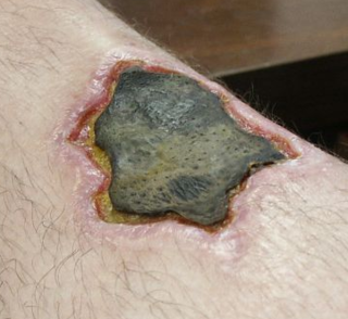
Loxoscelism is a condition occasionally produced by the bite of the recluse spiders. The area becomes dusky and a shallow open sore forms as the skin around the bite dies (necrosis). It is the only proven type of necrotic arachnidism in humans. While there is no known therapy effective for loxoscelism, there has been research on antibiotics, surgical timing, hyperbaric oxygen, potential antivenoms and vaccines. Because of the number of diseases that may mimic loxoscelism, it is frequently misdiagnosed by physicians.

Angiofibroma (AGF) is a descriptive term for a wide range of benign skin or mucous membrane lesions in which individuals have:
- benign papules, i.e. pinhead-sized elevations that lack visible evidence of containing fluid;
- nodules, i.e. small firm lumps usually > 1 mm in diameter; and/or
- tumors, i.e. masses often regarded as ~8 mm or larger.

Fat necrosis is necrosis affecting fat tissue. The term is well-established in medical terminology despite not denoting a specific pattern of necrosis. Fat necrosis may result from various injuries to adipose tissue, including: physical trauma, enzymatic digestion of adipocytes by lipases, radiation therapy, hypoxia, or inflammation of subcutaneous fat (panniculitis).
Infantile digital fibromatosis (IDF), also termed inclusion body fibromatosis or Reye's tumor, usually occurs as a single, small, asymptomatic, nodule in the dermis on a finger or toe of infants and young children. IMF is a rare disorder with approximately 200 cases reported in the medical literature as of 2021. The World Health Organization in 2020 classified these nodules as a specific benign tumor type in the category of fibroblastic and myofibroblastic tumors. IDF was first described by the Australian pathologist Douglas Reye in 1965.

Majocchi's granuloma is a skin condition characterized by deep, pustular plaques, and is a form of tinea corporis. It is a localized form of fungal folliculitis. Lesions often have a pink and scaly central component with pustules or folliculocentric papules at the periphery. The name comes from Domenico Majocchi, who discovered the disorder in 1883. Majocchi was a professor of dermatology at the University of Parma and later the University of Bologna. This disease is most commonly caused by filamentous fungi in the genus Trichophyton.
A coma blister, or coma bullae, is a skin lesion or blister that typically arises due to pressure in an individual with impaired consciousness. They vary in size, ranging from 4 to 5 centimeters in diameter, and may appear hemorrhagic or blood filled. Coma blisters are usually found in the extremities and trunk. These types of blisters have been associated with the overdose of central nervous system (CNS) depressants especially barbiturates, but also tricyclic antidepressants, hypnotics, benzodiazepines, opiates, antipsychotics, and alcohol. However, studies have found that coma blisters are not caused by the toxicity of these drugs, but due to hypoxia and external pressure on the comatose individual's skin from being immobilized. Coma blisters have been frequently found on individuals who have overdosed on drugs, but have also been found on individuals with chronic kidney failure, hypercalcemia, diabetic ketoacidosis, and a variety of neurologic conditions. Coma blisters are more frequent in adults and less common among children as demonstrated by the few cases published in literature.
Pulp necrosis is a clinical diagnostic category indicating the death of cells and tissues in the pulp chamber of a tooth with or without bacterial invasion. It is often the result of many cases of dental trauma, caries and irreversible pulpitis.

Palisaded encapsulated neuroma (PEN) is a rare, benign cutaneous condition characterized by small, firm, non-pigmented nodules or papules. They typically occur as a solitary (single) lesion near the mucocutaneous junction of the skin of the face, although they can occur elsewhere on the body.
Ultrasonography of liver tumors involves two stages: detection and characterization.
References
- 1 2 3 Ross, Casey L.; Ross, Nicholas A.; Lee, Jason B. (June 2021). "Mobile Encapsulated Fat Necrosis". Dermatologic Surgery. 47 (6): 880–882. doi:10.1097/DSS.0000000000002738. ISSN 1076-0512.
- 1 2 Sheehan, Connor; Tran, Theresa; Mollaee, Mehri; Hsu, Sylvia (2025-01-27). "A Case of Three Upper Extremity Lesions of Mobile Encapsulated Fat Necrosis in One Patient". Cureus. 17 (1). Springer Nature. doi: 10.7759/cureus.78080 . ISSN 2168-8184.
This article has not been added to any content categories . Please help out by adding categories to it so that it can be listed with similar articles. (January 2025) |