Modoc virus

Last updated
Modoc virus
DiGangi-Deermouse.jpg
Deer mouse, known host of Modoc virus
Virus classification OOjs UI icon edit-ltr.svg
(unranked): Virus
Realm: Riboviria
Kingdom: Orthornavirae
Phylum: Kitrinoviricota
Class: Flasuviricetes
Order: Amarillovirales
Family: Flaviviridae
Genus: Flavivirus
Species:
Modoc virus

Modoc virus (MODV) is a rodent-associated flavivirus. [1] Small and enveloped, MODV contains positive single-stranded RNA. [2] Taxonomically, MODV is part of the Flavivirus genus and Flaviviridae family. [1] [3] The Flavivirus genus includes nearly 80 viruses, [2] both vector-borne and no known vector (NKV) species. [4] Known flavivirus vector-borne viruses include Dengue virus, Yellow Fever virus, tick-borne encephalitis virus, and West Nile virus. [4]

Contents

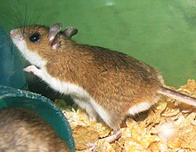
In 1958, MODV was first isolated from the mammary gland tissue of a white-footed deer mouse ( Peromyscus maniculatus ) captured in Modoc County, California. [1] [3] Since the first isolation, the MODV has also been isolated from deer mice in Oregon, Colorado, and Montana. [5] There are other anti-genetically and genetically related viruses which also have no known vector such as Jutiapa virus, the Cowbone Ridge virus, the Sal Vieja virus, and the San Perlita virus. [3] Little information is known about these viruses as well.

Structure

The MODV virus has a particle size of about 45 nm which is comparable with other flaviviruses particles that are about 40-60 nm in diameter. [6] A mature flavivirus has a spherical shape and contains multiple copies of three structural proteins (C, M, and E), a host-derived membrane bilayer, and a single copy of a positive-sense RNA genome of approximately 11,000 nucleotides. [7] The first structure of a flavivirus, the Dengue virus, was determined by using cryo-electron microscopy and an electron density map fitted with the known structure of glycoprotein E [8] (Fig.1).

Figure 1. Dengue virus three-dimensional cryo-electron microscopic reconstruction Image reconstructions from cryo-electron micrographs of flavivirus.png
Figure 1. Dengue virus three-dimensional cryo-electron microscopic reconstruction

Genome

Flaviviruses have positive (+) ssRNA genomes about 11kb in size. [4] The MODV genome is 10,600 nucleotides in length with a single open reading frame extending from nucleotides 110 to 10,234, encoding 3374 amino acids. [3] The ORF has the gene order C-prM-E-NS1-NS2A-NS2B-NS3-NS4A-NS4B-NS5 which is consistent with mosquito- and tick-borne flaviviruses [3] (Fig.2).

Figure 2. Flavivirus genome organization Genomic RNA of Flavivirus.png
Figure 2. Flavivirus genome organization

Replication cycle

Similar to most positive (+) ssRNA viruses, flaviviruses generate organelle-like structures in the endoplasmic reticulum (ER) of the host organism for replication. [9] Since the ER is involved in de novo biogenesis of some cell organelles, viruses take advantage of the replication location to take over some of the organelle functions for its own replication cycle. Viral genome replication in the ER occurs in structures called virus replication organelles. The organelles include two distinct subdomains, vesicle packets (VP) and convoluted membranes (CMs). [9] The site of viral genome replication is found within the vesicle packets which are clusters of small vesicle compartments. [9] The function of CMs is relatively unknown, but they are described as electron-dense amorphous structures near the VPs. [9] The large single polypeptide encoded by the genome is processed in the ER membrane by host or viral proteases. [9] The large polypeptide is divided into three structural proteins (capsid, prM, and E) and a group of non-structural proteins (NS1-NS5). [9] The viral genomic RNA forms a nucleocapsid complex with the capsid protein which aids in genome packaging into mature virus particles. [9] The prM and E proteins are considered significant components of the virus particle and can even form spherical virus particles. [9] The exact functions of NS proteins are relatively unknown, however, they are assumed to play a role in the formation of virus particle replication organelles. [9] The NS1 protein has a large ectodomain which is believed to function in the deformation of the ER membrane from the luminal side. [9] NS2B protein, a transmembrane protein, directly interacts with NS3 which is a soluble protein anchored to the membrane. [9] With its protease activity and RNA helicase activity, the NS3 protein is involved in viral polyprotein processing and viral RNA replication. [9] NS5 plays a role in the replication of the viral genomic RNA and the formation of the 5’-cap structure for protein translation with its RNA dependent and RNA polymerase (RdRp) activity and methyltransferase activity. [9] The 5′-end possesses a type I cap (m7GpppAmp) that is not seen in viruses of the other genera. [10] Proteins N2SA, NS4A, and NS4B are membrane-integrated proteins but have no clear function. [9]

Life cycle

Schematic model of single-stranded positive-sense RNA viruses Virus Entry, Replication, and Assembly.png
Schematic model of single-stranded positive-sense RNA viruses

Entry into cell

To enter the cell, MODV virus is assumed to invade the cell via steps similar to the Flavivirus family. Through endocytosis, the virus enters the host cell and then releases its positive (+) ssRNA genome into the cytoplasm through membrane fusion. [9]

Replication and transcription

The MDOV genome codes for a single ORF, which is processed by both cellular and viral proteases to form three structural proteins and at least seven non-structural proteins. [4]

On either side of the ORF, untranslated regions (UTRs) are present and fold into complex stem-loop structures required for replication. [4] The 5’ UTR consists of 109 nucleotides and the 3’ UTR consists of 366 nucleotides. [3] Comparing MODV to 20 other flaviviruses, several regions with high sequence similarity appeared. The regions corresponded to functionally important domains and conserved sites for proteolytic cleavage by viral and cellular proteases. [3] MDOV transcription proceeds according to the positive (+) RNA strand model.[ citation needed ]

Assembly and release

The rough endoplasmic reticulum is believed to be the site of viral assembly. [10] Following genome replication, the newly synthesized RNA interacts with the capsid and buds into the ER lumen along with immature prM and E proteins which undergo maturation in the Golgi and endosomes. [9] For instance, the prM protein is cleaved by furin or a furin-like cellular protease to generate mature virions. [10] The virion moves through the cytoplasm until it is released from the cell via exocytosis. [10]

Transmission and tropism

Since no vector has been identified for MODV, [3] the exact mode transmission is not known. However, the studied field strain of MODV showed persistent infection of the virus in deer mouse lungs that may be transmitted horizontally through close, prolonged contact of infected and susceptible individuals. [11] Direct contact (i.e., salivary secretions) or indirect contact (i.e., fomites, aerosols and urine) may enable viral spread. Rodent nests during the winter provide conditions suitable for the horizontal transmission of viruses. [12] In addition, transmission presumably occurs horizontally since attempts to infect ticks and mosquitoes as cultured cell lines or in vivo have been unsuccessful. [4] Other studies suggest cannibalism does not play a direct role in viral transmission, but the possibility of sexual transmission has yet to be explored. [12]

The molecular determinants of transmission are unknown, but comparison of the conserved sequence differences between the two groups suggests vector-borne conserved pentanucleotide sequence (CPS) nor variable region (VR) of the conserved sequences are required for vector-borne transmission. [4]

A transient viremia in deer mice ( Peromyscus maniculatus ) produced a measurable production of antibody titers and showed persistence in the lungs. [11] While deer mice are accepted as the main host organism, the presence of antibodies in chipmunks ( Tamias minimus ) and red squirrels (Tamias-ciurus hudsonicus) suggests the virus has multiple hosts. [11] Virus transmission in chipmunks and red squirrels was more successful than in deer mice, which may be due to a difference in viral shedding under experimental conditions and field conditions. [11] Field conditions may cause more stress due to cold temperatures and food shortages. [11]

Associated diseases

Modoc virus antigen in spinal cord and brain Modoc virus in tissues.png
Modoc virus antigen in spinal cord and brain

A rodent-associated virus, MODV has the potential to cause disease in humans similar to other flaviviruses. [11] It was determined that MODV was the responsible virus for a case of aseptic meningitis when the virus was first discovered in California. [3]

Typically, flaviviruses cause encephalitis in host organisms. MODV causes flavivirus-like encephalitis in SCID (severe combined immunodeficiency) mice and in hamsters with histopathological features reminiscent of flavivirus encephalitis in man. [3] Studies suggest envelope (E) proteins encoded for by the genome may play a dominant role as a determinant of flavivirus neurovirulence. [2] A single amino acid substitution was shown to cause major effects on neurovirulence. [2] The mechanisms and determinants involved in Flavivirus neuroinvasiveness remains unknown. [2] However, evidence suggests neuroinvasiveness depends entirely on envelope proteins E and prM (pre-membrane) [2]

In a study investigating the effects of MODV on hamsters, severe encephalitis, bilateral hindlimb paralysis, and complete paralysis with an intact corneal reflex were observed. [1] MODV-induced encephalitis in hamsters was characterized by movement of monocytes and lymphocytes into the cortex and bulbus olfactorius, causing massive destruction of the tissue structure. [6] In all surviving hamsters, IgM and HI antibodies to MODV were present in the blood after subcutaneous infection. [1] Of all infected hamsters, no substantial microscopic lesions were observed in the liver, spleen, lung, and heart. [1] However, rare focal portal inflammation of the liver and mild reactive lymphoid hyperplasia of the spleen were noted. Considerable pathological changes were observed in the brain and spinal cord during early infection. [1] During early infection, the lesions in the spinal cord were more severe than those observed in the brain. [1] In immunocompetent mice, MODV causes 100% morbidity and mortality when the virus was inoculated directly into the brain. [6] Alternatively, when inoculated via the intranasal route, 50% morbidity and mortality was observed [6]

Infectious MODV was also isolated from kidney tissue for at least eight months after infection, specifically the epithelium of the renal tubules. [1] Despite the presence of antibodies, infected hamsters continued to shed viruses in the urine for up to four months. [1] [3] Due to viral shedding in the urine, there is the potential use of urinalysis to monitor the effectiveness of therapy on viral replication by monitoring the viral RNA in the urine using quantitative RT-PCR assays [6]

Related Research Articles

<i>Flaviviridae</i> Family of viruses

Flaviviridae is a family of enveloped positive-strand RNA viruses which mainly infect mammals and birds. They are primarily spread through arthropod vectors. The family gets its name from the yellow fever virus; flavus is Latin for "yellow", and yellow fever in turn was named because of its propensity to cause jaundice in victims. There are 89 species in the family divided among four genera. Diseases associated with the group include: hepatitis (hepaciviruses), hemorrhagic syndromes, fatal mucosal disease (pestiviruses), hemorrhagic fever, encephalitis, and the birth defect microcephaly (flaviviruses).

<i>Flavivirus</i> Genus of viruses

Flavivirus, renamed Orthoflavivirus in 2023, is a genus of positive-strand RNA viruses in the family Flaviviridae. The genus includes the West Nile virus, dengue virus, tick-borne encephalitis virus, yellow fever virus, Zika virus and several other viruses which may cause encephalitis, as well as insect-specific flaviviruses (ISFs) such as cell fusing agent virus (CFAV), Palm Creek virus (PCV), and Parramatta River virus (PaRV). While dual-host flaviviruses can infect vertebrates as well as arthropods, insect-specific flaviviruses are restricted to their competent arthropods. The means by which flaviviruses establish persistent infection in their competent vectors and cause disease in humans depends upon several virus-host interactions, including the intricate interplay between flavivirus-encoded immune antagonists and the host antiviral innate immune effector molecules.

<i>Sin Nombre orthohantavirus</i> Prototypical agent of hantavirus cardiopulmonary syndrome

Sin Nombre orthohantavirus (SNV), a member of the genus Orthohantavirus, is the prototypical etiologic agent of hantavirus cardiopulmonary syndrome (HCPS).

<span class="mw-page-title-main">Plant virus</span> Virus that affects plants

Plant viruses are viruses that affect plants. Like all other viruses, plant viruses are obligate intracellular parasites that do not have the molecular machinery to replicate without a host. Plant viruses can be pathogenic to vascular plants.

<i>Tick-borne encephalitis virus</i> Species of virus

Tick-borne encephalitis virus (TBEV) is a positive-strand RNA virus associated with tick-borne encephalitis in the genus Flavivirus.

<i>Semliki Forest virus</i> Species of virus

The Semliki Forest virus is an alphavirus found in central, eastern, and southern Africa. It was first isolated from mosquitoes in the Semliki Forest, Uganda by the Uganda Virus Research Institute in 1942 and described by Smithburn and Haddow. It is known to cause disease in animals including humans.

<i>Alphavirus</i> Genus of viruses

Alphavirus is a genus of RNA viruses, the sole genus in the Togaviridae family. Alphaviruses belong to group IV of the Baltimore classification of viruses, with a positive-sense, single-stranded RNA genome. There are 32 alphaviruses, which infect various vertebrates such as humans, rodents, fish, birds, and larger mammals such as horses, as well as invertebrates. Alphaviruses that could infect both vertebrates and arthropods are referred dual-host alphaviruses, while insect-specific alphaviruses such as Eilat virus and Yada yada virus are restricted to their competent arthropod vector. Transmission between species and individuals occurs mainly via mosquitoes, making the alphaviruses a member of the collection of arboviruses – or arthropod-borne viruses. Alphavirus particles are enveloped, have a 70 nm diameter, tend to be spherical, and have a 40 nm isometric nucleocapsid.

<i>Pestivirus</i> Genus of viruses

Pestivirus is a genus of viruses, in the family Flaviviridae. Viruses in the genus Pestivirus infect mammals, including members of the family Bovidae and the family Suidae. There are 11 species in this genus. Diseases associated with this genus include: hemorrhagic syndromes, abortion, and fatal mucosal disease.

Powassan virus (POWV) is a Flavivirus transmitted by ticks, found in North America and in the Russian Far East. It is named after the town of Powassan, Ontario, where it was identified in a young boy who eventually died from it. It can cause encephalitis, inflammation of the brain. No approved vaccine or antiviral drug exists. Prevention of tick bites is the best precaution.

<i>Orthobunyavirus</i> Genus of viruses

Orthobunyavirus is a genus of the Peribunyaviridae family in the order Bunyavirales. There are currently ~170 viruses recognised in this genus. These have been assembled into 103 species and 20 serogroups.

<span class="mw-page-title-main">Veterinary virology</span> Study of viruses affecting animals

Veterinary virology is the study of viruses in non-human animals. It is an important branch of veterinary medicine.

Spondweni virus is an arbovirus, or arthropod-borne virus, which is a member of the family Flaviviridae and the genus Flavivirus. It is part of the Spondweni serogroup which consists of the Sponweni virus and the Zika virus (ZIKV). The Spondweni virus was first isolated in Nigeria in 1952, and ever since, SPONV transmission and activity have been reported throughout Africa. Its primary vector of transmission is the sylvatic mosquito Aedes circumluteolus, though it has been isolated from several different types of mosquito. Transmission of the virus into humans can lead to a viral infection known as Spondweni fever, with symptoms ranging from headache and nausea to myalgia and arthralgia. However, as SPONV is phylogenetically close to the ZIKV, it is commonly misdiagnosed as ZIKV along with other viral illnesses.

Royal Farm virus, previously known as Karshi virus, was not viewed as pathogenic or harmful to humans. Although infected people suffer with fever-like symptoms, some people in Uzbekistan have reported with severe disease such as encephalitis and other large outbreaks of fever illness connected infection with the virus.

<i>West Nile virus</i> Species of flavivirus causing West Nile fever

West Nile virus (WNV) is a single-stranded RNA virus that causes West Nile fever. It is a member of the family Flaviviridae, from the genus Flavivirus, which also contains the Zika virus, dengue virus, and yellow fever virus. The virus is primarily transmitted by mosquitoes, mostly species of Culex. The primary hosts of WNV are birds, so that the virus remains within a "bird–mosquito–bird" transmission cycle. The virus is genetically related to the Japanese encephalitis family of viruses. Humans and horses both exhibit disease symptoms from the virus, and symptoms rarely occur in other animals.

<span class="mw-page-title-main">Positive-strand RNA virus</span> Class of viruses in the Baltimore classification

Positive-strand RNA viruses are a group of related viruses that have positive-sense, single-stranded genomes made of ribonucleic acid. The positive-sense genome can act as messenger RNA (mRNA) and can be directly translated into viral proteins by the host cell's ribosomes. Positive-strand RNA viruses encode an RNA-dependent RNA polymerase (RdRp) which is used during replication of the genome to synthesize a negative-sense antigenome that is then used as a template to create a new positive-sense viral genome.

<span class="mw-page-title-main">Palm Creek virus</span> Species of virus

Palm Creek virus (PCV) is an insect virus belonging to the genus Flavivirus, of the family Flaviviridae. It was discovered in 2013 from the mosquito Coquillettidia xanthogaster. The female mosquitoes were originally collected in 2010 from Darwin, Katherine, Alice Springs, Alyangula, Groote Eylandt, Jabiru and the McArthur River Mine, and had since been preserved. The discovery was made by biologists at the University of Queensland. The virus is named after Palm Creek, near Darwin, from where it was originally isolated.

Yokose virus (YOKV) is in the genus Flavivirus of the family Flaviviridae. Flaviviridae are often found in arthropods, such as mosquitoes and ticks, and may also infect humans. The genus Flavivirus includes over 50 known viruses, including Yellow Fever, West Nile Virus, Zika Virus, and Japanese Encephalitis. Yokose virus is a new member of the Flavivirus family that has only been identified in a few bat species. Bats have been associated with several emerging zoonotic diseases such as Ebola and SARS.

<i>Sepik virus</i> Mosquito transmitted virus endemic to Papua New Guinea

Sepik virus (SEPV) is an arthropod-borne virus (arbovirus) of the genus Flavivirus and family Flaviviridae. Flaviviridae is one of the most well characterized viral families, as it contains many well-known viruses that cause diseases that have become very prevalent in the world, like Dengue virus. The genus Flavivirus is one of the largest viral genera and encompasses over 50 viral species, including tick and mosquito borne viruses like Yellow fever virus and West Nile virus. Sepik virus is much less well known and has not been as well-classified as other viruses because it has not been known of for very long. Sepik virus was first isolated in 1966 from the mosquito Mansoniaseptempunctata, and it derives its name from the Sepik River area in Papua New Guinea, where it was first found. The geographic range of Sepik virus is limited to Papua New Guinea, due to its isolation.

Rio Negro virus is an alphavirus that was first isolated in Argentina in 1980. The virus was first called Ag80-663 but was renamed to Rio Negro virus in 2005. It is a former member of the Venezuelan equine encephalitis complex (VEEC), which are a group of alphaviruses in the Americas that have the potential to emerge and cause disease. Río Negro virus was recently reclassified as a distinct species. Closely related viruses include Mucambo virus and Everglades virus.

Flavivirus 3' UTR are untranslated regions in the genome of viruses in the genus Flavivirus.

References

  1. 1 2 3 4 5 6 7 8 9 10 Adams, A. Paige; Travassos da Rosa, Amelia P. A.; Nunes, Marcio R.; Xiao, Shu-Yuan; Tesh, Robert B. (March 2013). "Pathogenesis of Modoc virus (Flaviviridae; Flavivirus) in persistently infected hamsters". The American Journal of Tropical Medicine and Hygiene. 88 (3): 455–460. doi:10.4269/ajtmh.12-0110. ISSN   1476-1645. PMC   3592524 . PMID   23358636.
  2. 1 2 3 4 5 6 Charlier, Nathalie; Molenkamp, Richard; Leyssen, Pieter; Paeshuyse, Jan; Drosten, Christian; Panning, Marcus; De Clercq, Erik; Bredenbeek, Peter J.; Neyts, Johan (July 2004). "Exchanging the yellow fever virus envelope proteins with Modoc virus prM and E proteins results in a chimeric virus that is neuroinvasive in SCID mice". Journal of Virology. 78 (14): 7418–7426. doi:10.1128/JVI.78.14.7418-7426.2004. ISSN   0022-538X. PMC   434118 . PMID   15220415.
  3. 1 2 3 4 5 6 7 8 9 10 11 Leyssen, Pieter; Charlier, Nathalie; Lemey, Philippe; Billoir, Frédérique; Vandamme, Anne-Mieke; De Clercq, Erik; de Lamballerie, Xavier; Neyts, Johan (2002-02-01). "Complete genome sequence, taxonomic assignment, and comparative analysis of the untranslated regions of the Modoc virus, a flavivirus with no known vector". Virology. 293 (1): 125–140. doi: 10.1006/viro.2001.1241 . ISSN   0042-6822. PMID   11853406.
  4. 1 2 3 4 5 6 7 Tumban, Ebenezer; Maes, Nyree E.; Schirtzinger, Erin E.; Young, Katherine I.; Hanson, Christopher T.; Whitehead, Stephen S.; Hanley, Kathryn A. (April 2013). "Replacement of conserved or variable sequences of the mosquito-borne dengue virus 3′ UTR with homologous sequences from Modoc virus does not change infectivity for mosquitoes". The Journal of General Virology. 94 (Pt 4): 783–788. doi:10.1099/vir.0.046664-0. ISSN   0022-1317. PMC   3709684 . PMID   23255623.
  5. Zarnke, Randall L.; Yuill, Thomas M. (April 1985). "Modoc-Like Virus Isolated from Wild Deer Mice (Peromyscus Maniculatus) in Alberta". Journal of Wildlife Diseases. 21 (2): 94–99. doi:10.7589/0090-3558-21.2.94. ISSN   0090-3558. PMID   2987550.
  6. 1 2 3 4 5 Leyssen, Pieter; Van Lommel, Alfons; Drosten, Christian; Schmitz, Herbert; De Clercq, Erik; Neyts, Johan (2001-01-05). "A Novel Model for the Study of the Therapy of Flavivirus Infections Using the Modoc Virus". Virology. 279 (1): 27–37. doi: 10.1006/viro.2000.0723 . ISSN   0042-6822. PMID   11145886.
  7. Jones, Christopher T.; Ma, Lixin; Burgner, John W.; Groesch, Teresa D.; Post, Carol B.; Kuhn, Richard J. (June 2003). "Flavivirus Capsid Is a Dimeric Alpha-Helical Protein". Journal of Virology. 77 (12): 7143–7149. doi:10.1128/JVI.77.12.7143-7149.2003. ISSN   0022-538X. PMC   156156 . PMID   12768036.
  8. Kuhn, Richard J.; Zhang, Wei; Rossmann, Michael G.; Pletnev, Sergei V.; Corver, Jeroen; Lenches, Edith; Jones, Christopher T.; Mukhopadhyay, Suchetana; Chipman, Paul R.; Strauss, Ellen G.; Baker, Timothy S. (2002-03-08). "Structure of Dengue Virus: Implications for Flavivirus Organization, Maturation, and Fusion". Cell. 108 (5): 717–725. doi:10.1016/S0092-8674(02)00660-8. ISSN   0092-8674. PMC   4152842 . PMID   11893341.
  9. 1 2 3 4 5 6 7 8 9 10 11 12 13 14 15 16 Arakawa, Masashi; Morita, Eiji (2019-05-11). "Flavivirus Replication Organelle Biogenesis in the Endoplasmic Reticulum: Comparison with Other Single-Stranded Positive-Sense RNA Viruses". International Journal of Molecular Sciences. 20 (9): 2336. doi: 10.3390/ijms20092336 . ISSN   1422-0067. PMC   6539296 . PMID   31083507.
  10. 1 2 3 4 "Genus: Flavivirus - Flaviviridae - Positive-sense RNA Viruses". International Committee on Taxonomy of Viruses (ICTV). Retrieved 2019-12-14.
  11. 1 2 3 4 5 6 Fairbrother, A.; Yuill, T. M. (April 1987). "Experimental infection and horizontal transmission of Modoc virus in deer mice (Peromyscus maniculatus)". Journal of Wildlife Diseases. 23 (2): 179–185. doi: 10.7589/0090-3558-23.2.179 . ISSN   0090-3558. PMID   3035240.
  12. 1 2 Blitvich, Bradley J.; Firth, Andrew E. (June 21, 2017). "A Review of Flaviviruses that Have No Known Arthropod Vector". Viruses. 9 (6): 154. doi: 10.3390/v9060154 . ISSN   1999-4915. PMC   5490829 . PMID   28635667.