Monoclonal gammopathy of renal significance

Last updated
Monoclonal gammopathy of renal significance
Specialty Nephrology, Pathology, Hematology
Complications Kidney failure,
Risk factors Presence of a monoclonal gammopathy, age greater than 50
Diagnostic method Kidney biopsy, serum protein electrophoresis with immunofixation, serum free light chain assay
Differential diagnosis Multiple myeloma, Waldenstrom's macroglobulinemia, Non-Hodgkin lymphoma, Chronic lymphocytic leukemia
TreatmentClone directed therapy (anti-cancer therapy), immunomodulating medications (secondary approach), autologous stem cell transplantation, kidney transplantation,

Monoclonal gammopathy of renal significance (MGRS) are a group of kidney disorders that present with kidney damage due to nephrotoxic monoclonal immunoglobulins (M proteins) secreted by clonal plasma cells or B cells. By definition, people with MGRS do not meet criteria for multiple myeloma or other hematologic malignancies. [1] The term MGRS was introduced in 2012 by the International Kidney and Monoclonal Gammopathy Research Group (IKMG). MGRS is associated with monoclonal gammopathy of undetermined significance (MGUS). People with MGUS have a monoclonal gammopathy (presence of monoclonal immunoglobulins) but does not meet the criteria for the clonal burden nor the presence of end organ damage seen in hematologic malignancies. In a population based study based on the NHANES III health survey; 6% of patients with MGUS were subsequently classified as having MGRS. [2] The prevalence and incidence of MGRS in the general population or in specific populations is not known but it is more prevalent in those over the age of 50 as there is a monoclonal protein (M-protein) present in 3% of those 50 and years older and 5% of those 70 years and older (the presence of MGUS), placing those 50 and older at increased risk of MGRS. [1]

Contents

MGRS disorders generally do not respond well to immunosuppressive treatment. [3] MGRS disorders also have a greater than 90% rate of recurrence if the monoclonal gammopathy is not eliminated either before or immediately after a renal transplant. [3] People with MGRS are at risk of progression to the corresponding hematologic malignancy. [3]

Pathophysiology

There are several separate conditions designated as MGRS which are associated with kidney damage (either directly or indirectly) due to monoclonal immunoglobulins (M proteins). Most kidney diseases associated with MGRS are glomerular disorders. [3]

AL amyloidosis is an immunoglobulin light chain associated amyloidosis that involves the deposition of misfolded amyloid light chain (AL) immunoglobulins. These AL light chains are misfolded leading to the disordered deposition of amyloid multimers and fibrils in the glomeruli and blood vessels of the kidney. [3] Other types of amyloidosis related MGRS disorders include heavy chain amyloidosis (deposition of only heavy amyloid chains in the kidney)(AH) and heavy and light chain amyloidosis (AHL)(deposition of heavy and light chains). Amyloidosis is the most common form of MGRS, with the AL subtype being the most common type. [3] Extra-renal manifestations, such as heart failure, gastrointestinal symptoms, secondary neuropathy (carpal tunnel syndrome), liver involvement and peripheral neuropathy are very common with amyloidosis related MGRS. [3] Light microscopy findings in AL, AH or AHL amyloidosis include acellular deposits in the glomeruli and blood vessels that stain pale eosinophilic, Congo red positive and the presence of apple green birefringence on polarized light. [1] Findings on electron microscopy include non-branching fibrils randomly arranged with thickness of 7-14 nm. [1]

Light chain proximal tubulopathy (LCPT) involves light chains with mutations of the hydrophobic residues of the variable domain causing them to have resistance to proteolysis; this causes the light chains to form intracytoplasmic aggregates in the proximal tubule of the nephron. [3] Light microscopy findings in LCPT include proximal tubular swelling with electron microscopy findings showing proximal tubule light chain crystals or lysosomal inclusions. [3]

Proliferative glomerulonephritis with monoclonal immunoglobulin deposits (PGNMID) involves monoclonal immunoglobulins (usually IgG) depositing in the glomeruli and activating complement leading to glomerular inflammation. [3] Light microscopy shows a membranoproliferative, endocapillary proliferative or membranous glomerulonephropathy with electron dense deposits in the glomeruli being present on electron microscopy. The lesions stain positive for the Ig (usually IgG) as well as complement; leading to granular immunofluorescent deposits in the mesangium and glomerular basement membrane. [3]

Monoclonal immunoglobulin deposition disease (MIDD) involves light chains with unusual characteristics of the variable domain (such as a positive charge, abnormal glycosylation, hydrophobic residues) depositing in the tubular, vascular or glomerular basement membranes of the nephron. [3] These immune deposits activate TGF-β which leads to matrix accumulation and remodeling of the mesangium. The heavy chain variant of MIDD occurs when a deletion of the first constant domain of immunoglobulin heavy chains renders them unable to bind to light chains to form a complete immunoglobulin; the heavy chains are then deposited in the glomeruli. [3] On light microscopy MIDD is seen as a nodular glomerulosclerosis with thickening of the tubular basement membrane with linear deposits along the glomerular, tubular vascular basement membranes seen on immunofluorescence. [3]

C3 glomerulopathy is characterized by glomerular deposits of C3 complement protein (with scant or absent Ig immunoglobulin deposits) due to mutations or inhibition of complement regulatory proteins causing dysregulation of the alternative complement pathway. [4] C3 glomerulopathy presents in light microscopy as mesangial proliferative, membranoproliferative or endocapillary proliferative glomerulonephritis. [4] Unlike other causes of MGRS, the monoclonal immunoglobulins in C3 glomerulopathy are not deposited in the kidney nor do they directly cause kidney damage. In C3 glomerulopathy monoclonal immunoglobulins inhibit factor H (a complement regulatory protein) causing uninhibited activation of the alternative complement pathway and C3 deposition in the kidneys. [4]

Cryoglobulinemic glomerulonephritis is characterized by glomerular involvement in those with cryoglobulinemia. In light microscopy it presents as a membranoproliferative or endocapillary proliferative glomerulonephritis with intracapillary monocytes and immune deposits that stain positive for PAS. [4] On immunofluorescence the deposits consist of monoclonal light and heavy chains (most commonly IgG) and complement. [4] Immunofluorescence can help distinguish the various subtypes of cryoglobulinemic glomerulonephritis. [4] In a subtype of cryoglobulinemic glomerulonephritis known as crystalglobulinemia; monoclonal immunoglobulins may precipitate in the small arterioles and capillaries of the glomeruli when exposed to colder temperatures causing endothelial injury and microthrombi. [3]

Diagnosis

Several clinical signs are associated with MGRS including declining kidney function, microscopic hematuria, proteinuria (ranging from sub-nephrotic to nephrotic syndrome range proteinuria) and proximal tubular dysfunction which may present as Fanconi Syndrome. [1] Once MGRS is suspected, it is critical to identify the monoclonal protein responsible for kidney toxicity or the cell line secreting the monoclonal protein. [3] Usually MGRS disorders have small levels of circulating M-proteins (due to the small clonal burden of the underlying B-cell or plasma cell disorder) thus decreasing the sensitivity of serum protein electrophoresis (SPEP). [1] Serum immunofixation electrophoresis is 10 times more sensitive than SPEP in the identification of M-proteins in MGRS. [1] Serum protein electrophoresis, urine protein electrophoresis (UPEP), serum and urine immunofixation, serum free light chain assay, serum free light chain ratios and a bone marrow biopsy are required for the identification of the cell population secreting the pathologic monoclonal protein. A kidney biopsy should be performed in all cases of MGRS to confirm the M-protein's causal relationship with regards to kidney disease. [1] The only exception is AL amyloidosis which can be diagnosed if AL deposits are detected in other tissues, such as peripheral fat. [5] Overt hematologic malignancies such as multiple myeloma should be excluded. [1]

Treatment

The optimal treatment in MGRS is a clone directed therapy; where treatment is directed specifically to the cell line responsible for the pathologic monoclonal immunoglobulin or M-protein. [3] The goal of therapy is to preserve kidney function, reduce the risk of MGRS recurrence after kidney transplant and maintain a sustained hematologic response. The difference of involved-uninvolved free light chains less than 4 or greater than a 90% reduction of involved free light chains are the minimum hematologic responses needed for the preservation of kidney function. [3]

Plasma cell dyscrasias are best treated with the proteasome inhibitor bortezomib, often given with the steroid dexamethasone. [1] Bortezomib can also be given before and after an autologous stem cell transplant (ASCT) with high dose of the chemotherapy medication mephalan given before ASCT to eradicate the monoclonal gammopathy prior to transplant and prevent recurrence. [1] Alternative treatment options include immunomodulatory drugs such as thalidomide or lenalidomide. [1] The monoclonal antibody against the plasma cell surface protein CD38 daratumumab may also be used with very high efficacy against AL amyloidosis. [3]

B-cell dyscrasias associated with MGRS have less evidence for specific medications that are effective in achieving a hematologic response. With CD20 expressing B-cells and lymphoplasmacytic clones; rituximab is the preferred treatment and it can be combined with dexamethasone and cyclophosphamide. [1] The benefit of ASCT in B-cell related MGRS is less established. [1]

Prognosis

Overall mortality of the various MGRS disorders is generally less than that of multiple myeloma, but light chain amyloidosis (AL type) with cardiac involvement is associated with rapid progression to death. [5]

Related Research Articles

<span class="mw-page-title-main">Serum protein electrophoresis</span> Laboratory test

Serum protein electrophoresis is a laboratory test that examines specific proteins in the blood called globulins. The most common indications for a serum protein electrophoresis test are to diagnose or monitor multiple myeloma, a monoclonal gammopathy of uncertain significance (MGUS), or further investigate a discrepancy between a low albumin and a relatively high total protein. Unexplained bone pain, anemia, proteinuria, chronic kidney disease, and hypercalcemia are also signs of multiple myeloma, and indications for SPE. Blood must first be collected, usually into an airtight vial or syringe. Electrophoresis is a laboratory technique in which the blood serum is applied to either an acetate membrane soaked in a liquid buffer, or to a buffered agarose gel matrix, or into liquid in a capillary tube, and exposed to an electric current to separate the serum protein components into five major fractions by size and electrical charge: serum albumin, alpha-1 globulins, alpha-2 globulins, beta 1 and 2 globulins, and gamma globulins.

<span class="mw-page-title-main">IgA nephropathy</span> Disease of the kidney

IgA nephropathy (IgAN), also known as Berger's disease, or synpharyngitic glomerulonephritis, is a disease of the kidney and the immune system; specifically it is a form of glomerulonephritis or an inflammation of the glomeruli of the kidney. Aggressive Berger's disease can attack other major organs, such as the liver, skin and heart.

<span class="mw-page-title-main">Glomerulonephritis</span> Term for several kidney diseases

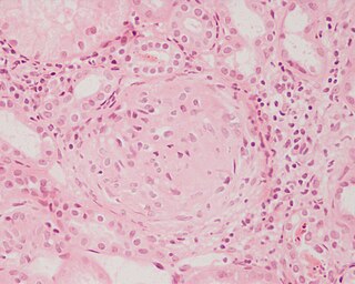
Glomerulonephritis (GN) is a term used to refer to several kidney diseases. Many of the diseases are characterised by inflammation either of the glomeruli or of the small blood vessels in the kidneys, hence the name, but not all diseases necessarily have an inflammatory component.

<span class="mw-page-title-main">Cryoglobulinemia</span> Medical condition

Cryoglobulinemia is a medical condition in which the blood contains large amounts of pathological cold sensitive antibodies called cryoglobulins – proteins that become insoluble at reduced temperatures. This should be contrasted with cold agglutinins, which cause agglutination of red blood cells.

<span class="mw-page-title-main">Membranous glomerulonephritis</span> Medical condition

Membranous glomerulonephritis (MGN) is a slowly progressive disease of the kidney affecting mostly people between ages of 30 and 50 years, usually white people.

<span class="mw-page-title-main">Monoclonal gammopathy of undetermined significance</span> Medical condition

Monoclonal gammopathy of undetermined significance (MGUS) is a plasma cell dyscrasia in which plasma cells or other types of antibody-producing cells secrete a myeloma protein, i.e. an abnormal antibody, into the blood; this abnormal protein is usually found during standard laboratory blood or urine tests. MGUS resembles multiple myeloma and similar diseases, but the levels of antibodies are lower, the number of plasma cells in the bone marrow is lower, and it rarely has symptoms or major problems. However, since MGUS can lead to multiple myeloma, which develops at the rate of about 1.5% a year, or other symptomatic conditions, yearly monitoring is recommended.

<span class="mw-page-title-main">Monoclonal gammopathy</span> Excess myeloma protein or monoclonal gamma globulin in the blood

Monoclonal gammopathy, also known as paraproteinemia, is the presence of excessive amounts of myeloma protein or monoclonal gamma globulin in the blood. It is usually due to an underlying immunoproliferative disorder or hematologic neoplasms, especially multiple myeloma. It is sometimes considered equivalent to plasma cell dyscrasia. The most common form of the disease is monoclonal gammopathy of undetermined significance.

<span class="mw-page-title-main">Rapidly progressive glomerulonephritis</span> Medical condition

Rapidly progressive glomerulonephritis (RPGN) is a syndrome of the kidney that is characterized by a rapid loss of kidney function, with glomerular crescent formation seen in at least 50% or 75% of glomeruli seen on kidney biopsies. If left untreated, it rapidly progresses into acute kidney failure and death within months. In 50% of cases, RPGN is associated with an underlying disease such as Goodpasture syndrome, systemic lupus erythematosus or granulomatosis with polyangiitis; the remaining cases are idiopathic. Regardless of the underlying cause, RPGN involves severe injury to the kidneys' glomeruli, with many of the glomeruli containing characteristic glomerular crescents.

<span class="mw-page-title-main">Membranoproliferative glomerulonephritis</span> Medical condition

Membranoproliferative glomerulonephritis (MPGN) is a type of glomerulonephritis caused by deposits in the kidney glomerular mesangium and basement membrane (GBM) thickening, activating the complement system and damaging the glomeruli.

<span class="mw-page-title-main">Myeloma protein</span> Abnormal immunoglobulin fragment

A myeloma protein is an abnormal antibody (immunoglobulin) or a fragment thereof, such as an immunoglobulin light chain, that is produced in excess by an abnormal monoclonal proliferation of plasma cells, typically in multiple myeloma or Monoclonal gammopathy of undetermined significance. Other terms for such a protein are monoclonal protein, M protein, M component, M spike, spike protein, or paraprotein. This proliferation of the myeloma protein has several deleterious effects on the body, including impaired immune function, abnormally high blood viscosity, and kidney damage.

In hematology, plasma cell dyscrasias are a spectrum of progressively more severe monoclonal gammopathies in which a clone or multiple clones of pre-malignant or malignant plasma cells over-produce and secrete into the blood stream a myeloma protein, i.e. an abnormal monoclonal antibody or portion thereof. The exception to this rule is the disorder termed non-secretory multiple myeloma; this disorder is a form of plasma cell dyscrasia in which no myeloma protein is detected in serum or urine of individuals who have clear evidence of an increase in clonal bone marrow plasma cells and/or evidence of clonal plasma cell-mediated tissue injury. Here, a clone of plasma cells refers to group of plasma cells that are abnormal in that they have an identical genetic identity and therefore are descendants of a single genetically distinct ancestor cell.

<span class="mw-page-title-main">Mesangial proliferative glomerulonephritis</span> Medical condition

Mesangial proliferative glomerulonephritis (MesPGN) is a morphological pattern characterized by a numerical increase in mesangial cells and expansion of the extracellular matrix within the mesangium of the glomerulus. The increase in the number of mesangial cells can be diffuse or local and immunoglobulin and/or complement deposition can also occur. MesPGN is associated with a variety of disease processes affecting the glomerulus, though can be idiopathic. The clinical presentation of MesPGN usually consists of hematuria or nephrotic syndrome. Treatment is often consistent with the histologic pattern of and/or disease process contributing to mesangial proliferative glomerulonephritis, and usually involves some form of immunosuppressant.

Diffuse proliferative glomerulonephritis (DPGN) is a type of glomerulonephritis that is the most serious form of renal lesions in SLE and is also the most common, occurring in 35% to 60% of patients. In absence of SLE, DPGN pathology looks more like Membranoproliferative glomerulonephritis

Free light chains (FLCs) are immunoglobulin light chains that are found in the serum (blood) in an unbound (free) state. In recent decades, measuring the amount of free light chains (FLCs) in the blood has become a practical clinical test. FLC tests can be used to diagnose and monitor diseases like multiple myeloma and amyloidosis.

Complement factor H-related protein 5 (CFHR5) nephropathy is a form of inherited kidney disease which is endemic in Cyprus and is caused by a mutation in the gene CFHR5. It is thought to affect up to 1:6000 Cypriots but has not been reported in anybody who is not of Cypriot descent.

<span class="mw-page-title-main">Light chain deposition disease</span> Medical condition

Light chain deposition disease (LCDD) is a rare blood cell disease which is characterized by deposition of fragments of infection-fighting immunoglobulins, called light chains (LCs), in the body. LCs are normally cleared by the kidneys, but in LCDD, these light chain deposits damage organs and cause disease. The kidneys are almost always affected and this often leads to kidney failure. About half of people with light chain deposition disease also have a plasma cell dyscrasia, a spectrum of diseases that includes multiple myeloma, Waldenström's macroglobulinemia, and the monoclonal gammopathy of undetermined significance premalignant stages of these two diseases. Unlike in AL amyloidosis, in which light chains are laid down in characteristic amyloid deposits, in LCDD, light chains are deposited in non-amyloid granules.

Onconephrology is a specialty in nephrology that deals with the study of kidney diseases in cancer patients. A nephrologist who takes care of patients with cancer and kidney disease is called an onconephrologist. This branch of nephrology encompasses nephrotoxicity associated with existing and novel chemotherapeutics, kidney disease as it pertains to stem cell transplant, paraneoplastic kidney disorders, paraproteinemias, electrolyte disorders associated with cancer, and more as discussed below.

<span class="mw-page-title-main">LECT2 amyloidosis</span> Medical condition

LECT2 Amyloidosis (ALECT2) is a form of amyloidosis caused by the LECT2 protein. It was found to be the third most common cause of amyloidosis in a set of more than 4,000 individuals studied at the Mayo Clinic; the first and second most common forms the disorder were AL amyloidosis and AA amyloidosis, respectively. Amyloidosis is a disorder in which the abnormal deposition of a protein in organs and/or tissues gradually leads to organ failure and/or tissue injury.

Monoclonal immunoglobulin deposition disease, or MIDD, is a disease characterised by the deposition of monoclonal immunoglobulins on the basement membrane of the kidney. Monoclonal immunoglobulins are produced by monoclonal plasma cells, which are found in a variety of plasma cell dyscrasias. The deposition of monoclonal immunoglobulins on the basement membrane of the kidney causes renal impairment. As well as the kidney, MIDD may also affect the liver, heart, peripheral nerves, lung and skin.

Crystal-storing histiocytosis is a form of histiocytosis which mostly occurs in people with monoclonal gammopathies. Histiocytosis is an excessive number of histiocytes. In the vast majority of crystal-storing histiocytosis cases, immunoglobulins accumulate within the cytoplasm of histiocytes; in rare cases clofazimine, cystine, silica, or Charcot–Leyden crystals may be found in the histiocytes instead. Non-immunoglobulin crystal-storing histiocytosis is mostly associated with non-malignant disorders, such as chronic inflammation or autoimmune abnormality conditions such as rheumatoid arthritis, Crohn's disease, or Helicobacter pylori gastritis. It may be a localised or generalised disease. Examples of locations where histiocytosis may occur include the lungs, pleura, stomach, kidney, bone marrow, thyroid, thymus, and parotid gland. The disease is described as generalised if two or more unrelated sites are involved.

References

  1. 1 2 3 4 5 6 7 8 9 10 11 12 13 14 Amaador, K; Peeters, H; Minnema, MC; Nguyen, TQ; Dendooven, A; Vos, JMI; Croockewit, AJ; van de Donk, NWCJ; Jacobs, JFM; Wetzels, JFM; Sprangers, B; Abrahams, AC (September 2019). "Monoclonal gammopathy of renal significance (MGRS) histopathologic classification, diagnostic workup, and therapeutic options". The Netherlands Journal of Medicine. 77 (7): 243–254. PMID   31582582.
  2. Shaik, Mohammed; Al-Janadi, Anas (6 December 2014). "Long Term Survival of Monoclonal Gammopathy of Renal Significance (MGRS): An Analysis of Nhanes III". Blood. 124 (21): 4849. doi:10.1182/blood.V124.21.4849.4849.
  3. 1 2 3 4 5 6 7 8 9 10 11 12 13 14 15 16 17 18 19 Leung, Nelson; Bridoux, Frank; Nasr, Samih H. (20 May 2021). "Monoclonal Gammopathy of Renal Significance". New England Journal of Medicine. 384 (20): 1931–1941. doi:10.1056/NEJMra1810907. PMID   34010532. S2CID   234791002.
  4. 1 2 3 4 5 6 Bridoux, Frank; Leung, Nelson; Hutchison, Colin A.; Touchard, Guy; Sethi, Sanjeev; Fermand, Jean-Paul; Picken, Maria M.; Herrera, Guillermo A.; Kastritis, Efstathios; Merlini, Giampaolo; Roussel, Murielle; Fervenza, Fernando C.; Dispenzieri, Angela; Kyle, Robert A.; Nasr, Samih H. (April 2015). "Diagnosis of monoclonal gammopathy of renal significance". Kidney International. 87 (4): 698–711. doi: 10.1038/ki.2014.408 . PMID   25607108.
  5. 1 2 Fermand, Jean-Paul; Bridoux, Frank; Kyle, Robert A.; Kastritis, Efstathios; Weiss, Brendan M.; Cook, Mark A.; Drayson, Mark T.; Dispenzieri, Angela; Leung, Nelson (21 November 2013). "How I treat monoclonal gammopathy of renal significance (MGRS)". Blood. 122 (22): 3583–3590. doi: 10.1182/blood-2013-05-495929 . PMID   24108460. S2CID   6636184.