PANX1

Last updated
PANX1
Identifiers
Aliases PANX1 , MRS1, PX1, UNQ2529, pannexin 1, OOMD7, Pannexin1
External IDs OMIM: 608420 MGI: 1860055 HomoloGene: 49416 GeneCards: PANX1
Orthologs
SpeciesHumanMouse
Entrez
Ensembl
UniProt
RefSeq (mRNA)

NM_015368

NM_019482

RefSeq (protein)

NP_056183

NP_062355

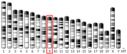
Location (UCSC) Chr 11: 94.13 – 94.18 Mb Chr 9: 14.91 – 14.96 Mb
PubMed search [3] [4]
Wikidata
View/Edit Human View/Edit Mouse

Pannexin 1 is a protein in humans that is encoded by the PANX1 gene. [5]

Contents

The protein encoded by this gene belongs to the innexin family. Innexin family members are the structural components of gap junctions. This protein and pannexin 2 are abundantly expressed in central nerve system (CNS) and are coexpressed in various neuronal populations. Studies in Xenopus oocytes suggest that this protein alone and in combination with pannexin 2 may form cell type-specific gap junctions with distinct properties. [5]

Clinical relevance

Truncating mutations in this gene have been shown to promote breast cancer metastasis to the lungs by allowing cancer cells to survive mechanical stretch in the microcirculation. [6]

Disruptions of this gene have been associated to melanoma tumor progression. [7]

Pannexin 1 is also an important component of membrane channels involved in the formation of thin plasma membrane extensions called apoptopodia and beaded apoptopodia during apoptosis. [8] [9]

Related Research Articles

<span class="mw-page-title-main">Gap junction</span> Cell-cell junction composed of innexins or connexins,

Gap junctions are one of four broad categories of intercellular connections that form between a multitude of animal cell types. First photographed around 1952, it wasn't until 1969 that gap junctions were referred to as "gap junctions". Named after the 2-4 nm gap they bridged between cell membranes, they had been characterised in more detail by 1967.

<span class="mw-page-title-main">Connexin</span> Group of proteins which form the intermembrane channels of gap junctions

Connexins (Cx), or gap junction proteins, are structurally related transmembrane proteins that assemble to form vertebrate gap junctions. An entirely different family of proteins, the innexins, form gap junctions in invertebrates. Each gap junction is composed of two hemichannels, or connexons, which consist of homo- or heterohexameric arrays of connexins, and the connexon in one plasma membrane docks end-to-end with a connexon in the membrane of a closely opposed cell. The hemichannel is made of six connexin subunits, each of which consist of four transmembrane segments. Gap junctions are essential for many physiological processes, such as the coordinated depolarization of cardiac muscle, proper embryonic development, and the conducted response in microvasculature. Connexins also have non-channel dependant functions relating to cytoskeleton and cell migration. For these reasons, mutations in connexin-encoding genes can lead to functional and developmental abnormalities.

<span class="mw-page-title-main">Pannexin</span>

Pannexins are a family of vertebrate proteins identified by their homology to the invertebrate innexins. While innexins are responsible for forming gap junctions in invertebrates, the pannexins have been shown to predominantly exist as large transmembrane channels connecting the intracellular and extracellular space, allowing the passage of ions and small molecules between these compartments.

Innexins are transmembrane proteins that form gap junctions in invertebrates. Gap junctions are composed of membrane proteins that form a channel permeable to ions and small molecules connecting the cytoplasm of adjacent cells. Although gap junctions provide similar functions in all multicellular organisms, it was not known what proteins invertebrates used for this purpose until the late 1990s. While the connexin family of gap junction proteins was well-characterized in vertebrates, no homologues were found in non-chordates.

<span class="mw-page-title-main">Bcl-2 homologous antagonist killer</span> Protein-coding gene in the species Homo sapiens

Bcl-2 homologous antagonist/killer is a protein that in humans is encoded by the BAK1 gene on chromosome 6. The protein encoded by this gene belongs to the BCL2 protein family. BCL2 family members form oligomers or heterodimers and act as anti- or pro-apoptotic regulators that are involved in a wide variety of cellular activities. This protein localizes to mitochondria, and functions to induce apoptosis. It interacts with and accelerates the opening of the mitochondrial voltage-dependent anion channel, which leads to a loss in membrane potential and the release of cytochrome c. This protein also interacts with the tumor suppressor P53 after exposure to cell stress.

<span class="mw-page-title-main">CDC42</span> Protein-coding gene in the species Homo sapiens

Cell division control protein 42 homolog is a protein that in humans is encoded by the CDC42 gene. Cdc42 is involved in regulation of the cell cycle. It was originally identified in S. cerevisiae (yeast) as a mediator of cell division, and is now known to influence a variety of signaling events and cellular processes in a variety of organisms from yeast to mammals.

<span class="mw-page-title-main">PRKCB1</span> Protein-coding gene in the species Homo sapiens

Protein kinase C beta type is an enzyme that in humans is encoded by the PRKCB gene.

<span class="mw-page-title-main">Tight junction protein 1</span> Protein found in humans

Zonula occludens-1 ZO-1, also known as Tight junction protein-1 is a 220-kD peripheral membrane protein that is encoded by the TJP1 gene in humans. It belongs to the family of zonula occludens proteins, which are tight junction-associated proteins and of which, ZO-1 is the first to be cloned. It was first isolated in 1986 by Stevenson and Goodenough using a monoclonal antibody raised in rodent liver to recognise a 225-kD polypeptide in whole liver homogenates and in tight junction-enriched membrane fractions. It has a role as a scaffold protein which cross-links and anchors Tight Junction (TJ) strand proteins, which are fibril-like structures within the lipid bilayer, to the actin cytoskeleton.

<span class="mw-page-title-main">CASK</span> Protein-coding gene in humans

Peripheral plasma membrane protein CASK is a protein that in humans is encoded by the CASK gene. This gene is also known by several other names: CMG 2, calcium/calmodulin-dependent serine protein kinase 3 and membrane-associated guanylate kinase 2. CASK gene mutations are the cause of XL-ID with or without nystagmus and MICPCH, an X-linked neurological disorder.

<span class="mw-page-title-main">T-cell lymphoma invasion and metastasis-inducing protein 1</span> Protein-coding gene in the species Homo sapiens

Rho guanine nucleotide exchange factor TIAM1 is a protein that in humans is encoded by the TIAM1 gene.

<span class="mw-page-title-main">BNIP3</span> Protein-coding gene in the species Homo sapiens

BCL2/adenovirus E1B 19 kDa protein-interacting protein 3 is a protein found in humans that is encoded by the BNIP3 gene.

<span class="mw-page-title-main">PAWR</span> Protein-coding gene in humans

PRKC apoptosis WT1 regulator protein, or Prostate apoptosis response-4, is a tumor-suppressor protein coded for in the human by the PAWR gene, that induces apoptosis in cancer cells, but not in normal cells.

<span class="mw-page-title-main">MAGI1</span> Protein-coding gene in the species Homo sapiens

Membrane-associated guanylate kinase, WW and PDZ domain-containing protein 1 is an enzyme that in humans is encoded by the MAGI1 gene.

<span class="mw-page-title-main">RAB3B</span> Protein-coding gene in the species Homo sapiens

Ras-related protein Rab-3B is a protein that in humans is encoded by the RAB3B gene.

<span class="mw-page-title-main">TAOK2</span> Protein-coding gene in the species Homo sapiens

Serine/threonine-protein kinase TAO2 is an enzyme that in humans is encoded by the TAOK2 gene.

<span class="mw-page-title-main">SEC22B</span> Protein-coding gene in the species Homo sapiens

Vesicle-trafficking protein SEC22b is a protein that in humans is encoded by the SEC22B gene.

<span class="mw-page-title-main">AIFM2</span> Protein-coding gene in the species Homo sapiens

Apoptosis-inducing factor 2 (AIFM2), also known as ferroptosis suppressor protein 1 (FSP1), apoptosis-inducing factor-homologous mitochondrion-associated inducer of death (AMID), is a protein that in humans is encoded by the AIFM2 gene, also known as p53-responsive gene 3 (PRG3), on chromosome 10.

<span class="mw-page-title-main">TXN2</span> Protein-coding gene in the species Homo sapiens

Thioredoxin, mitochondrial also known as thioredoxin-2 is a protein that in humans is encoded by the TXN2 gene on chromosome 22. This nuclear gene encodes a mitochondrial member of the thioredoxin family, a group of small multifunctional redox-active proteins. The encoded protein may play important roles in the regulation of the mitochondrial membrane potential and in protection against oxidant-induced apoptosis.

<span class="mw-page-title-main">VDAC3</span> Protein-coding gene in the species Homo sapiens

Voltage-dependent anion-selective channel protein 3 (VDAC3) is a protein that in humans is encoded by the VDAC3 gene on chromosome 8. The protein encoded by this gene is a voltage-dependent anion channel and shares high structural homology with the other VDAC isoforms. Nonetheless, VDAC3 demonstrates limited pore-forming ability and, instead, interacts with other proteins to perform its biological functions, including sperm flagella assembly and centriole assembly. Mutations in VDAC3 have been linked to male infertility, as well as Parkinson’s disease.

Vinnexin is a transmembrane protein whose DNA code is held in a virus genome. When the virus genome is expressed in a cell the vinnexin gene from the virus is made into a functioning protein by the infected cell. The vinnexin protein is then incorporated into the host's cell membranes to alter the way the hosts cells communicate with each other. The altered communication aids the transmission and replication of the virus in complex ways. The communication structure that the vinnexin is involved in is the gap junction and vinnexin forms part of a wider family of proteins that are innexin homologues referred to as pannexins. So far Vinnexins have only been found in Adenovirus and the way they affect the functioning of innexins is being studied in great detail.

References

  1. 1 2 3 GRCh38: Ensembl release 89: ENSG00000110218 - Ensembl, May 2017
  2. 1 2 3 GRCm38: Ensembl release 89: ENSMUSG00000031934 - Ensembl, May 2017
  3. "Human PubMed Reference:". National Center for Biotechnology Information, U.S. National Library of Medicine.
  4. "Mouse PubMed Reference:". National Center for Biotechnology Information, U.S. National Library of Medicine.
  5. 1 2 "Entrez Gene: Pannexin 1" . Retrieved 2012-04-11.
  6. Furlow PW, Zhang S, Soong TD, Halberg N, Goodarzi H, Mangrum C, Wu YG, Elemento O, Tavazoie SF (July 2015). "Mechanosensitive pannexin-1 channels mediate microvascular metastatic cell survival". Nature Cell Biology. 17 (7): 943–952. doi:10.1038/ncb3194. PMC   5310712 . PMID   26098574.
  7. Penuela S, Gyenis L, Ablack A, Churko JM, Berger AC, Litchfield DW, Lewis JD, Laird DW (Aug 2012). "Loss of pannexin 1 attenuates melanoma progression by reversion to a melanocytic phenotype". The Journal of Biological Chemistry. 287 (34): 29184–93. doi: 10.1074/jbc.M112.377176 . PMC   3436541 . PMID   22753409.
  8. Poon IK, Chiu YH, Armstrong AJ, Kinchen JM, Juncadella IJ, Bayliss DA, Ravichandran KS (2014). "Unexpected link between an antibiotic, pannexin channels and apoptosis". Nature. 507 (7492): 329–34. Bibcode:2014Natur.507..329P. doi:10.1038/nature13147. PMC   4078991 . PMID   24646995.
  9. Atkin-Smith GK, Tixeira R, Paone S, Mathivanan S, Collins C, Liem M, Goodall KJ, Ravichandran KS, Hulett MD, Poon IK (2015). "A novel mechanism of generating extracellular vesicles during apoptosis via a beads-on-a-string membrane structure". Nat Commun. 6: 7439. Bibcode:2015NatCo...6.7439A. doi:10.1038/ncomms8439. PMC   4490561 . PMID   26074490.

Further reading