Phase problem

Last updated

In physics, the phase problem is the problem of loss of information concerning the phase that can occur when making a physical measurement. The name comes from the field of X-ray crystallography, where the phase problem has to be solved for the determination of a structure from diffraction data. [1] The phase problem is also met in the fields of imaging and signal processing. [2] Various approaches of phase retrieval have been developed over the years.

Contents

Overview

Light detectors, such as photographic plates or CCDs, measure only the intensity of the light that hits them. This measurement is incomplete (even when neglecting other degrees of freedom such as polarization and angle of incidence) because a light wave has not only an amplitude (related to the intensity), but also a phase (related to the direction), and polarization which are systematically lost in a measurement. [2] In diffraction or microscopy experiments, the phase part of the wave often contains valuable information on the studied specimen. The phase problem constitutes a fundamental limitation ultimately related to the nature of measurement in quantum mechanics.

In X-ray crystallography, the diffraction data when properly assembled gives the amplitude of the 3D Fourier transform of the molecule's electron density in the unit cell. [1] If the phases are known, the electron density can be simply obtained by Fourier synthesis. This Fourier transform relation also holds for two-dimensional far-field diffraction patterns (also called Fraunhofer diffraction) giving rise to a similar type of phase problem.

Phase retrieval

There are several ways to retrieve the lost phases. The phase problem must be solved in x-ray crystallography, [1] neutron crystallography, [3] and electron crystallography. [4] [5] [6]

Not all of the methods of phase retrieval work with every wavelength (x-ray, neutron, and electron) used in crystallography.

Direct (ab initio) methods

If the crystal diffracts to high resolution (<1.2 Å), the initial phases can be estimated using direct methods. [1] Direct methods can be used in x-ray crystallography, [1] neutron crystallography, [7] and electron crystallography. [4] [5]

A number of initial phases are tested and selected by this method. The other is the Patterson method, which directly determines the positions of heavy atoms. The Patterson function gives a large value in a position which corresponds to interatomic vectors. This method can be applied only when the crystal contains heavy atoms or when a significant fraction of the structure is already known.

For molecules whose crystals provide reflections in the sub-Ångström range, it is possible to determine phases by brute force methods, testing a series of phase values until spherical structures are observed in the resultant electron density map. This works because atoms have a characteristic structure when viewed in the sub-Ångström range. The technique is limited by processing power and data quality. For practical purposes, it is limited to "small molecules" and peptides because they consistently provide high-quality diffraction with very few reflections.

Molecular replacement (MR)

Phases can also be inferred by using a process called molecular replacement, where a similar molecule's already-known phases are grafted onto the intensities of the molecule at hand, which are observationally determined. These phases can be obtained experimentally from a homologous molecule or if the phases are known for the same molecule but in a different crystal, by simulating the molecule's packing in the crystal and obtaining theoretical phases. Generally, these techniques are less desirable since they can severely bias the solution of the structure. They are useful, however, for ligand binding studies, or between molecules with small differences and relatively rigid structures (for example derivatizing a small molecule).

Isomorphous replacement

Multiple isomorphous replacement (MIR)

Multiple isomorphous replacement (MIR), where heavy atoms are inserted into structure (usually by synthesizing proteins with analogs or by soaking)

Anomalous scattering

Single-wavelength anomalous dispersion (SAD).

Multi-wavelength anomalous dispersion (MAD)

A powerful solution is the multi-wavelength anomalous dispersion (MAD) method. In this technique, atoms' inner electrons[ clarification needed ] absorb X-rays of particular wavelengths, and reemit the X-rays after a delay, inducing a phase shift in all of the reflections, known as the anomalous dispersion effect. Analysis of this phase shift (which may be different for individual reflections) results in a solution for the phases. Since X-ray fluorescence techniques (like this one) require excitation at very specific wavelengths, it is necessary to use synchrotron radiation when using the MAD method.

Phase improvement

Refining initial phases

In many cases, an initial set of phases are determined, and the electron density map for the diffraction pattern is calculated. Then the map is used to determine portions of the structure, which portions are used to simulate a new set of phases. This new set of phases is known as a refinement. These phases are reapplied to the original amplitudes, and an improved electron density map is derived, from which the structure is corrected. This process is repeated until an error term (usually ) has stabilized to a satisfactory value. Because of the phenomenon of phase bias, it is possible for an incorrect initial assignment to propagate through successive refinements, so satisfactory conditions for a structure assignment are still a matter of debate. Indeed, some spectacular incorrect assignments have been reported, including a protein where the entire sequence was threaded backwards. [8]

Density modification (phase improvement)

Solvent flattening

Histogram matching

Non-crystallographic symmetry averaging

Partial structure

Phase extension

See also

Related Research Articles

<span class="mw-page-title-main">Crystallography</span> Scientific study of crystal structures

Crystallography is the experimental science of determining the arrangement of atoms in crystalline solids. Crystallography is a fundamental subject in the fields of materials science and solid-state physics. The word crystallography is derived from the Ancient Greek word κρύσταλλος, and γράφειν. In July 2012, the United Nations recognised the importance of the science of crystallography by proclaiming that 2014 would be the International Year of Crystallography.

<span class="mw-page-title-main">X-ray crystallography</span> Technique used for determining crystal structures and identifying mineral compounds

X-ray crystallography is the experimental science determining the atomic and molecular structure of a crystal, in which the crystalline structure causes a beam of incident X-rays to diffract into many specific directions. By measuring the angles and intensities of these diffracted beams, a crystallographer can produce a three-dimensional picture of the density of electrons within the crystal. From this electron density, the positions of the atoms in the crystal can be determined, as well as their chemical bonds, crystallographic disorder, and various other information.

<span class="mw-page-title-main">Electron diffraction</span> Bending of electron beams due to electrostatic interactions with matter

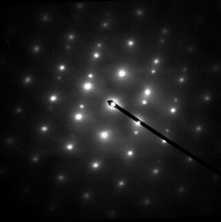
Electron diffraction is a generic term for phenomena associated with changes in the direction of electron beams due to elastic interactions with atoms. It occurs due to elastic scattering, when there is no change in the energy of the electrons. The negatively charged electrons are scattered due to Coulomb forces when they interact with both the positively charged atomic core and the negatively charged electrons around the atoms. The resulting map of the directions of the electrons far from the sample is called a diffraction pattern, see for instance Figure 1. Beyond patterns showing the directions of electrons, electron diffraction also plays a major role in the contrast of images in electron microscopes.

<span class="mw-page-title-main">Chemical structure</span> Organized way in which molecules are ordered and sorted

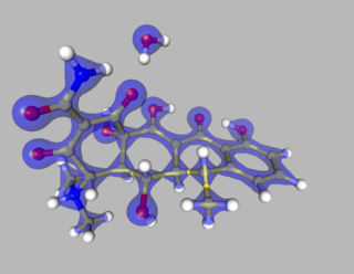
A chemical structure of a molecule is a spatial arrangement of its atoms and their chemical bonds. Its determination includes a chemist's specifying the molecular geometry and, when feasible and necessary, the electronic structure of the target molecule or other solid. Molecular geometry refers to the spatial arrangement of atoms in a molecule and the chemical bonds that hold the atoms together and can be represented using structural formulae and by molecular models; complete electronic structure descriptions include specifying the occupation of a molecule's molecular orbitals. Structure determination can be applied to a range of targets from very simple molecules to very complex ones.

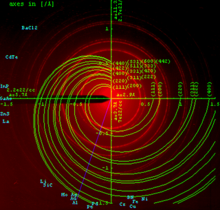
The Ewald sphere is a geometric construction used in electron, neutron, and x-ray diffraction which shows the relationship between:
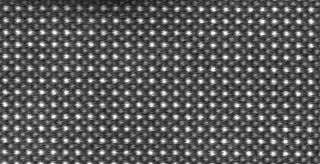
Electron crystallography is a method to determine the arrangement of atoms in solids using a transmission electron microscope (TEM). It can involve the use of high-resolution transmission electron microscopy images, electron diffraction patterns including convergent-beam electron diffraction or combinations of these. It has been successful in determining some bulk structures, and also surface structures. Two related methods are low-energy electron diffraction which has solved the structure of many surfaces, and reflection high-energy electron diffraction which is used to monitor surfaces often during growth.

<span class="mw-page-title-main">Powder diffraction</span> Experimental method in X-ray diffraction

Powder diffraction is a scientific technique using X-ray, neutron, or electron diffraction on powder or microcrystalline samples for structural characterization of materials. An instrument dedicated to performing such powder measurements is called a powder diffractometer.

In X-ray crystallography, a difference density map or Fo–Fc map shows the spatial distribution of the difference between the measured electron density of the crystal and the electron density explained by the current model.

Multi-wavelength anomalous diffraction is a technique used in X-ray crystallography that facilitates the determination of the three-dimensional structure of biological macromolecules via solution of the phase problem.

Diffraction topography is a imaging technique based on Bragg diffraction. Diffraction topographic images ("topographies") record the intensity profile of a beam of X-rays diffracted by a crystal. A topography thus represents a two-dimensional spatial intensity mapping of reflected X-rays, i.e. the spatial fine structure of a Laue reflection. This intensity mapping reflects the distribution of scattering power inside the crystal; topographs therefore reveal the irregularities in a non-ideal crystal lattice. X-ray diffraction topography is one variant of X-ray imaging, making use of diffraction contrast rather than absorption contrast which is usually used in radiography and computed tomography (CT). Topography is exploited to a lesser extends with neutrons, and has similarities to dark field imaging in the electron microscope community.

<span class="mw-page-title-main">Coherent diffraction imaging</span>

Coherent diffractive imaging (CDI) is a "lensless" technique for 2D or 3D reconstruction of the image of nanoscale structures such as nanotubes, nanocrystals, porous nanocrystalline layers, defects, potentially proteins, and more. In CDI, a highly coherent beam of X-rays, electrons or other wavelike particle or photon is incident on an object.

Isomorphous replacement (IR) is historically the most common approach to solving the phase problem in X-ray crystallography studies of proteins. For protein crystals this method is conducted by soaking the crystal of a sample to be analyzed with a heavy atom solution or co-crystallization with the heavy atom. The addition of the heavy atom (or ion) to the structure should not affect the crystal formation or unit cell dimensions in comparison to its native form, hence, they should be isomorphic.

Resolution in the context of structural biology is the ability to distinguish the presence or absence of atoms or groups of atoms in a biomolecular structure. Usually, the structure originates from methods such as X-ray crystallography, electron crystallography, or cryo-electron microscopy. The resolution is measured of the "map" of the structure produced from experiment, where an atomic model would then be fit into. Due to their different natures and interactions with matter, in X-ray methods the map produced is of the electron density of the system, whereas in electron methods the map is of the electrostatic potential of the system. In both cases, atomic positions are assumed similarly.

Single-wavelength anomalous diffraction (SAD) is a technique used in X-ray crystallography that facilitates the determination of the structure of proteins or other biological macromolecules by allowing the solution of the phase problem. In contrast to multi-wavelength anomalous diffraction (MAD), SAD uses a single dataset at a single appropriate wavelength.

A crystallographic database is a database specifically designed to store information about the structure of molecules and crystals. Crystals are solids having, in all three dimensions of space, a regularly repeating arrangement of atoms, ions, or molecules. They are characterized by symmetry, morphology, and directionally dependent physical properties. A crystal structure describes the arrangement of atoms, ions, or molecules in a crystal.

<span class="mw-page-title-main">Crystallographic image processing</span>

Crystallographic image processing (CIP) is traditionally understood as being a set of key steps in the determination of the atomic structure of crystalline matter from high-resolution electron microscopy (HREM) images obtained in a transmission electron microscope (TEM) that is run in the parallel illumination mode. The term was created in the research group of Sven Hovmöller at Stockholm University during the early 1980s and became rapidly a label for the "3D crystal structure from 2D transmission/projection images" approach. Since the late 1990s, analogous and complementary image processing techniques that are directed towards the achieving of goals with are either complementary or entirely beyond the scope of the original inception of CIP have been developed independently by members of the computational symmetry/geometry, scanning transmission electron microscopy, scanning probe microscopy communities, and applied crystallography communities.

<span class="mw-page-title-main">Multipole density formalism</span>

The Multipole Density Formalism is an X-ray crystallography method of electron density modelling proposed by Niels K. Hansen and Philip Coppens in 1978. Unlike the commonly used Independent Atom Model, the Hansen-Coppens Formalism presents an aspherical approach, allowing one to model the electron distribution around a nucleus separately in different directions and therefore describe numerous chemical features of a molecule inside the unit cell of an examined crystal in detail.

In crystallography, direct methods is a set of techniques used for structure determination using diffraction data and a priori information. It is a solution to the crystallographic phase problem, where phase information is lost during a diffraction measurement. Direct methods provides a method of estimating the phase information by establishing statistical relationships between the recorded amplitude information and phases of strong reflections.

Quantum crystallography is a branch of crystallography that investigates crystalline materials within the framework of quantum mechanics, with analysis and representation, in position or in momentum space, of quantities like wave function, electron charge and spin density, density matrices and all properties related to them. Like the quantum chemistry, Quantum crystallography involves both experimental and computational work. The theoretical part of quantum crystallography is based on quantum mechanical calculations of atomic/molecular/crystal wave functions, density matrices or density models, used to simulate the electronic structure of a crystalline material. While in quantum chemistry, the experimental works mainly rely on spectroscopy, in quantum crystallography the scattering techniques play the central role, although spectroscopy as well as atomic microscopy are also sources of information.

This is a timeline of crystallography.

References

  1. 1 2 3 4 5 Taylor, Garry (2003-11-01). "The phase problem". Acta Crystallographica Section D. 59 (11): 1881–1890. doi: 10.1107/S0907444903017815 . PMID   14573942.
  2. 1 2 Shechtman, Yoav; Eldar, Yonina C.; Cohen, Oren; Chapman, Henry N.; Miao, Jianwei; Segev, Mordechai (2014-02-28). "Phase Retrieval with Application to Optical Imaging". arXiv: 1402.7350 [cs.IT].
  3. Hauptman, Herbert A.; Langs, David A. (2003-05-01). "The phase problem in neutron crystallography". Acta Crystallographica Section A. 59 (3): 250–254. doi:10.1107/S010876730300521X. PMID   12714776.
  4. 1 2 Dorset, D. L. (1997-03-04). "Direct phase determination in protein electron crystallography: The pseudo-atom approximation". Proceedings of the National Academy of Sciences. 94 (5): 1791–1794. Bibcode:1997PNAS...94.1791D. doi: 10.1073/pnas.94.5.1791 . PMC   19995 . PMID   9050857.
  5. 1 2 Dorset, D. L. (1996-05-01). "Direct Phasing in Protein Electron Crystallography – Phase Extension and the Prospects for Ab Initio Determinations". Acta Crystallographica Section A. 52 (3): 480–489. doi: 10.1107/S0108767396001420 . PMID   8694993.
  6. Henderson, R.; Baldwin, J. M.; Downing, K. H.; Lepault, J.; Zemlin, F. (1986-01-01). "Structure of purple membrane from halobacterium halobium: recording, measurement and evaluation of electron micrographs at 3.5 Å resolution". Ultramicroscopy. 19 (2): 147–178. doi:10.1016/0304-3991(86)90203-2.
  7. Hauptman, H. (1976-09-01). "Probabilistic theory of the structure invariants: extension to the unequal atom case with application to neutron diffraction". Acta Crystallographica Section A. 32 (5): 877–882. Bibcode:1976AcCrA..32..877H. doi: 10.1107/S0567739476001757 .
  8. Kleywegt, Gerard J. (2000). "Validation of protein crystal structures". Acta Crystallographica Section D. 56 (3): 249–265. doi: 10.1107/S0907444999016364 . PMID   10713511.