
In anatomy, the atlas (C1) is the most superior (first) cervical vertebra of the spine and is located in the neck.

The suboccipital nerve is the dorsal primary ramus of the first cervical nerve (C1). It exits the spinal cord between the skull and the first cervical vertebra, the atlas.

In neuroanatomy, dura mater is a thick membrane made of dense irregular connective tissue that surrounds the brain and spinal cord. It is the outermost of the three layers of membrane called the meninges that protect the central nervous system. The other two meningeal layers are the arachnoid mater and the pia mater. It envelops the arachnoid mater, which is responsible for keeping in the cerebrospinal fluid. It is derived primarily from the neural crest cell population, with postnatal contributions of the paraxial mesoderm.

In tetrapods, cervical vertebrae are the vertebrae of the neck, immediately below the skull. Truncal vertebrae lie caudal of cervical vertebrae. In sauropsid species, the cervical vertebrae bear cervical ribs. In lizards and saurischian dinosaurs, the cervical ribs are large; in birds, they are small and completely fused to the vertebrae. The vertebral transverse processes of mammals are homologous to the cervical ribs of other amniotes. Most mammals have seven cervical vertebrae, with the only three known exceptions being the manatee with six, the two-toed sloth with five or six, and the three-toed sloth with nine.

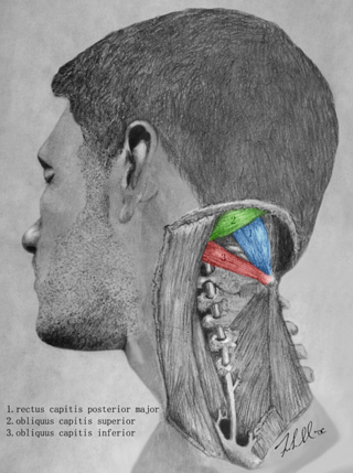
The obliquus capitis inferior muscle is a muscle in the upper back of the neck. It is one of the suboccipital muscles. Its inferior attachment is at the spinous process of the axis; its superior attachment is at the transverse process of the atlas. It is innervated by the suboccipital nerve. The muscle rotates the head to its side.

The obliquus capitis superior muscle is a small muscle in the upper back part of the neck. It is one of the suboccipital muscles. It attaches inferiorly at the transverse process of the atlas ; it attaches superiorly at the external surface of the occipital bone. The muscle is innervated by the suboccipital nerve.

The vertebral arteries are major arteries of the neck. Typically, the vertebral arteries originate from the subclavian arteries. Each vessel courses superiorly along each side of the neck, merging within the skull to form the single, midline basilar artery. As the supplying component of the vertebrobasilar vascular system, the vertebral arteries supply blood to the upper spinal cord, brainstem, cerebellum, and posterior part of brain.

The rectus capitis posterior major is a muscle in the upper back part of the neck. It is one of the suboccipital muscles. Its inferior attachment is at the spinous process of the axis ; its superior attachment is onto the outer surface of the occipital bone on and around the side part of the inferior nuchal line. The muscle is innervated by the suboccipital nerve. The muscle acts to extend the head and rorate the head to its side.

The rectus capitis posterior minor is a muscle in the upper back part of the neck. It is one of the suboccipital muscles. Its inferior attachment is at the posterior arch of atlas; its superior attachment is onto the occipital bone at and below the inferior nuchal line. The muscle is innervated by the suboccipital nerve. The muscle acts as a weak extensor of the head.

The occipital artery is a branch of the external carotid artery that provides arterial supply to the back of the scalp, sternocleidomastoid muscles, and deep muscles of the back and neck.

The inferior thyroid artery is an artery in the neck. It arises from the thyrocervical trunk and passes upward, in front of the vertebral artery and longus colli muscle. It then turns medially behind the carotid sheath and its contents, and also behind the sympathetic trunk, the middle cervical ganglion resting upon the vessel.

The ascending pharyngeal artery is an artery of the neck that supplies the pharynx.

The deep cervical vein is the vena comitans of the deep cervical artery. The vein is formed in the suboccipital region by the convergence of communicating branches of the occipital vein, veins draining the suboccipital muscles, and veins from the venous plexuses that surround cervical nerves. The vein and corresponding artery then pass in between the semispinalis capitis muscle and the semispinalis colli muscle. The vein passes anterior-ward in between the transverse process of the 7th cervical vertebra and the neck of the first rib to terminate in the vertebral vein.
The posterior meningeal artery is one of the meningeal branches of the ascending pharyngeal artery. It passes through the jugular foramen to enter the posterior cranial fossa. It is the largest vessel supplying the dura of the posterior cranial fossa.
The suboccipital triangle is a region of the neck bounded by the following three muscles of the suboccipital group of muscles:

In anatomy, the transverse ligament of the atlas is a broad, tough ligament which arches across the ring of the atlas posterior to the dens to keep the dens in contact with the atlas. It forms the transverse component of the cruciform ligament of atlas.

The tectorial membrane of atlanto-axial joint is a tough membrane/broad, strong band representing the superior-ward prolongation of the posterior longitudinal ligament.
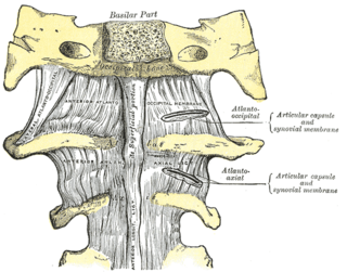
The anterior atlantooccipital membrane is a broad, dense membrane extending between the anterior margin of the foramen magnum (superiorly), and the anterior arch of atlas (inferiorly).

The posterior branches of cervical nerves branch from the dorsal rami of the cervical nerves.
The myodural bridge or miodural ligament is a bridge of connective tissue that extends between the suboccipital muscles and the cervical spinal dura mater, the outer membrane that envelops the spinal cord. It provides a physical connection between the musculoskeletal and nervous systems, and the circulation of cerebrospinal fluid. Its importance has been highlighted by various authors.

