Pregnancy Outcome Prediction study

Last updated
Logo of the POP study. POPS logo tiff.tif
Logo of the POP study.

The Pregnancy Outcome Prediction (POP) Study [1] is a prospective cohort study of 4,512 women who have never given birth, recruited at the Rosie Hospital (Cambridge, UK) between January 2008 and July 2012. [2] [3]

Contents

Staff and funding

The Principal Investigator is Gordon Smith [4] of the Department of Obstetrics and Gynaecology (University of Cambridge, UK). Lab work using the biobank of samples is led by Stephen Charnock-Jones [5] of the same department. The POP study is supported by the Women’s Health and Paediatrics [6] theme of the National Institute for Health Research (NIHR) Cambridge Biomedical Research Centre (BRC), [7] which is based at the Cambridge Biomedical Campus (UK). Other funding sources for the projects related to the POP study are the Medical Research Council (UK), the NIHR, the Wellcome Trust, the British Heart Foundation, and the Stillbirth and Neonatal Death Society (now known as Sands), as well as Industry supported studies.

Background

In 2007, Cambridge (UK) received funding from NIHR to establish a comprehensive Biomedical Research Centre (BRC). The Cambridge BRC Women’s Health and Paediatrics theme focuses on tackling the origins, treatment and prevention of many of the health issues that affect women during their reproductive years. The bulk of the Cambridge BRC funding for the first five years of the theme supported the POP study.

Over recent years, we have witnessed a great improvement in population-based screening methods for fetal abnormalities as a result of technological developments in ultrasound, biochemical screening and molecular genetics. However, the methods for screening the low risk population for other complications of pregnancy such as fetal growth restriction (FGR), pre-eclampsia and stillbirth have remained largely unchanged for the past 20-30 years. The UK National Institute for Clinical Excellence (NICE) has highlighted a number of research priorities in their guidelines for pregnancy care. These include a focus on improving the screening of apparently healthy pregnant women and identify those at high risk of complications. This area is the main focus of the POP study.

Wiki-POPS protocol.tif

Design and aims

Nulliparous women with a viable singleton pregnancy were recruited to the POP study when attending their dating ultrasound scan at The Rosie Hospital (Cambridge, UK). The only clinical exclusion criterion was multiple pregnancy. Participants had serial ultrasound scans and blood obtained at recruitment, 20, 28 and 36 weeks of gestation. DNA samples and height/weight measurements of the partners were also collected. After delivery, biopsies of the placenta, placental membranes, umbilical cord, and a sample of cord blood were collected. [8] Thorough design of the POP study and careful sample collection allowed the creation of an extensive, optimally phenotyped biobank of complicated and healthy pregnancies, including approximately 230,000 blood and tissue samples stored in -80°C freezers, 24,000 formalin fixed placental biopsies, and 4,000 paternal DNA samples.

The types of samples collected during the POP study reflect, first of all, the focus on the role of the placenta and its altered development and function in several major complications of pregnancy. Second, they reflect the interest in circulating biomarkers since, despite years of intensive research, screening for these conditions is still largely based upon clinical grounds rather than ultrasonic and/or biochemical assessment of placental function. Therefore the projects based on the samples from the POP study have the following aims:

  1. understanding the mechanisms leading to placentally-related complications, such as pre-eclampsia, FGR and stillbirth;
  2. studying the association between pregnancy complications and the presence of infectious agents in the placenta;
  3. evaluating the performance of known biomarkers and serial ultrasonography in assessing maternal and fetal well-being;
  4. discovering novel, placentally-derived circulating biomarkers which reflect the underlying pathology and are highly sensitive and specific in predicting the associated disease.
Wiki-POPS samples.tif

Key findings

Multiple projects based on this data have been published and many are currently on-going[ as of? ]. The key findings obtained so far studying the samples from the POP study are (see paragraph “Research papers” for a complete list of publications):

To externally validate the POP study findings, some of the above projects also involved collaboration with investigators from the Born in Bradford study. [17] [20] Moreover, additional collaborations included the Cambridge University School of Biological Sciences (in particular, the Centre for Trophoblast Research, [22] a major international centre for placental research including >20 research groups), the Cancer Research UK (CRUK) Cambridge Institut and the Wellcome Sanger Institute. These studies also benefited from collaborations with industrial partners such as GE Healthcare and Roche.

Follow-up work

Logo of the POPS2. POPS2 logo tiff.tif
Logo of the POPS2.

POPS2

The Pregnancy Outcome Prediction study 2 (POPS2) is a prospective cohort study funded by the Wellcome Trust. Recruitment to the study started in January 2020. The first POP study generated a simple screening test which is strongly predictive for pre-eclampsia and FGR at term. The test combines maternal risk factors, ultrasound measurements and levels of placentally-derived proteins in maternal serum (sFlt-1:PlGF ratio). The goal of POPS2 is to collect similar data and samples from approximately 4,500 unselected nulliparous women to address the following aims:

  1. to assess whether the test generated by the POP study works in an independent cohort;
  2. to nest a randomised controlled trial (RCT) of screening and intervention within the cohort in order to evaluate whether pregnancy outcome can be improved by inducing labour early (from 37 weeks) in women who screen positive;
  3. to collect more blood samples in order to develop and validate new predictive tests to detect a bigger proportion of pregnancy complications and to identify them earlier in pregnancy.
Logo of the POPStar study. Popstar logo including text cropped black.tif
Logo of the POPStar study.

POPStar

Pregnancy Outcome Prediction Study: transgenerational and adults review (POPStar) is a follow-up study of the original POP study, conducted by the Department of Obstetrics and Gynaecology in collaboration with the Department of Paediatrics (both University of Cambridge). The study is funded by the National Institute for Health Research (NIHR) Cambridge Biomedical Research Centre (BRC) and the children’s charity, Action Medical Research.

POPStar looks at the long-term health and development of the children and the mothers of the POP study cohort. POPStar started in June 2020, when the children were between 8 and 12 year old. The study is based on the link between routinely-collected data (e.g. from the UK National Health Service and the Department for Education) and the results from POP study in order to determine which pregnancy parameters could predict a high risk of health or developmental problems. Using the existing and follow-up data collected during the POP and POPStar studies, the research team aims to make connections between conditions during pregnancy and current maternal and children health.

Related Research Articles

<span class="mw-page-title-main">Intrauterine growth restriction</span> Medical condition

Intrauterine growth restriction (IUGR), or fetal growth restriction, refers to poor growth of a fetus while in the womb during pregnancy. IUGR is defined by clinical features of malnutrition and evidence of reduced growth regardless of an infant's birth weight percentile. The causes of IUGR are broad and may involve maternal, fetal, or placental complications.

<span class="mw-page-title-main">Eclampsia</span> Pre-eclampsia characterized by the presence of seizures

Eclampsia is the onset of seizures (convulsions) in a woman with pre-eclampsia. Pre-eclampsia is a hypertensive disorder of pregnancy that presents with three main features: new onset of high blood pressure, large amounts of protein in the urine or other organ dysfunction, and edema. If left untreated, pre-eclampsia can result in long-term consequences for the mother, namely increased risk of cardiovascular diseases and associated complications. In more severe cases, it may be fatal for both the mother and the fetus. The diagnostic criteria for pre-eclampsia is high blood pressure occurring after 20 weeks gestation or during the second half of pregnancy. Most often it occurs during the 3rd trimester of pregnancy and may occur before, during, or after delivery. The seizures are of the tonic–clonic type and typically last about a minute. Following the seizure, there is either a period of confusion or coma. Other complications include aspiration pneumonia, cerebral hemorrhage, kidney failure, pulmonary edema, HELLP syndrome, coagulopathy, placental abruption and cardiac arrest.

<span class="mw-page-title-main">Pre-eclampsia</span> Hypertension occurring during pregnancy

Pre-eclampsia is a multi-system disorder specific to pregnancy, characterized by the onset of high blood pressure and often a significant amount of protein in the urine. When it arises, the condition begins after 20 weeks of pregnancy. In severe cases of the disease there may be red blood cell breakdown, a low blood platelet count, impaired liver function, kidney dysfunction, swelling, shortness of breath due to fluid in the lungs, or visual disturbances. Pre-eclampsia increases the risk of undesirable as well as lethal outcomes for both the mother and the fetus including preterm labor. If left untreated, it may result in seizures at which point it is known as eclampsia.

<span class="mw-page-title-main">Prenatal testing</span> Testing for diseases or conditions in a fetus

Prenatal testing is a tool that can be used to detect some of these abnormalities at various stages prior to birth. Prenatal testing consists of prenatal screening and prenatal diagnosis, which are aspects of prenatal care that focus on detecting problems with the pregnancy as early as possible. These may be anatomic and physiologic problems with the health of the zygote, embryo, or fetus, either before gestation even starts or as early in gestation as practicable. Screening can detect problems such as neural tube defects, chromosome abnormalities, and gene mutations that would lead to genetic disorders and birth defects, such as spina bifida, cleft palate, Down syndrome, Tay–Sachs disease, sickle cell anemia, thalassemia, cystic fibrosis, muscular dystrophy, and fragile X syndrome. Some tests are designed to discover problems which primarily affect the health of the mother, such as PAPP-A to detect pre-eclampsia or glucose tolerance tests to diagnose gestational diabetes. Screening can also detect anatomical defects such as hydrocephalus, anencephaly, heart defects, and amniotic band syndrome.

<span class="mw-page-title-main">Placenta praevia</span> Medical condition

Placenta praevia is when the placenta attaches inside the uterus but in a position near or over the cervical opening. Symptoms include vaginal bleeding in the second half of pregnancy. The bleeding is bright red and tends not to be associated with pain. Complications may include placenta accreta, dangerously low blood pressure, or bleeding after delivery. Complications for the baby may include fetal growth restriction.

<span class="mw-page-title-main">Gestational hypertension</span> Medical condition

Gestational hypertension or pregnancy-induced hypertension (PIH) is the development of new hypertension in a pregnant woman after 20 weeks' gestation without the presence of protein in the urine or other signs of pre-eclampsia. Gestational hypertension is defined as having a blood pressure greater than 140/90 on two occasions at least 6 hours apart.

<span class="mw-page-title-main">Placental abruption</span> Medical condition

Placental abruption is when the placenta separates early from the uterus, in other words separates before childbirth. It occurs most commonly around 25 weeks of pregnancy. Symptoms may include vaginal bleeding, lower abdominal pain, and dangerously low blood pressure. Complications for the mother can include disseminated intravascular coagulopathy and kidney failure. Complications for the baby can include fetal distress, low birthweight, preterm delivery, and stillbirth.

<span class="mw-page-title-main">Complications of pregnancy</span> Medical condition

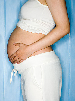
Complications of pregnancy are health problems that are related to, or arise during pregnancy. Complications that occur primarily during childbirth are termed obstetric labor complications, and problems that occur primarily after childbirth are termed puerperal disorders. While some complications improve or are fully resolved after pregnancy, some may lead to lasting effects, morbidity, or in the most severe cases, maternal or fetal mortality.

<span class="mw-page-title-main">Abdominal pregnancy</span> Medical condition

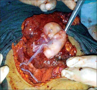
An abdominal pregnancy is a rare type of ectopic pregnancy where the embryo or fetus is growing and developing outside the womb in the abdomen, but not in the Fallopian tube, ovary or broad ligament.

<span class="mw-page-title-main">Intrauterine hypoxia</span> Medical condition when the fetus is deprived of sufficient oxygen

Intrauterine hypoxia occurs when the fetus is deprived of an adequate supply of oxygen. It may be due to a variety of reasons such as prolapse or occlusion of the umbilical cord, placental infarction, maternal diabetes and maternal smoking. Intrauterine growth restriction may cause or be the result of hypoxia. Intrauterine hypoxia can cause cellular damage that occurs within the central nervous system. This results in an increased mortality rate, including an increased risk of sudden infant death syndrome (SIDS). Oxygen deprivation in the fetus and neonate have been implicated as either a primary or as a contributing risk factor in numerous neurological and neuropsychiatric disorders such as epilepsy, attention deficit hyperactivity disorder, eating disorders and cerebral palsy.

Placental insufficiency or utero-placental insufficiency is the failure of the placenta to deliver sufficient nutrients to the fetus during pregnancy, and is often a result of insufficient blood flow to the placenta. The term is also sometimes used to designate late decelerations of fetal heart rate as measured by cardiotocography or an NST, even if there is no other evidence of reduced blood flow to the placenta, normal uterine blood flow rate being 600mL/min.

<span class="mw-page-title-main">Velamentous cord insertion</span> Velamentous placenta

Velamentous cord insertion is a complication of pregnancy where the umbilical cord is inserted in the fetal membranes. It is a major cause of antepartum hemorrhage that leads to loss of fetal blood and associated with high perinatal mortality. In normal pregnancies, the umbilical cord inserts into the middle of the placental mass and is completely encased by the amniotic sac. The vessels are hence normally protected by Wharton's jelly, which prevents rupture during pregnancy and labor. In velamentous cord insertion, the vessels of the umbilical cord are improperly inserted in the chorioamniotic membrane, and hence the vessels traverse between the amnion and the chorion towards the placenta. Without Wharton's jelly protecting the vessels, the exposed vessels are susceptible to compression and rupture.

<span class="mw-page-title-main">Circumvallate placenta</span> Medical condition

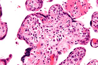
Circumvallate placenta is a rare condition affecting about 1-2% of pregnancies, in which the amnion and chorion fetal membranes essentially "double back" on the fetal side around the edges of the placenta. After delivery, a circumvallate placenta has a thick ring of membranes on its fetal surface. Circumvallate placenta is a placental morphological abnormality associated with increased fetal morbidity and mortality due to the restricted availability of nutrients and oxygen to the developing fetus.

<span class="mw-page-title-main">Prenatal nutrition</span>

Prenatal nutrition addresses nutrient recommendations before and during pregnancy. Nutrition and weight management before and during pregnancy has a profound effect on the development of infants. This is a rather critical time for healthy development since infants rely heavily on maternal stores and nutrient for optimal growth and health outcome later in life.

Thyroid disease in pregnancy can affect the health of the mother as well as the child before and after delivery. Thyroid disorders are prevalent in women of child-bearing age and for this reason commonly present as a pre-existing disease in pregnancy, or after childbirth. Uncorrected thyroid dysfunction in pregnancy has adverse effects on fetal and maternal well-being. The deleterious effects of thyroid dysfunction can also extend beyond pregnancy and delivery to affect neurointellectual development in the early life of the child. Due to an increase in thyroxine binding globulin, an increase in placental type 3 deioidinase and the placental transfer of maternal thyroxine to the fetus, the demand for thyroid hormones is increased during pregnancy. The necessary increase in thyroid hormone production is facilitated by high human chorionic gonadotropin (hCG) concentrations, which bind the TSH receptor and stimulate the maternal thyroid to increase maternal thyroid hormone concentrations by roughly 50%. If the necessary increase in thyroid function cannot be met, this may cause a previously unnoticed (mild) thyroid disorder to worsen and become evident as gestational thyroid disease. Currently, there is not enough evidence to suggest that screening for thyroid dysfunction is beneficial, especially since treatment thyroid hormone supplementation may come with a risk of overtreatment. After women give birth, about 5% develop postpartum thyroiditis which can occur up to nine months afterwards. This is characterized by a short period of hyperthyroidism followed by a period of hypothyroidism; 20–40% remain permanently hypothyroid.

<span class="mw-page-title-main">High-risk pregnancy</span> Medical condition

A high-risk pregnancy is one where the mother or the fetus has an increased risk of adverse outcomes compared to uncomplicated pregnancies. No concrete guidelines currently exist for distinguishing “high-risk” pregnancies from “low-risk” pregnancies; however, there are certain studied conditions that have been shown to put the mother or fetus at a higher risk of poor outcomes. These conditions can be classified into three main categories: health problems in the mother that occur before she becomes pregnant, health problems in the mother that occur during pregnancy, and certain health conditions with the fetus.

A pre-existing disease in pregnancy is a disease that is not directly caused by the pregnancy, in contrast to various complications of pregnancy, but which may become worse or be a potential risk to the pregnancy. A major component of this risk can result from necessary use of drugs in pregnancy to manage the disease.

The anomaly scan, also sometimes called the anatomy scan, 20-week ultrasound, or level 2 ultrasound, evaluates anatomic structures of the fetus, placenta, and maternal pelvic organs. This scan is an important and common component of routine prenatal care. The function of the ultrasound is to measure the fetus so that growth abnormalities can be recognized quickly later in pregnancy, to assess for congenital malformations and multiple pregnancies, and to plan method of delivery.

<span class="mw-page-title-main">Lesley McCowan</span> New Zealand medical researcher

Lesley Margaret Elizabeth McCowan is a New Zealand medical researcher and academic specialising in maternal health. She is currently a full professor and head of obstetrics and gynaecology at the University of Auckland.

Fetal programming, also known as prenatal programming, is the theory that environmental cues experienced during fetal development play a seminal role in determining health trajectories across the lifespan.

References

  1. "The Pregnancy Outcome Prediction (POP) Study".
  2. Pasupathy D, Dacey A, Cook E, Charnock-Jones DS, White IR, Smith GC (November 2008). "Study protocol. A prospective cohort study of unselected primiparous women: the pregnancy outcome prediction study". BMC Pregnancy and Childbirth. 8 (1): 51. doi: 10.1186/1471-2393-8-51 . PMC   2611961 . PMID   19019223.
  3. Gaccioli F, Lager S, Sovio U, Charnock-Jones DS, Smith GC (2017-11-01). "The pregnancy outcome prediction (POP) study: Investigating the relationship between serial prenatal ultrasonography, biomarkers, placental phenotype and adverse pregnancy outcomes". Placenta. 59: S17–S25. doi: 10.1016/j.placenta.2016.10.011 .
  4. "Professor Gordon Smith".
  5. "Professor Stephen Charnock-Jones".
  6. "Women's Health and Paediatrics".
  7. "National Institute for Health Research (NIHR) Cambridge Biomedical Research Centre (BRC)".
  8. "Visual placental sampling demonstration". YouTube .
  9. Sovio U, White IR, Dacey A, Pasupathy D, Smith GC (November 2015). "Screening for fetal growth restriction with universal third trimester ultrasonography in nulliparous women in the Pregnancy Outcome Prediction (POP) study: a prospective cohort study". Lancet. 386 (10008): 2089–2097. doi:10.1016/S0140-6736(15)00131-2. PMC   4655320 . PMID   26360240.
  10. Cleaton MA, Dent CL, Howard M, Corish JA, Gutteridge I, Sovio U, et al. (December 2016). "Fetus-derived DLK1 is required for maternal metabolic adaptations to pregnancy and is associated with fetal growth restriction". Nature Genetics. 48 (12): 1473–1480. doi:10.1038/ng.3699. PMC   5373434 . PMID   27776119.
  11. Sovio U, Murphy HR, Smith GC (June 2016). "Accelerated Fetal Growth Prior to Diagnosis of Gestational Diabetes Mellitus: A Prospective Cohort Study of Nulliparous Women". Diabetes Care. 39 (6): 982–7. doi: 10.2337/dc16-0160 . PMID   27208333.
  12. Sovio U, Smith GC (July 2018). "Blinded ultrasound fetal biometry at 36 weeks and risk of emergency Cesarean delivery in a prospective cohort study of low-risk nulliparous women". Ultrasound in Obstetrics & Gynecology. 52 (1): 78–86. doi: 10.1002/uog.17513 . PMID   28452133.
  13. Sovio U, Moraitis AA, Wong HS, Smith GC (June 2018). "Universal vs selective ultrasonography to screen for large-for-gestational-age infants and associated morbidity". Ultrasound in Obstetrics & Gynecology. 51 (6): 783–791. doi: 10.1002/uog.17491 . PMID   28425156.
  14. Sovio U, Gaccioli F, Cook E, Hund M, Charnock-Jones DS, Smith GC (April 2017). "Prediction of Preeclampsia Using the Soluble fms-Like Tyrosine Kinase 1 to Placental Growth Factor Ratio: A Prospective Cohort Study of Unselected Nulliparous Women". Hypertension. 69 (4): 731–738. doi:10.1161/HYPERTENSIONAHA.116.08620. PMC   5344177 . PMID   28167687.
  15. Gaccioli F, Sovio U, Cook E, Hund M, Charnock-Jones DS, Smith GC (August 2018). "Screening for fetal growth restriction using ultrasound and the sFLT1/PlGF ratio in nulliparous women: a prospective cohort study". The Lancet. Child & Adolescent Health. 2 (8): 569–581. doi:10.1016/S2352-4642(18)30129-9. PMC   6473551 . PMID   30119716.
  16. Gong S, Sovio U, Aye IL, Gaccioli F, Dopierala J, Johnson MD, et al. (July 2018). "Placental polyamine metabolism differs by fetal sex, fetal growth restriction, and preeclampsia". JCI Insight. 3 (13). doi:10.1172/jci.insight.120723. PMC   6124516 . PMID   29997303.
  17. 1 2 Sovio U, McBride N, Wood AM, Masconi KL, Cook E, Gaccioli F, et al. (February 2020). "4-Hydroxyglutamate is a novel predictor of pre-eclampsia". International Journal of Epidemiology. 49 (1): 301–311. doi:10.1093/ije/dyz098. PMC   7124498 . PMID   31098639.
  18. Wastlund D, Moraitis AA, Dacey A, Sovio U, Wilson EC, Smith GC (April 2019). "Screening for breech presentation using universal late-pregnancy ultrasonography: A prospective cohort study and cost effectiveness analysis". PLOS Medicine. 16 (4): e1002778. doi: 10.1371/journal.pmed.1002778 . PMC   6467368 . PMID   30990808.
  19. de Goffau MC, Lager S, Sovio U, Gaccioli F, Cook E, Peacock SJ, et al. (August 2019). "Human placenta has no microbiome but can contain potential pathogens". Nature. 572 (7769): 329–334. Bibcode:2019Natur.572..329D. doi:10.1038/s41586-019-1451-5. PMC   6697540 . PMID   31367035.
  20. 1 2 Sovio U, Goulding N, McBride N, Cook E, Gaccioli F, Charnock-Jones DS, et al. (March 2020). "A maternal serum metabolite ratio predicts fetal growth restriction at term" (PDF). Nature Medicine. 26 (3): 348–353. doi:10.1038/s41591-020-0804-9. hdl: 1983/7dd6b8f2-7ef5-4d7f-b71d-df9f9e32067e . PMID   32161413. S2CID   212668904.
  21. Gaccioli F, Lager S, de Goffau MC, Sovio U, Dopierala J, Gong S, et al. (May 2020). "Fetal inheritance of chromosomally integrated human herpesvirus 6 predisposes the mother to pre-eclampsia". Nature Microbiology. 5 (7): 901–908. doi:10.1038/s41564-020-0711-3. PMC   7610361 . PMID   32367053.
  22. "Centre for Trophoblast Research".